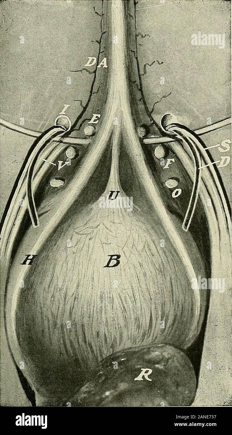
A text-book of clinical anatomy : for students and practitioners . Fig. 78.—Location of various forms of abdominal hernia; (diagrammatic). U,Umbilical hernia. D, Direct inguinal hernia. B, Indirect incomplete inguinal hernia.O, Complete or scrotal inguinal hernia. F, Femoral hernia. 241. - *.-*¥& Fig. 79.—View of inner aspect of anterior wall of abdomen to show internal orifices ofinguinal, femoral,, and obturator hernias. DA, Deep epigastric artery. E, Middle in-guinal fossa, corresponding externally to external abdominal ring. A direct inguinalhernia passes directly outward through this de

Image details
Contributor:
The Reading Room / Alamy Stock PhotoImage ID:
2ANE737File size:
7.1 MB (702.1 KB Compressed download)Releases:
Model - no | Property - noDo I need a release?Dimensions:
1196 x 2089 px | 20.3 x 35.4 cm | 8 x 13.9 inches | 150dpiMore information:
This image is a public domain image, which means either that copyright has expired in the image or the copyright holder has waived their copyright. Alamy charges you a fee for access to the high resolution copy of the image.
This image could have imperfections as it’s either historical or reportage.
A text-book of clinical anatomy : for students and practitioners . Fig. 78.—Location of various forms of abdominal hernia; (diagrammatic). U, Umbilical hernia. D, Direct inguinal hernia. B, Indirect incomplete inguinal hernia.O, Complete or scrotal inguinal hernia. F, Femoral hernia. 241. _ - *.-*¥& Fig. 79.—View of inner aspect of anterior wall of abdomen to show internal orifices ofinguinal, femoral, , and obturator hernias. DA, Deep epigastric artery. E, Middle in-guinal fossa, corresponding externally to external abdominal ring. A direct inguinalhernia passes directly outward through this depression without traversing the inguinalcanal from I to E. This fossa is situated at the lower portion of Hesselbachs triangle.This triangle is formed by the deep epigastric artery on the outer side, Pouparts ligamentbelow, and the remains of the hypogastric artery (H) and fold of peritoneum covering iton the inner side. Between H and U the internal inguinal fossa is to be seen. U, Re-mains of urachus, passing from anterior surface of bladder to umbilicus. I, Internalabdominal ring. Across its lower edge on either side is to be seen the spermatic arteryand vein (S), and the vas deferens (D), passing into the inguinal canal, all lying extraperi-toneally. V, External iliac a