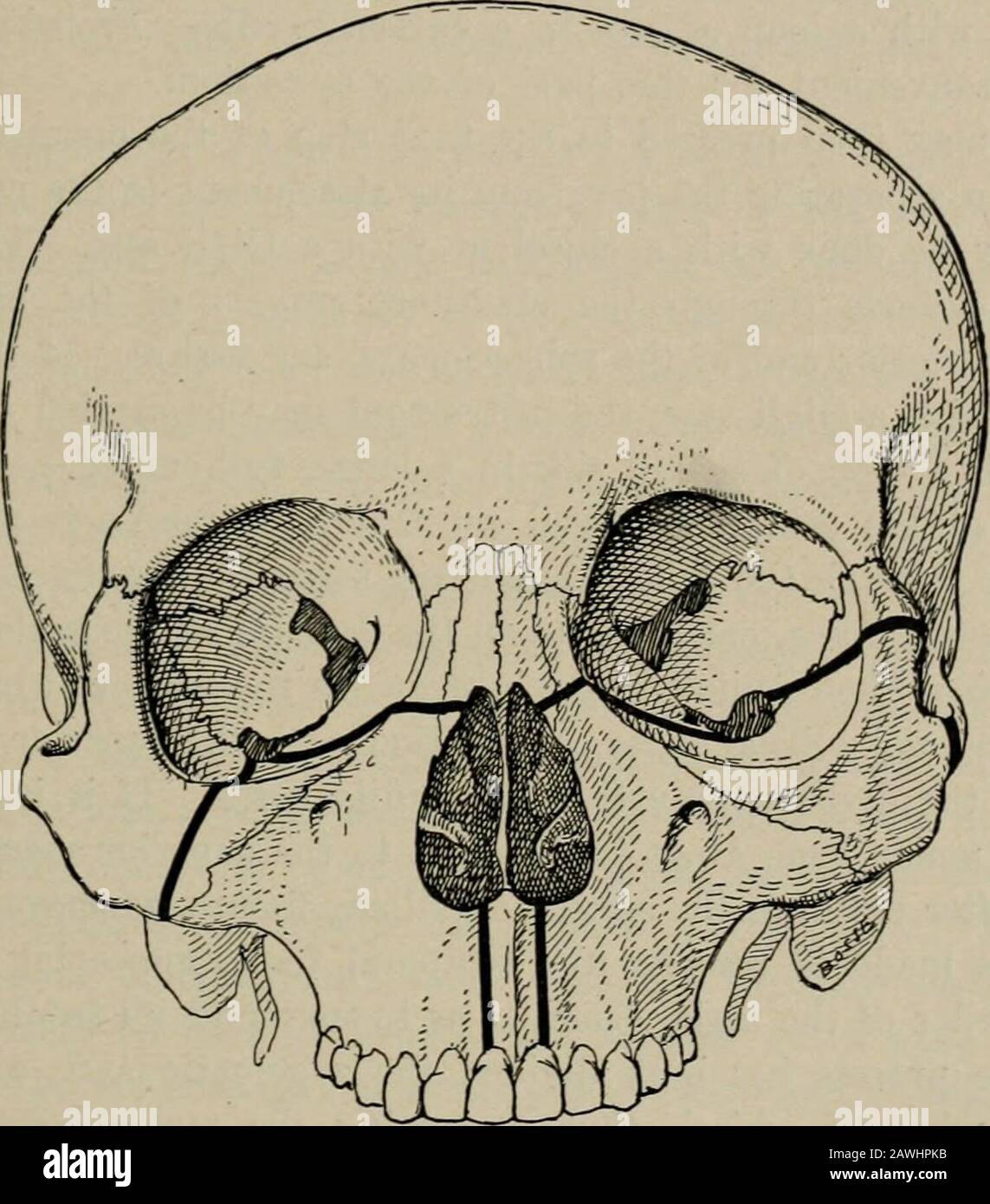
Operative surgery, for students and practitioners . Fig. 75.—Resection of Upper Jaw. L, Langenbeck incision; V, Velpeauincision; W, Weber incision. the lower border of the lobe of the ear, and is then carried upwardto a point over the prominence of the cheek-bone. This incisiondoes not divide the lip, but it will be necessary later to separatethe lip from its attachment to the jaw-bone. It divides some branchesof the facial nerve, which is a disadvantage. The front surface ofthe bone is exposed by reflecting the flap upward, subperiosteally, if OPERATIONS UPON THE FACE. 145 the conditions perm

Image details
Contributor:
The Reading Room / Alamy Stock PhotoImage ID:
2AWHPKBFile size:
7.2 MB (306.1 KB Compressed download)Releases:
Model - no | Property - noDo I need a release?Dimensions:
1483 x 1686 px | 25.1 x 28.5 cm | 9.9 x 11.2 inches | 150dpiMore information:
This image is a public domain image, which means either that copyright has expired in the image or the copyright holder has waived their copyright. Alamy charges you a fee for access to the high resolution copy of the image.
This image could have imperfections as it’s either historical or reportage.
Operative surgery, for students and practitioners . Fig. 75.—Resection of Upper Jaw. L, Langenbeck incision; V, Velpeauincision; W, Weber incision. the lower border of the lobe of the ear, and is then carried upwardto a point over the prominence of the cheek-bone. This incisiondoes not divide the lip, but it will be necessary later to separatethe lip from its attachment to the jaw-bone. It divides some branchesof the facial nerve, which is a disadvantage. The front surface ofthe bone is exposed by reflecting the flap upward, subperiosteally, if OPERATIONS UPON THE FACE. 145 the conditions permit. In raisino- the flap from the bone the infra-orbital vessels and nerve are divided. In making either of these incisions the facial artery is dividedand must be clamped and ligated. After the soft parts have been detached from the bone the carti-lage of the nose is separated from the nasal notch, and the soft parts.. Fig. 76.—Resection of Upper Jaw. When it is desired to leave the majorpart of the malar bone, the line of section through the bone should be asIndicated upon the right side of the skull. If the malar bone is to be removedtogether with the superior maxillary, the section through the bone should be asis represented upon the left side of the skull, the line of division passing throughthe frontal process of the malar and the zygoma. corresponding to the lower margin of the orbit, raised from the bone, and the tarso-orbital fascia cut along the margin of the orbit. Thefloor of the orbit being thus exposed, the contents of the orbit areraised out of the way with a blunt retractor. We are then ready tocut through the nasal process of the superior maxillary. This di-vision extends from the margin of the nasal notch, across the nasalprocess, as far as the lacrymal groove or fossa. It is necessary to 10 146 HEAD AND FACE. avoid injury to the lacrymal sac^ the upper expanded part of thelacrjona