Quick filters:
Curvularia geniculata Stock Photos and Images
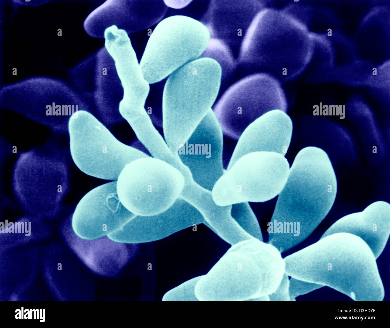 CURVULARIA GENICULATA Stock Photohttps://www.alamy.com/image-license-details/?v=1https://www.alamy.com/stock-photo-curvularia-geniculata-53859219.html
RMD3HDYF–CURVULARIA GENICULATA
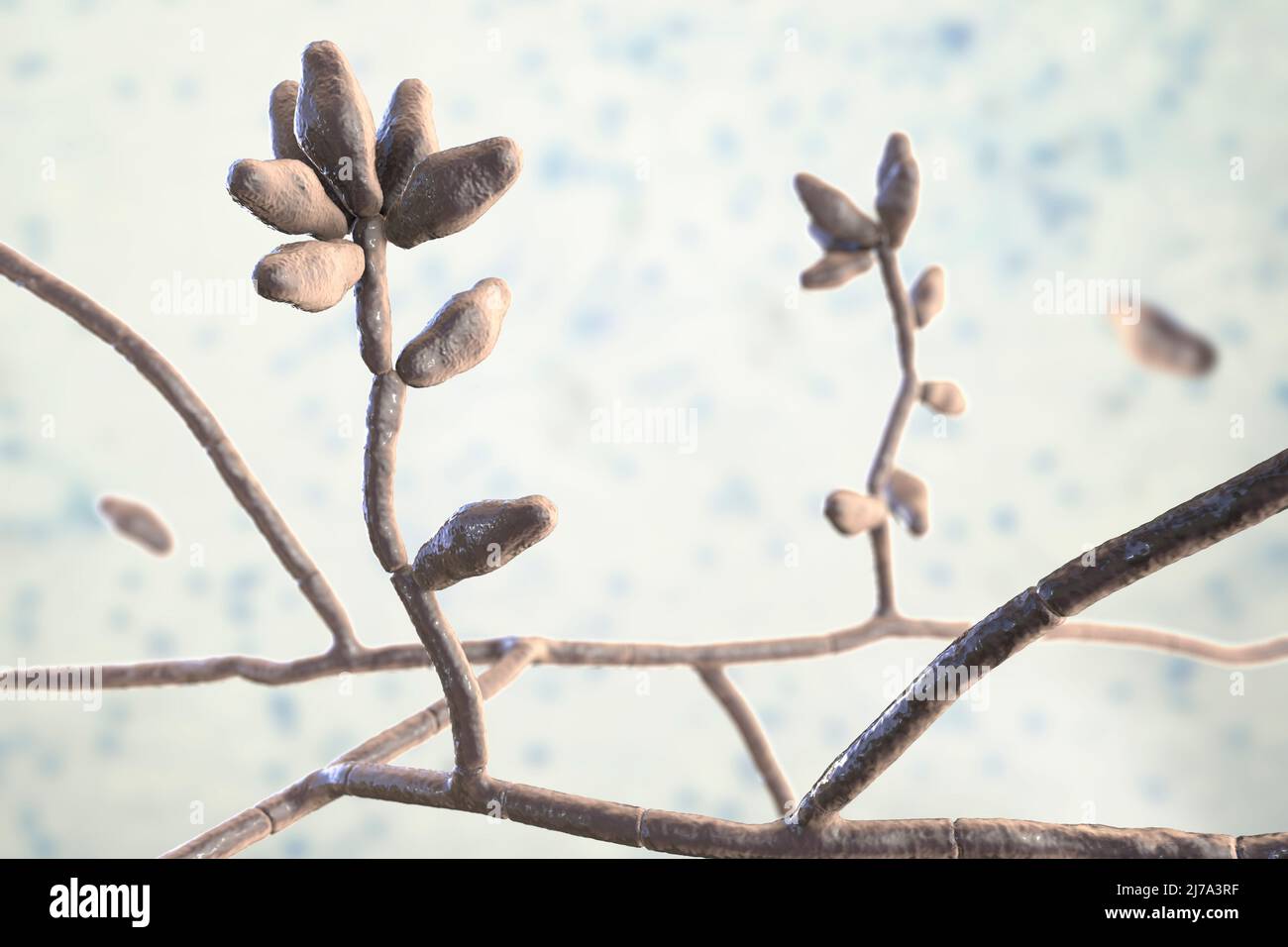 Curvularia mould fungus, illustration Stock Photohttps://www.alamy.com/image-license-details/?v=1https://www.alamy.com/curvularia-mould-fungus-illustration-image469205059.html
RF2J7A3RF–Curvularia mould fungus, illustration
 Curvularia geniculata Stock Photohttps://www.alamy.com/image-license-details/?v=1https://www.alamy.com/stock-photo-curvularia-geniculata-134986785.html
RMHRH50H–Curvularia geniculata
 Scanning Electron Micrograph of Curvularia geniculata. Image courtesy CDC/Robert Simmons, Janice Haney Carr. 1984. Stock Photohttps://www.alamy.com/image-license-details/?v=1https://www.alamy.com/scanning-electron-micrograph-of-curvularia-geniculata-image-courtesy-image155848581.html
RMK1FECN–Scanning Electron Micrograph of Curvularia geniculata. Image courtesy CDC/Robert Simmons, Janice Haney Carr. 1984.
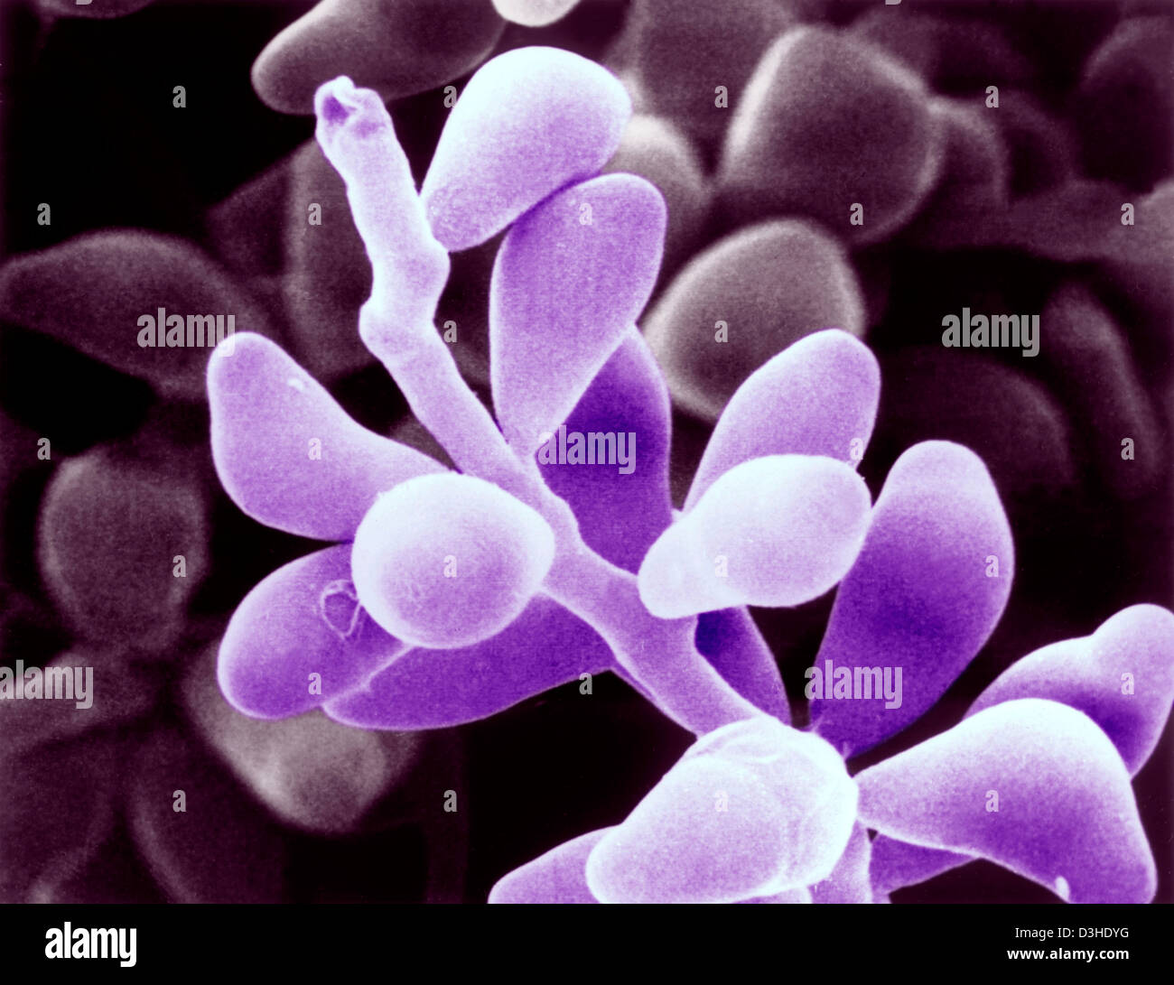 CURVULARIA GENICULATA Stock Photohttps://www.alamy.com/image-license-details/?v=1https://www.alamy.com/stock-photo-curvularia-geniculata-53859220.html
RMD3HDYG–CURVULARIA GENICULATA
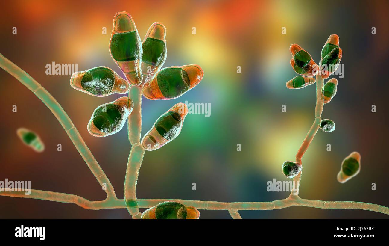 Curvularia mould fungus, illustration Stock Photohttps://www.alamy.com/image-license-details/?v=1https://www.alamy.com/curvularia-mould-fungus-illustration-image469205063.html
RF2J7A3RK–Curvularia mould fungus, illustration
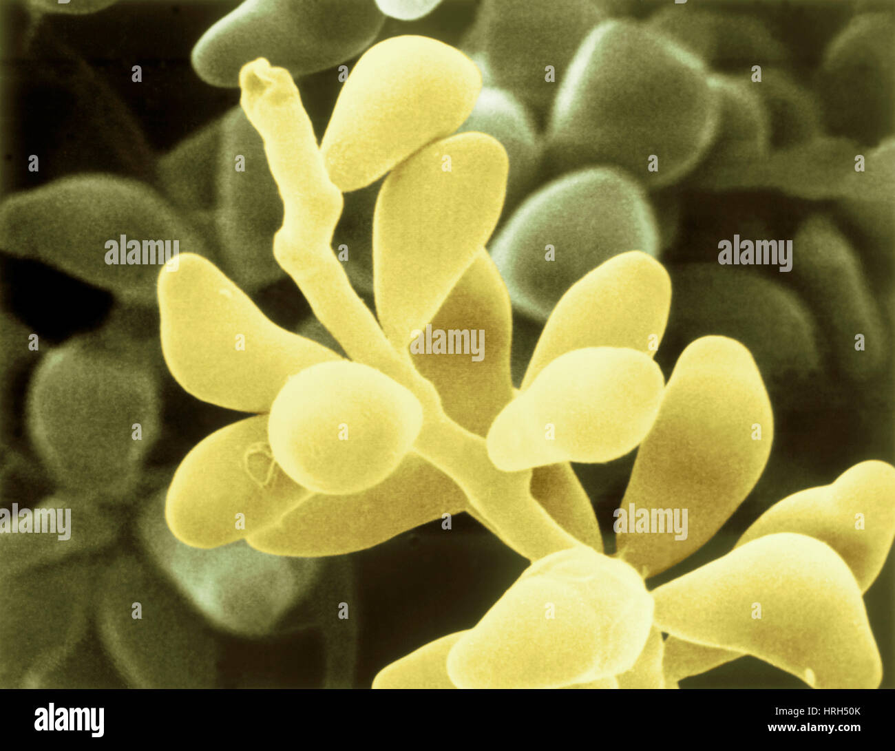 Curvularia geniculata Stock Photohttps://www.alamy.com/image-license-details/?v=1https://www.alamy.com/stock-photo-curvularia-geniculata-134986787.html
RMHRH50K–Curvularia geniculata
 Scanning Electron Micrograph (SEM) of Curvularia geniculata, Scanning Electron Micrograph of Curvularia geniculata. Image courtesy CDC/Robert Simmons/Janice Haney Carr. 1984. Stock Photohttps://www.alamy.com/image-license-details/?v=1https://www.alamy.com/scanning-electron-micrograph-sem-of-curvularia-geniculata-scanning-image155848578.html
RMK1FECJ–Scanning Electron Micrograph (SEM) of Curvularia geniculata, Scanning Electron Micrograph of Curvularia geniculata. Image courtesy CDC/Robert Simmons/Janice Haney Carr. 1984.
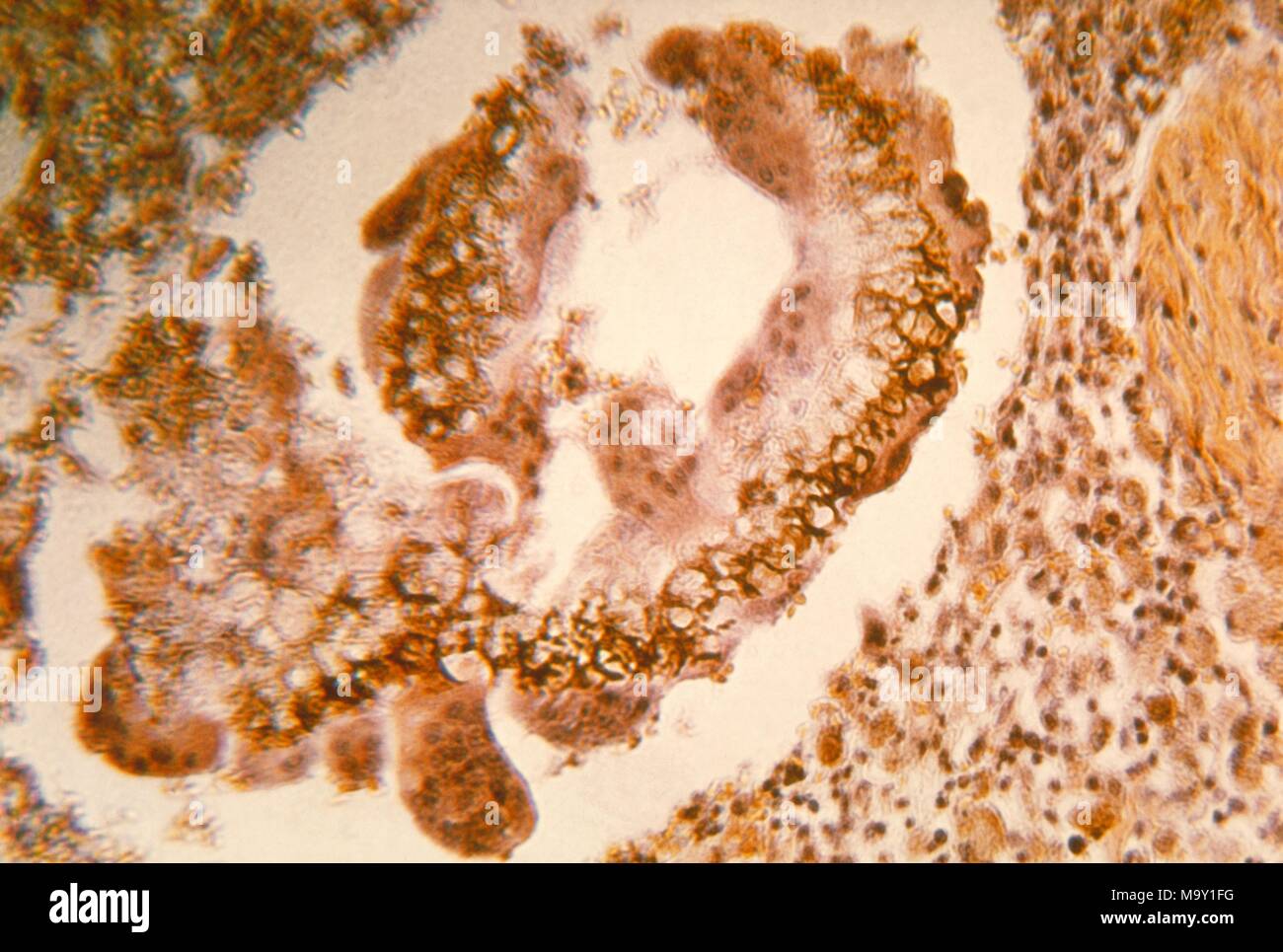 Histopathologic changes found in black grain mycetoma in a dog due to Curvularia geniculata, revealed in the micrograph film, 1964. Image courtesy Centers for Disease Control (CDC) / Dr Lucille K Georg. Image courtesy CDC. () Stock Photohttps://www.alamy.com/image-license-details/?v=1https://www.alamy.com/histopathologic-changes-found-in-black-grain-mycetoma-in-a-dog-due-to-curvularia-geniculata-revealed-in-the-micrograph-film-1964-image-courtesy-centers-for-disease-control-cdc-dr-lucille-k-georg-image-courtesy-cdc-image178229508.html
RMM9Y1FG–Histopathologic changes found in black grain mycetoma in a dog due to Curvularia geniculata, revealed in the micrograph film, 1964. Image courtesy Centers for Disease Control (CDC) / Dr Lucille K Georg. Image courtesy CDC. ()
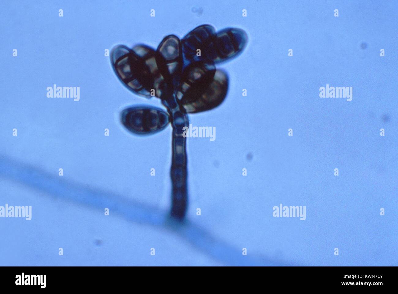 This photomicrograph shows a conidiophore of the fungus Curvularia geniculata, which can be dangerous, and is associated with various human infections such as onychomycosis, keratitis, sinusitis, mycetoma, pneumonia, endocarditis, cerebral abscess, and disseminated infection, 1963. Image courtesy CDC/Dr. Libero Ajello. Stock Photohttps://www.alamy.com/image-license-details/?v=1https://www.alamy.com/stock-photo-this-photomicrograph-shows-a-conidiophore-of-the-fungus-curvularia-170726555.html
RMKWN7CY–This photomicrograph shows a conidiophore of the fungus Curvularia geniculata, which can be dangerous, and is associated with various human infections such as onychomycosis, keratitis, sinusitis, mycetoma, pneumonia, endocarditis, cerebral abscess, and disseminated infection, 1963. Image courtesy CDC/Dr. Libero Ajello.
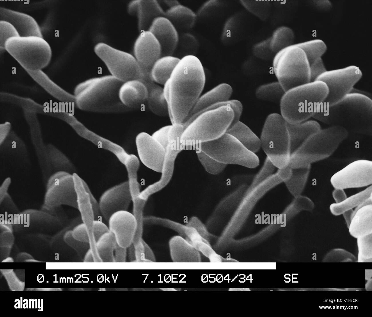 This scanning electron micrograph (SEM) depicts a magnified view of a colony of the dematiaceous filamentous fungus Curvularia geniculata, revealing the morphologic details of the organism?s hyphae, and conidiophores topped with spore-containing conidia. Though normally found living in soil or decaying vegetation, Curvularia geniculata is pathogenic to humans, causing wound infections known as phaeohyphomycosis, which are fungal infections that can involve a number of bodily structures including the skin, respiratory tract, and brain, to name a few. Image courtesy CDC/Robert Simmons, Janice Ha Stock Photohttps://www.alamy.com/image-license-details/?v=1https://www.alamy.com/this-scanning-electron-micrograph-sem-depicts-a-magnified-view-of-image155848583.html
RMK1FECR–This scanning electron micrograph (SEM) depicts a magnified view of a colony of the dematiaceous filamentous fungus Curvularia geniculata, revealing the morphologic details of the organism?s hyphae, and conidiophores topped with spore-containing conidia. Though normally found living in soil or decaying vegetation, Curvularia geniculata is pathogenic to humans, causing wound infections known as phaeohyphomycosis, which are fungal infections that can involve a number of bodily structures including the skin, respiratory tract, and brain, to name a few. Image courtesy CDC/Robert Simmons, Janice Ha
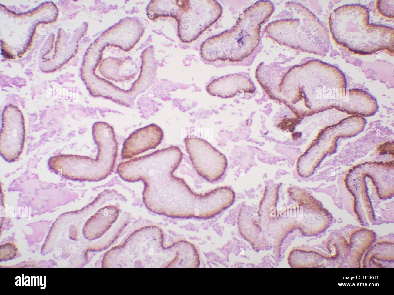 Black grain eumycotic mycetoma of subcutaneous tissue, caused by the fungus Curvularia geniculata, 1973. Image courtesy CDC/Dr. William Kaplan. Stock Photohttps://www.alamy.com/image-license-details/?v=1https://www.alamy.com/stock-photo-black-grain-eumycotic-mycetoma-of-subcutaneous-tissue-caused-by-the-135479032.html
RMHTBGTT–Black grain eumycotic mycetoma of subcutaneous tissue, caused by the fungus Curvularia geniculata, 1973. Image courtesy CDC/Dr. William Kaplan.
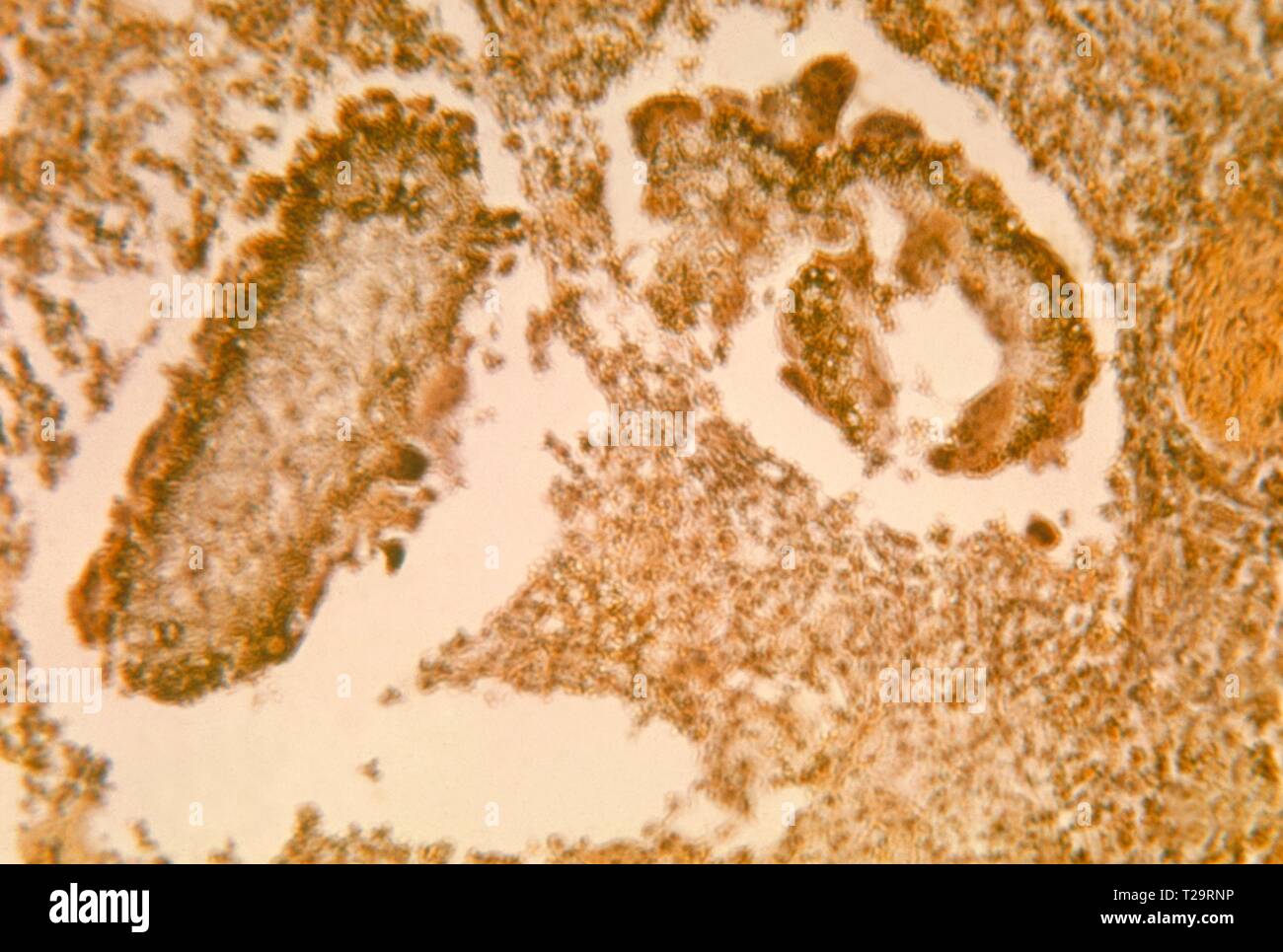 Photomicrograph of the histopathologic changes found in black grain mycetoma in a dog, caused by Curvularia geniculata, 1964. Image courtesy Centers for Disease Control and Prevention (CDC) / Dr Lucille K. Georg. () Stock Photohttps://www.alamy.com/image-license-details/?v=1https://www.alamy.com/photomicrograph-of-the-histopathologic-changes-found-in-black-grain-mycetoma-in-a-dog-caused-by-curvularia-geniculata-1964-image-courtesy-centers-for-disease-control-and-prevention-cdc-dr-lucille-k-georg-image242390674.html
RMT29RNP–Photomicrograph of the histopathologic changes found in black grain mycetoma in a dog, caused by Curvularia geniculata, 1964. Image courtesy Centers for Disease Control and Prevention (CDC) / Dr Lucille K. Georg. ()
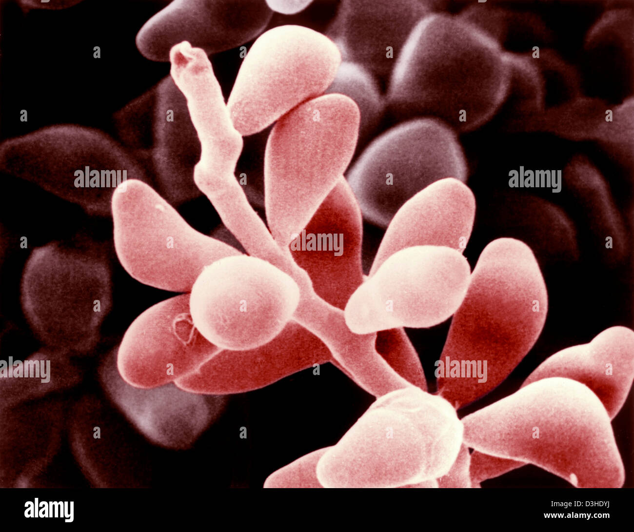 CURVULARIA GENICULATA Stock Photohttps://www.alamy.com/image-license-details/?v=1https://www.alamy.com/stock-photo-curvularia-geniculata-53859222.html
RMD3HDYJ–CURVULARIA GENICULATA
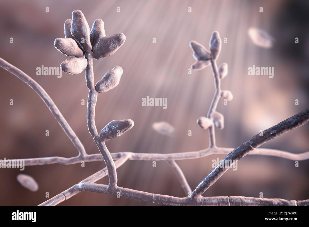 Curvularia mould fungus, illustration Stock Photohttps://www.alamy.com/image-license-details/?v=1https://www.alamy.com/curvularia-mould-fungus-illustration-image469205056.html
RF2J7A3RC–Curvularia mould fungus, illustration
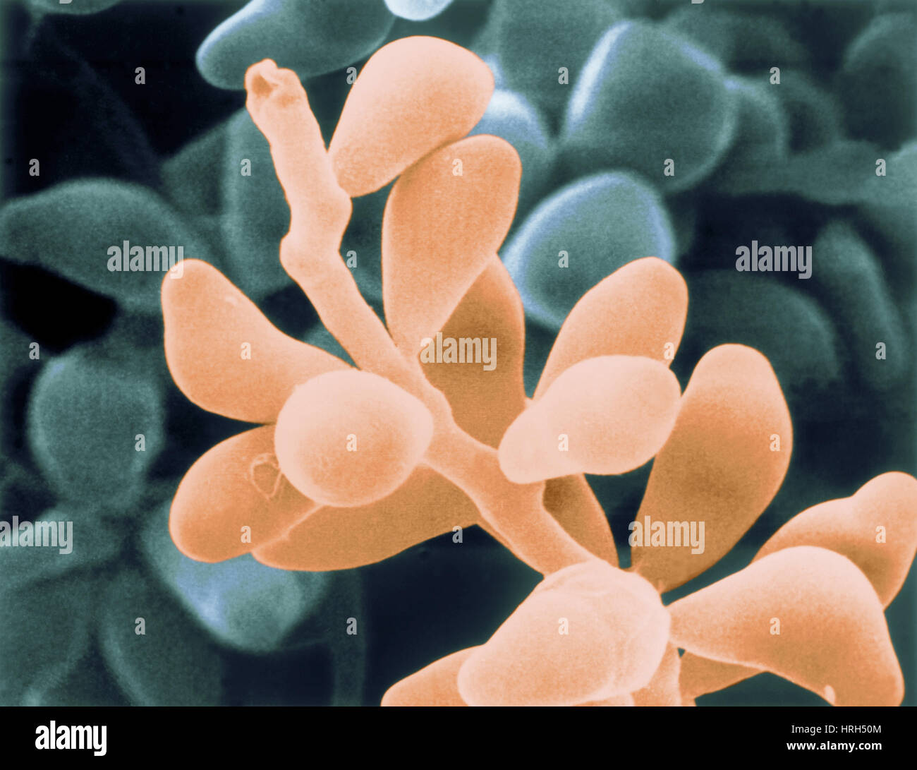 Curvularia geniculata Stock Photohttps://www.alamy.com/image-license-details/?v=1https://www.alamy.com/stock-photo-curvularia-geniculata-134986788.html
RMHRH50M–Curvularia geniculata
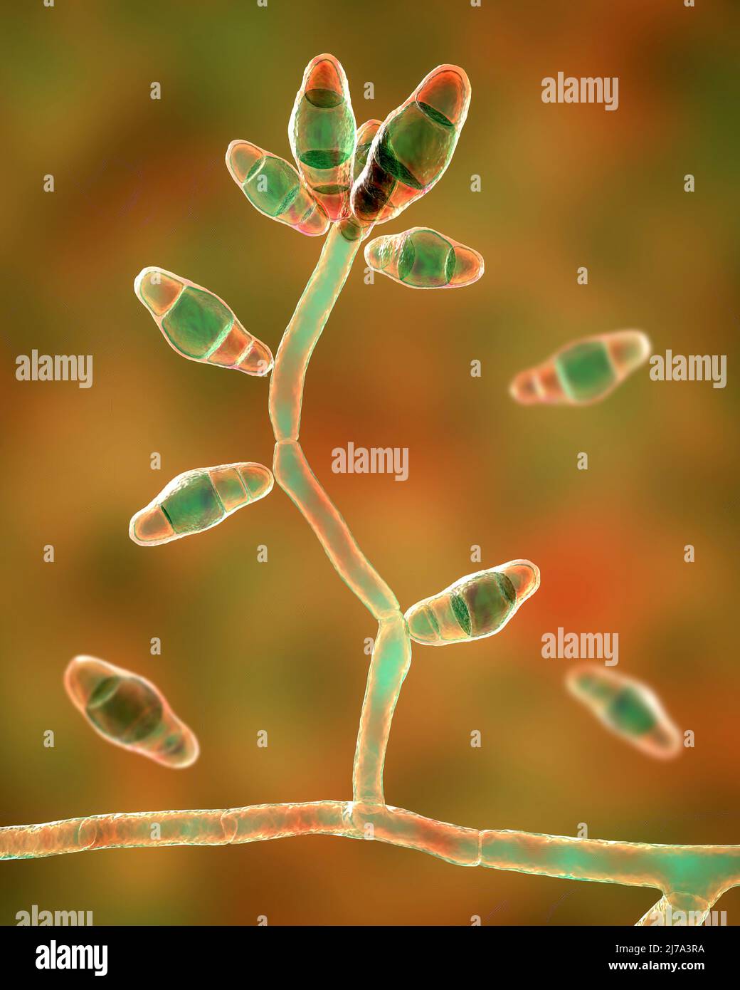 Curvularia mould fungus, illustration Stock Photohttps://www.alamy.com/image-license-details/?v=1https://www.alamy.com/curvularia-mould-fungus-illustration-image469205054.html
RF2J7A3RA–Curvularia mould fungus, illustration
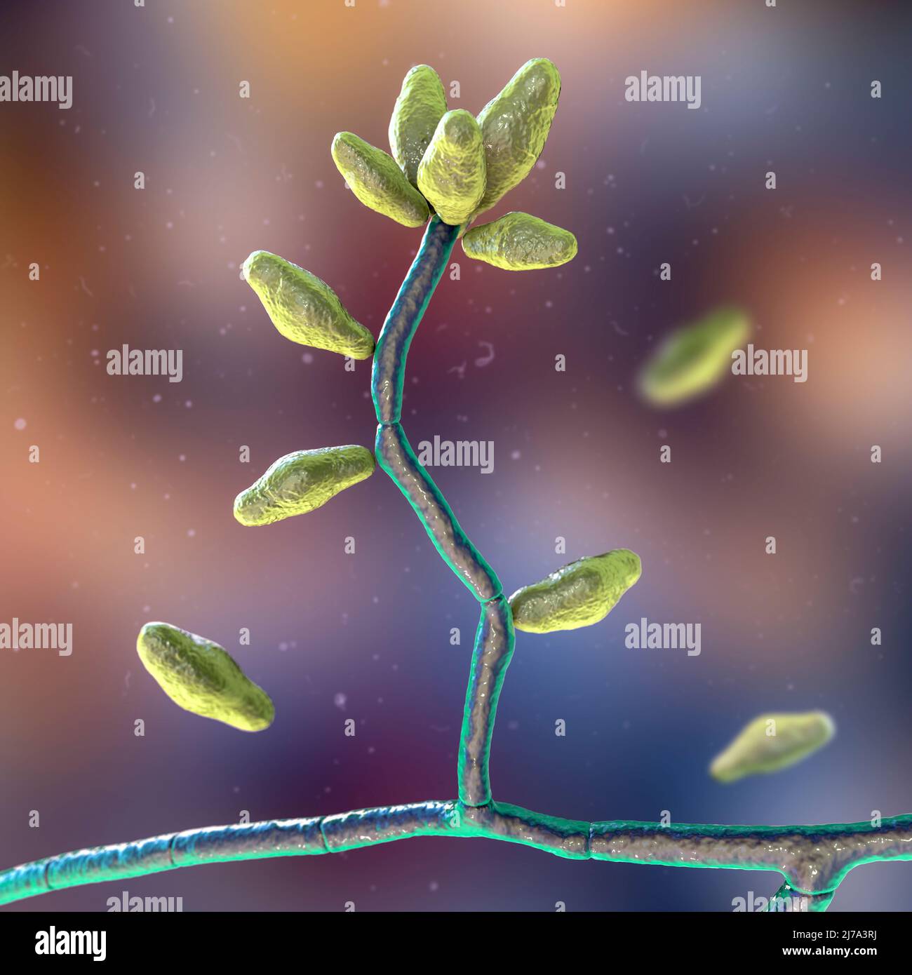 Curvularia mould fungus, illustration Stock Photohttps://www.alamy.com/image-license-details/?v=1https://www.alamy.com/curvularia-mould-fungus-illustration-image469205062.html
RF2J7A3RJ–Curvularia mould fungus, illustration
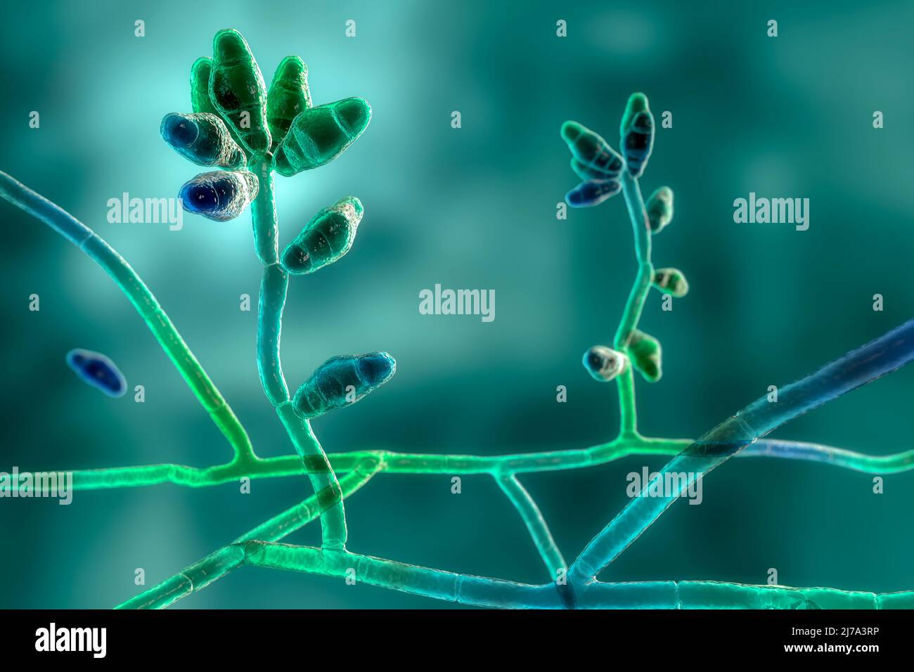 Curvularia mould fungus, illustration Stock Photohttps://www.alamy.com/image-license-details/?v=1https://www.alamy.com/curvularia-mould-fungus-illustration-image469205066.html
RF2J7A3RP–Curvularia mould fungus, illustration
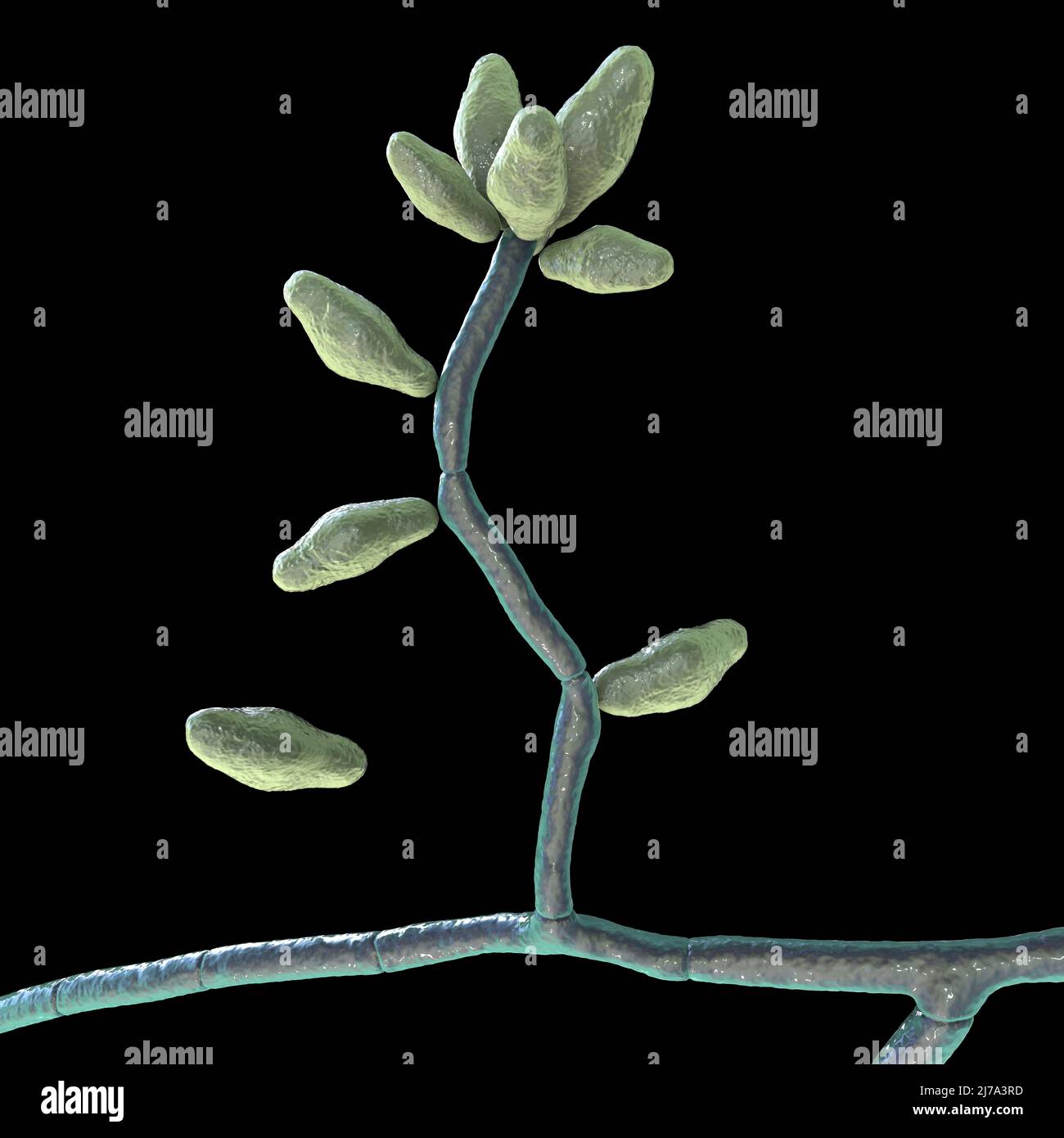 Curvularia mould fungus, illustration Stock Photohttps://www.alamy.com/image-license-details/?v=1https://www.alamy.com/curvularia-mould-fungus-illustration-image469205057.html
RF2J7A3RD–Curvularia mould fungus, illustration
 Curvularia mould fungus, illustration Stock Photohttps://www.alamy.com/image-license-details/?v=1https://www.alamy.com/curvularia-mould-fungus-illustration-image469205053.html
RF2J7A3R9–Curvularia mould fungus, illustration
 Curvularia mould fungus, illustration Stock Photohttps://www.alamy.com/image-license-details/?v=1https://www.alamy.com/curvularia-mould-fungus-illustration-image469205058.html
RF2J7A3RE–Curvularia mould fungus, illustration
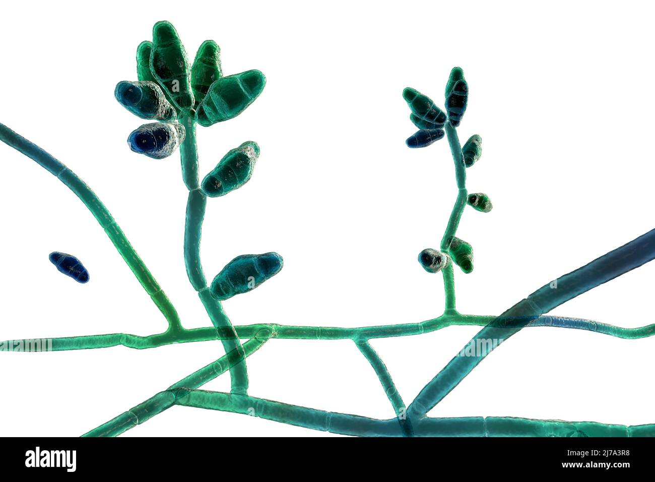 Curvularia mould fungus, illustration Stock Photohttps://www.alamy.com/image-license-details/?v=1https://www.alamy.com/curvularia-mould-fungus-illustration-image469205052.html
RF2J7A3R8–Curvularia mould fungus, illustration
