Quick filters:
Diatoma Stock Photos and Images
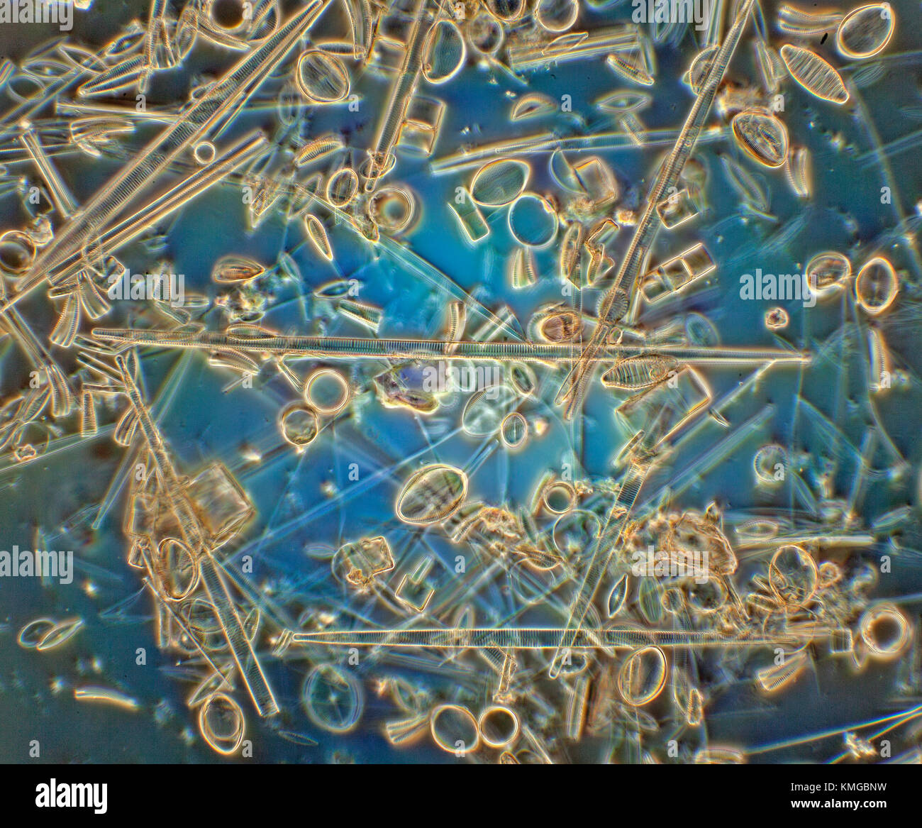 Diatoms photomicrograph, Cocconeis placentula, Synedra, Melosira, Cyclotella, Diatoma Stock Photohttps://www.alamy.com/image-license-details/?v=1https://www.alamy.com/stock-image-diatoms-photomicrograph-cocconeis-placentula-synedra-melosira-cyclotella-167546901.html
RMKMGBNW–Diatoms photomicrograph, Cocconeis placentula, Synedra, Melosira, Cyclotella, Diatoma
 Diatoma marinum, on Ceramium rubrum, Anna Atkins, United Kingdom, c. 1843 - c. 1853, photographic support, cyanotype, height 250 mm × width 200 mm Stock Photohttps://www.alamy.com/image-license-details/?v=1https://www.alamy.com/diatoma-marinum-on-ceramium-rubrum-anna-atkins-united-kingdom-c-1843-c-1853-photographic-support-cyanotype-height-250-mm-width-200-mm-image454397060.html
RM2HB7G2C–Diatoma marinum, on Ceramium rubrum, Anna Atkins, United Kingdom, c. 1843 - c. 1853, photographic support, cyanotype, height 250 mm × width 200 mm
 Inspired by Diatoma marinum, on Ceramium rubrum, Anna Atkins, United Kingdom, c. 1843 - c. 1853, cyanotype, height 250 mm × width 200 mm, Reimagined by Artotop. Classic art reinvented with a modern twist. Design of warm cheerful glowing of brightness and light ray radiance. Photography inspired by surrealism and futurism, embracing dynamic energy of modern technology, movement, speed and revolutionize culture Stock Photohttps://www.alamy.com/image-license-details/?v=1https://www.alamy.com/inspired-by-diatoma-marinum-on-ceramium-rubrum-anna-atkins-united-kingdom-c-1843-c-1853-cyanotype-height-250-mm-width-200-mm-reimagined-by-artotop-classic-art-reinvented-with-a-modern-twist-design-of-warm-cheerful-glowing-of-brightness-and-light-ray-radiance-photography-inspired-by-surrealism-and-futurism-embracing-dynamic-energy-of-modern-technology-movement-speed-and-revolutionize-culture-image459455151.html
RF2HKDYMF–Inspired by Diatoma marinum, on Ceramium rubrum, Anna Atkins, United Kingdom, c. 1843 - c. 1853, cyanotype, height 250 mm × width 200 mm, Reimagined by Artotop. Classic art reinvented with a modern twist. Design of warm cheerful glowing of brightness and light ray radiance. Photography inspired by surrealism and futurism, embracing dynamic energy of modern technology, movement, speed and revolutionize culture
 Diatoma vulgare (Diatoma vulgare), with phase-contrast MRI Stock Photohttps://www.alamy.com/image-license-details/?v=1https://www.alamy.com/diatoma-vulgare-diatoma-vulgare-with-phase-contrast-mri-image9524969.html
RMAWXBXA–Diatoma vulgare (Diatoma vulgare), with phase-contrast MRI
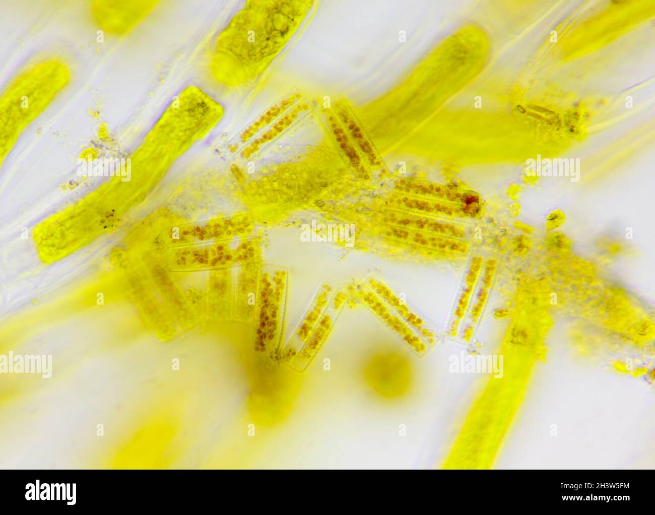 Microscopic view of a diatoms (Diatoma) between algae cells. Brightfield illumination. Stock Photohttps://www.alamy.com/image-license-details/?v=1https://www.alamy.com/microscopic-view-of-a-diatoms-diatoma-between-algae-cells-brightfield-illumination-image449866696.html
RF2H3W5FM–Microscopic view of a diatoms (Diatoma) between algae cells. Brightfield illumination.
 Gruene Schiffchenalge, Navicula viridis (3. von oben Mitte), Staebchenalge, Diatoma flocculosum (4. von oben Mitte), Stueckchenalge, Frustulia (2. von unten Mitte), Spiralbandalge, Spirogyra quinina (oben Mitte), Einzellige Alge, Protococcus viridis (2.von oben Mitte), Flachsalge, Conferva linum (2. von oben rechts), Blasentang, Braunalge, Fucus vesiculosus (rechts unten), Beerentang, Sargassum bacciferum), Gemeine Froschlaichalge, Batrachospermum moniliforme (2. und 3. unten von links), Roter Knorpeltang, Gelidium corneum (rechts oben), Purpurner Kammtang, Plocamium coccineum (2. und 3. von Stock Photohttps://www.alamy.com/image-license-details/?v=1https://www.alamy.com/stock-photo-gruene-schiffchenalge-navicula-viridis-3-von-oben-mitte-staebchenalge-121257313.html
RFH17MX9–Gruene Schiffchenalge, Navicula viridis (3. von oben Mitte), Staebchenalge, Diatoma flocculosum (4. von oben Mitte), Stueckchenalge, Frustulia (2. von unten Mitte), Spiralbandalge, Spirogyra quinina (oben Mitte), Einzellige Alge, Protococcus viridis (2.von oben Mitte), Flachsalge, Conferva linum (2. von oben rechts), Blasentang, Braunalge, Fucus vesiculosus (rechts unten), Beerentang, Sargassum bacciferum), Gemeine Froschlaichalge, Batrachospermum moniliforme (2. und 3. unten von links), Roter Knorpeltang, Gelidium corneum (rechts oben), Purpurner Kammtang, Plocamium coccineum (2. und 3. von
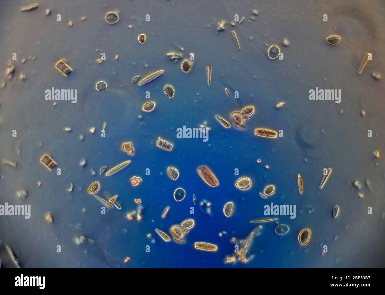 Diatom diversity, Amphora, Cocconeis, Cyclotella, Diatoma, Cymatopleura, Synedra, Melosira, Meridon, Navicula, Nitzschia, Stock Photohttps://www.alamy.com/image-license-details/?v=1https://www.alamy.com/diatom-diversity-amphora-cocconeis-cyclotella-diatoma-cymatopleura-synedra-melosira-meridon-navicula-nitzschia-image351085707.html
RM2BB59B7–Diatom diversity, Amphora, Cocconeis, Cyclotella, Diatoma, Cymatopleura, Synedra, Melosira, Meridon, Navicula, Nitzschia,
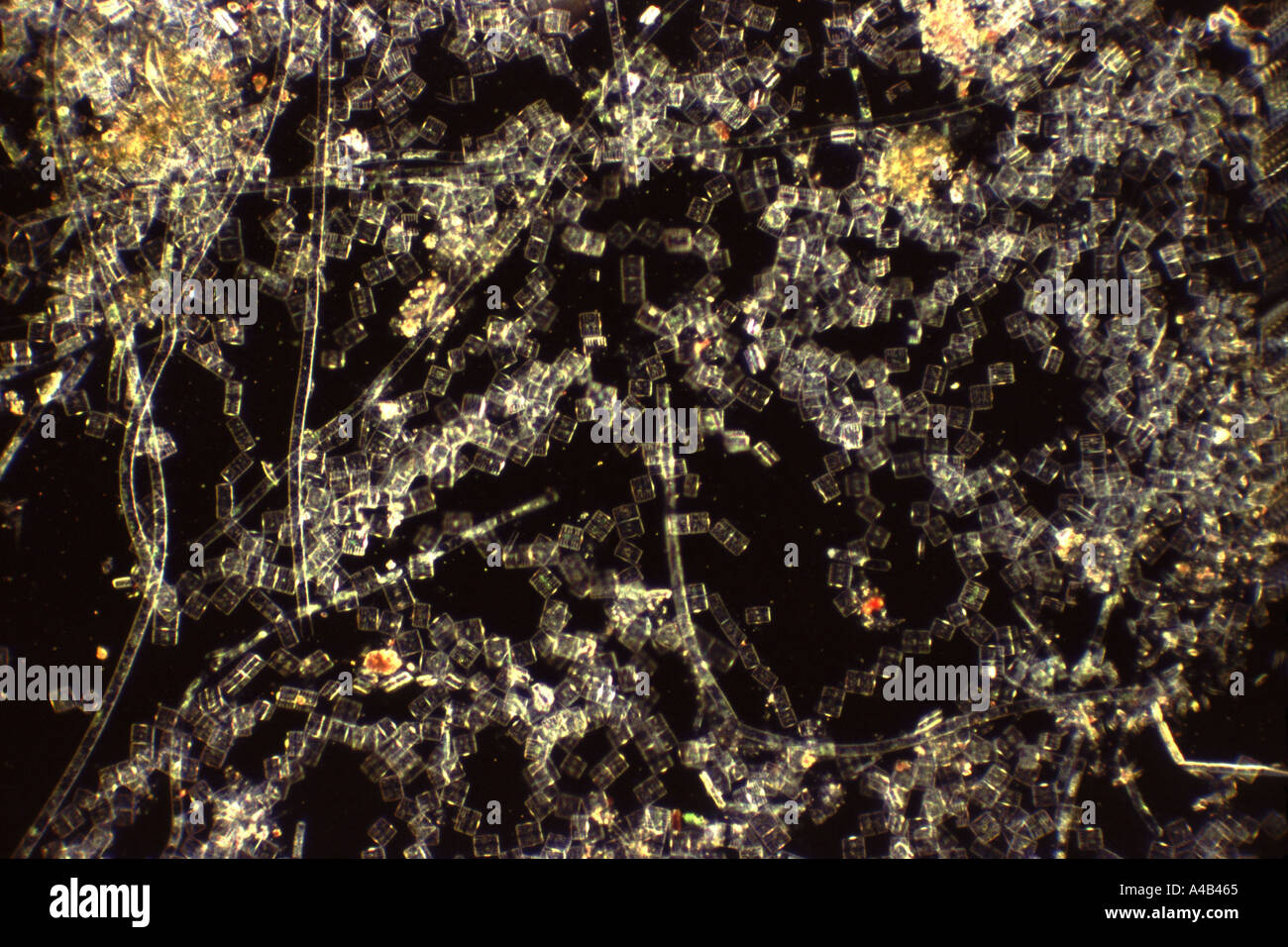 Photomicrograph of living golden algae diatoma species of diatoms Stock Photohttps://www.alamy.com/image-license-details/?v=1https://www.alamy.com/photomicrograph-of-living-golden-algae-diatoma-species-of-diatoms-image308325.html
RMA4B465–Photomicrograph of living golden algae diatoma species of diatoms
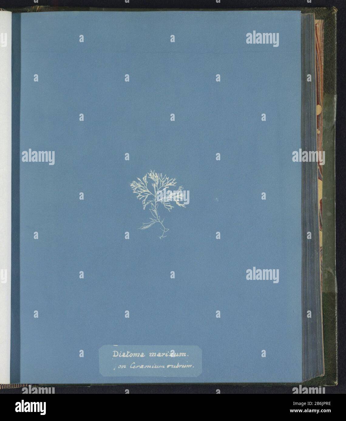 Diatoma marinum on Ceramium rubrum Diatoma marinum / on Ceramium rubrum Object Type : photo page Item number: RP-F 2016-133-146 Manufacturer : photographer Anna Atkins Place manufacture: United Kingdom Dating: ca. 1843 - ca. 1853 Material: paper Technique: cyanotypie Dimensions: page: h 250 mm × W 200 mm Subject: algae, seaweed Stock Photohttps://www.alamy.com/image-license-details/?v=1https://www.alamy.com/diatoma-marinum-on-ceramium-rubrum-diatoma-marinum-on-ceramium-rubrum-object-type-photo-page-item-number-rp-f-2016-133-146-manufacturer-photographer-anna-atkins-place-manufacture-united-kingdom-dating-ca-1843-ca-1853-material-paper-technique-cyanotypie-dimensions-page-h-250-mm-w-200-mm-subject-algae-seaweed-image348308338.html
RM2B6JPRE–Diatoma marinum on Ceramium rubrum Diatoma marinum / on Ceramium rubrum Object Type : photo page Item number: RP-F 2016-133-146 Manufacturer : photographer Anna Atkins Place manufacture: United Kingdom Dating: ca. 1843 - ca. 1853 Material: paper Technique: cyanotypie Dimensions: page: h 250 mm × W 200 mm Subject: algae, seaweed
 Diatoma Marine / Ceramium Red, Anna Atkins, c. 1843 - c. 1853 photograph United Kingdom photographic support cyanotype algae, seaweed Stock Photohttps://www.alamy.com/image-license-details/?v=1https://www.alamy.com/diatoma-marine-ceramium-red-anna-atkins-c-1843-c-1853-photograph-united-kingdom-photographic-support-cyanotype-algae-seaweed-image592522081.html
RM2WBYKXW–Diatoma Marine / Ceramium Red, Anna Atkins, c. 1843 - c. 1853 photograph United Kingdom photographic support cyanotype algae, seaweed
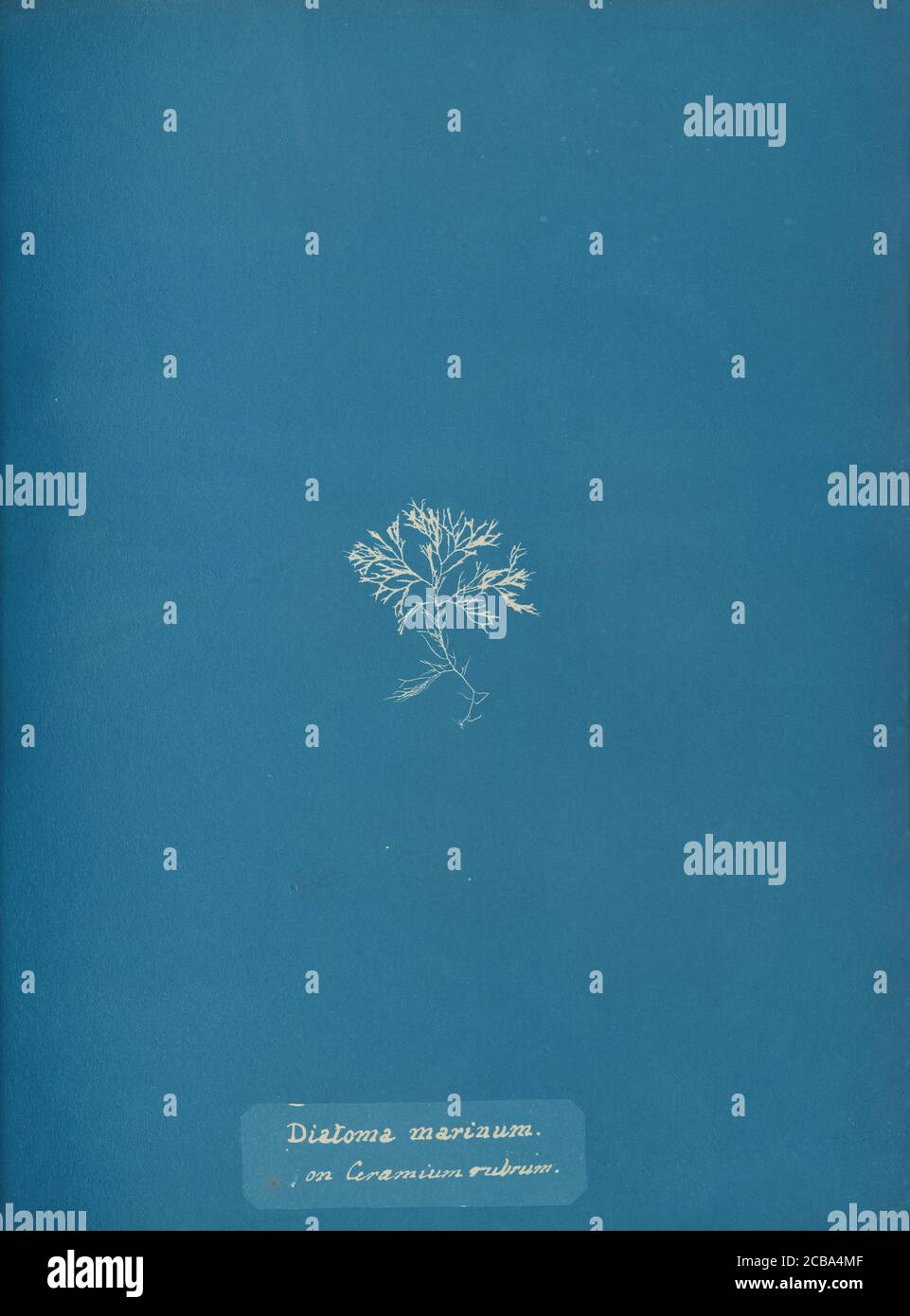 Diatoma marinum on Ceramium rubrum, ca. 1853. Stock Photohttps://www.alamy.com/image-license-details/?v=1https://www.alamy.com/diatoma-marinum-on-ceramium-rubrum-ca-1853-image368402175.html
RM2CBA4MF–Diatoma marinum on Ceramium rubrum, ca. 1853.
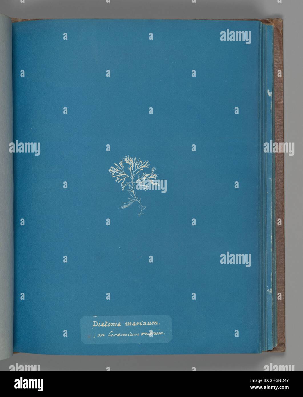 Diatoma marinum on Ceramium rubrum ca. 1853 Anna Atkins British. Diatoma marinum on Ceramium rubrum. Anna Atkins (British, 1799–1871). ca. 1853. Cyanotype. Photographs Stock Photohttps://www.alamy.com/image-license-details/?v=1https://www.alamy.com/diatoma-marinum-on-ceramium-rubrum-ca-1853-anna-atkins-british-diatoma-marinum-on-ceramium-rubrum-anna-atkins-british-17991871-ca-1853-cyanotype-photographs-image457775387.html
RM2HGND4Y–Diatoma marinum on Ceramium rubrum ca. 1853 Anna Atkins British. Diatoma marinum on Ceramium rubrum. Anna Atkins (British, 1799–1871). ca. 1853. Cyanotype. Photographs
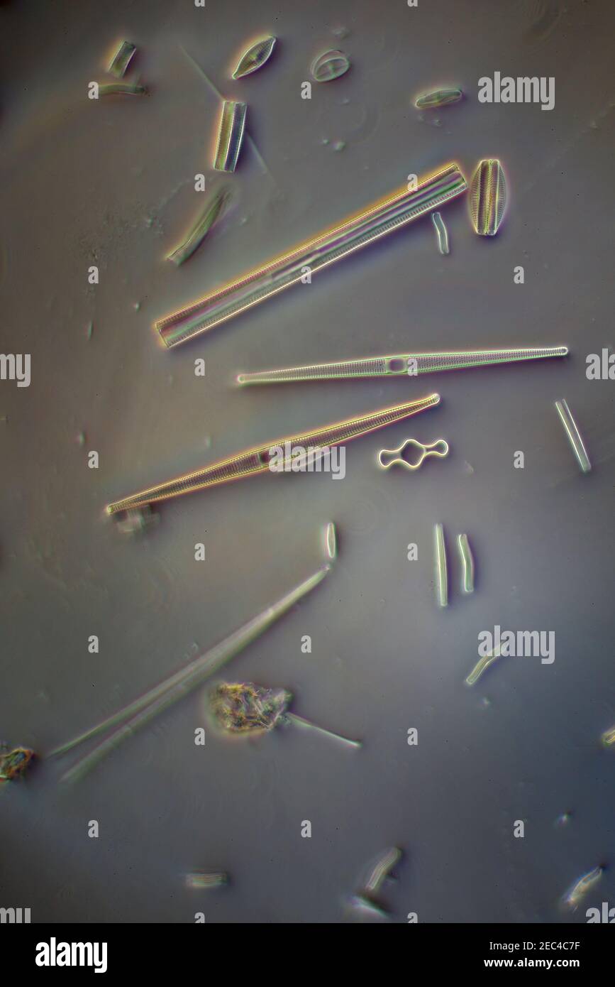 Diatoms, Yorkshire, UK. Oblique illumination, photomicrograph Stock Photohttps://www.alamy.com/image-license-details/?v=1https://www.alamy.com/diatoms-yorkshire-uk-oblique-illumination-photomicrograph-image403311763.html
RM2EC4C7F–Diatoms, Yorkshire, UK. Oblique illumination, photomicrograph
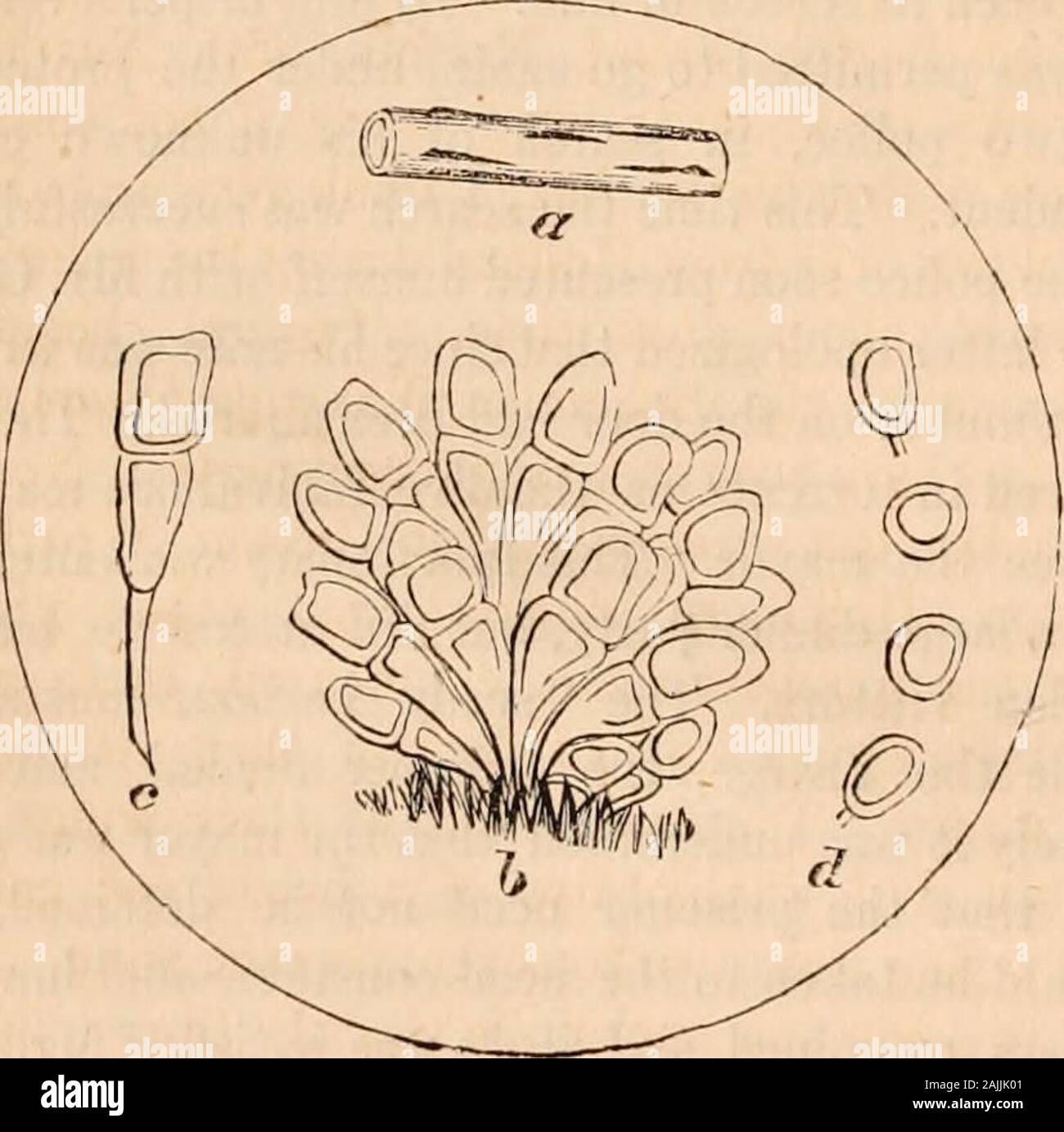 Hardwicke's science-gossip : an illustrated medium of interchange and gossip for students and lovers of nature . the word was Diatoma-ceous earth, and if he required to know further, hemust refer to Pritchards Infusoria or SmithsDiatomaceae. He professed to know all that, butstill he was certain there was something about itthat required to be explained! Suffice it to saythat on the return of the head-constable, his ex-anination of the papers, &c, after some altercation,to the great annoyance and chagrin of the harbour-constable, Mr. Gray obtained his liberty againbetween 10 and 11 oclock at ni Stock Photohttps://www.alamy.com/image-license-details/?v=1https://www.alamy.com/hardwickes-science-gossip-an-illustrated-medium-of-interchange-and-gossip-for-students-and-lovers-of-nature-the-word-was-diatoma-ceous-earth-and-if-he-required-to-know-further-hemust-refer-to-pritchards-infusoria-or-smithsdiatomaceae-he-professed-to-know-all-that-butstill-he-was-certain-there-was-something-about-itthat-required-to-be-explained!-suffice-it-to-saythat-on-the-return-of-the-head-constable-his-ex-anination-of-the-papers-c-after-some-altercationto-the-great-annoyance-and-chagrin-of-the-harbour-constable-mr-gray-obtained-his-liberty-againbetween-10-and-11-oclock-at-ni-image338470833.html
RM2AJJK01–Hardwicke's science-gossip : an illustrated medium of interchange and gossip for students and lovers of nature . the word was Diatoma-ceous earth, and if he required to know further, hemust refer to Pritchards Infusoria or SmithsDiatomaceae. He professed to know all that, butstill he was certain there was something about itthat required to be explained! Suffice it to saythat on the return of the head-constable, his ex-anination of the papers, &c, after some altercation,to the great annoyance and chagrin of the harbour-constable, Mr. Gray obtained his liberty againbetween 10 and 11 oclock at ni
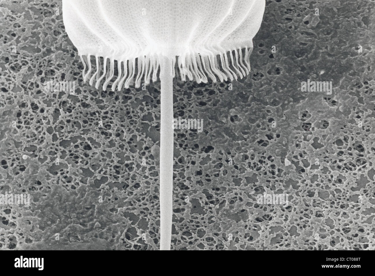 DIATOM, SEM Stock Photohttps://www.alamy.com/image-license-details/?v=1https://www.alamy.com/stock-photo-diatom-sem-49179000.html
RMCT088T–DIATOM, SEM
 . Diatomées marines de la côte occidentale d'Afrique. Diatomées delà Côte Occidle d Afrique PL. VI. Please note that these images are extracted from scanned page images that may have been digitally enhanced for readability - coloration and appearance of these illustrations may not perfectly resemble the original work.. Leuduger-Fortmorel, Georges, 1803-1902. Saint-Brieuc, F. Guyon Stock Photohttps://www.alamy.com/image-license-details/?v=1https://www.alamy.com/diatomes-marines-de-la-cte-occidentale-dafrique-diatomes-del-cte-occidle-d-afrique-pl-vi-please-note-that-these-images-are-extracted-from-scanned-page-images-that-may-have-been-digitally-enhanced-for-readability-coloration-and-appearance-of-these-illustrations-may-not-perfectly-resemble-the-original-work-leuduger-fortmorel-georges-1803-1902-saint-brieuc-f-guyon-image215893542.html
RMPF6PBJ–. Diatomées marines de la côte occidentale d'Afrique. Diatomées delà Côte Occidle d Afrique PL. VI. Please note that these images are extracted from scanned page images that may have been digitally enhanced for readability - coloration and appearance of these illustrations may not perfectly resemble the original work.. Leuduger-Fortmorel, Georges, 1803-1902. Saint-Brieuc, F. Guyon
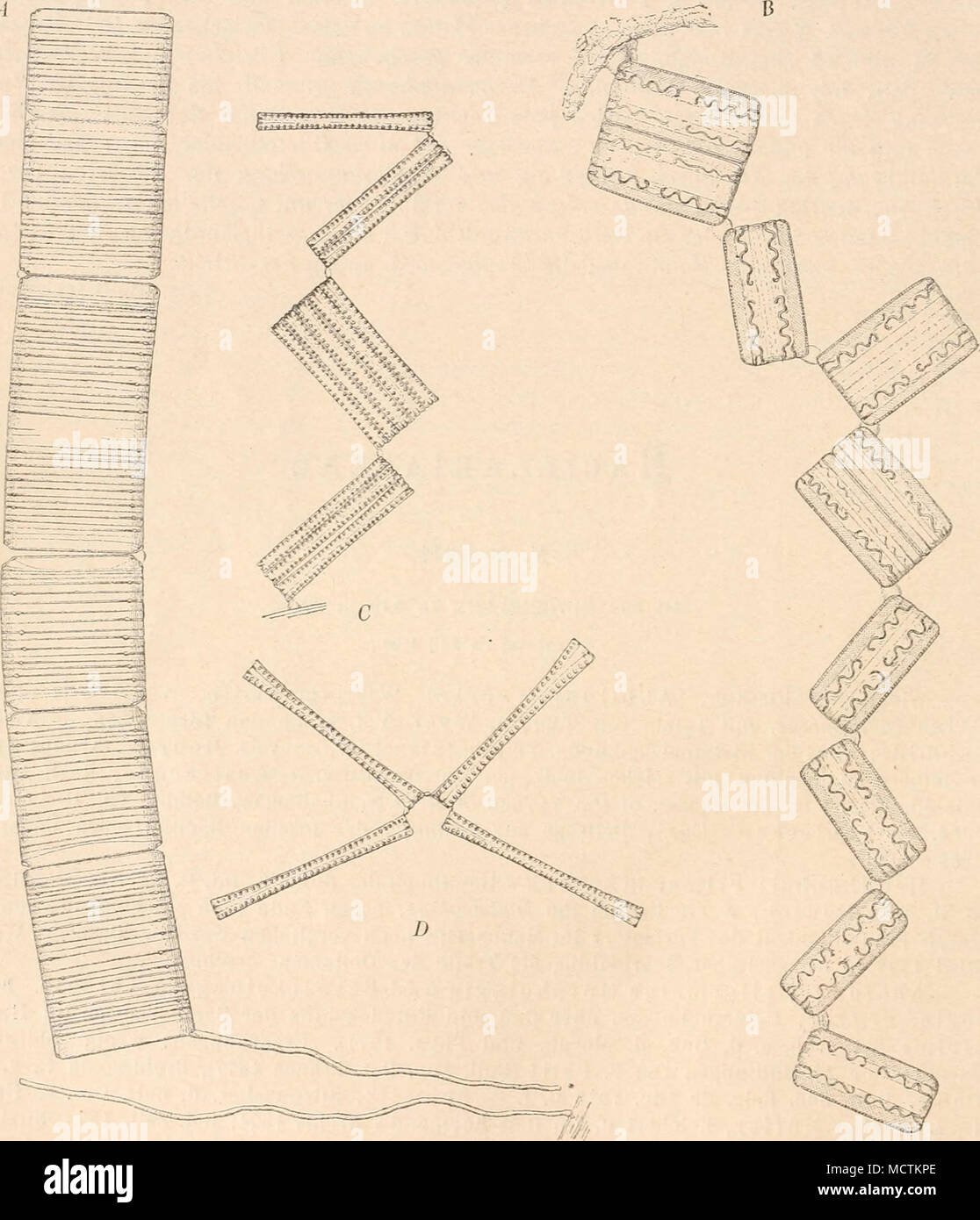 . Fig. 45. VerschiedenekettenartigeColonien. A gerade, gestielte Kette von Tahellaria (Striatellu) unipunc- tuta (Ag.) F. S., die ganzen Schalenflächen der henachbarten Zellen sind an einander gekittet. — i? Zickzackkette von Grammatophora serpentina Ralfs, die Zellen haften mittelst Gallertpolster mit je einer Ecke an einander. — C ge- mischte, gerade und Zickzackkette und D Sternkette, heide von Diatoma tlongatum Ag. (Alles nach W. Smith; 400/1). Stock Photohttps://www.alamy.com/image-license-details/?v=1https://www.alamy.com/fig-45-verschiedenekettenartigecolonien-a-gerade-gestielte-kette-von-tahellaria-striatellu-unipunc-tuta-ag-f-s-die-ganzen-schalenflchen-der-henachbarten-zellen-sind-an-einander-gekittet-i-zickzackkette-von-grammatophora-serpentina-ralfs-die-zellen-haften-mittelst-gallertpolster-mit-je-einer-ecke-an-einander-c-ge-mischte-gerade-und-zickzackkette-und-d-sternkette-heide-von-diatoma-tlongatum-ag-alles-nach-w-smith-4001-image180021926.html
RMMCTKPE–. Fig. 45. VerschiedenekettenartigeColonien. A gerade, gestielte Kette von Tahellaria (Striatellu) unipunc- tuta (Ag.) F. S., die ganzen Schalenflächen der henachbarten Zellen sind an einander gekittet. — i? Zickzackkette von Grammatophora serpentina Ralfs, die Zellen haften mittelst Gallertpolster mit je einer Ecke an einander. — C ge- mischte, gerade und Zickzackkette und D Sternkette, heide von Diatoma tlongatum Ag. (Alles nach W. Smith; 400/1).
 Art inspired by Diatoma marinum, on Ceramium rubrum, Anna Atkins, United Kingdom, c. 1843 - c. 1853, photographic support, cyanotype, height 250 mm × width 200 mm, Classic works modernized by Artotop with a splash of modernity. Shapes, color and value, eye-catching visual impact on art. Emotions through freedom of artworks in a contemporary way. A timeless message pursuing a wildly creative new direction. Artists turning to the digital medium and creating the Artotop NFT Stock Photohttps://www.alamy.com/image-license-details/?v=1https://www.alamy.com/art-inspired-by-diatoma-marinum-on-ceramium-rubrum-anna-atkins-united-kingdom-c-1843-c-1853-photographic-support-cyanotype-height-250-mm-width-200-mm-classic-works-modernized-by-artotop-with-a-splash-of-modernity-shapes-color-and-value-eye-catching-visual-impact-on-art-emotions-through-freedom-of-artworks-in-a-contemporary-way-a-timeless-message-pursuing-a-wildly-creative-new-direction-artists-turning-to-the-digital-medium-and-creating-the-artotop-nft-image459761887.html
RF2HKYXYB–Art inspired by Diatoma marinum, on Ceramium rubrum, Anna Atkins, United Kingdom, c. 1843 - c. 1853, photographic support, cyanotype, height 250 mm × width 200 mm, Classic works modernized by Artotop with a splash of modernity. Shapes, color and value, eye-catching visual impact on art. Emotions through freedom of artworks in a contemporary way. A timeless message pursuing a wildly creative new direction. Artists turning to the digital medium and creating the Artotop NFT
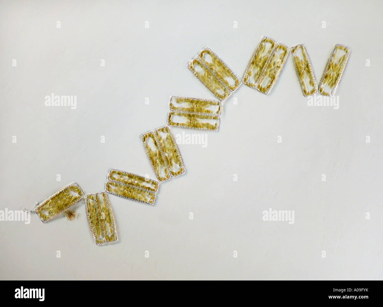 Diatoma vulgare (Diatoma vulgare), with light microscopy, COL Stock Photohttps://www.alamy.com/image-license-details/?v=1https://www.alamy.com/diatoma-vulgare-diatoma-vulgare-with-light-microscopy-col-image9924534.html
RMA09FYK–Diatoma vulgare (Diatoma vulgare), with light microscopy, COL
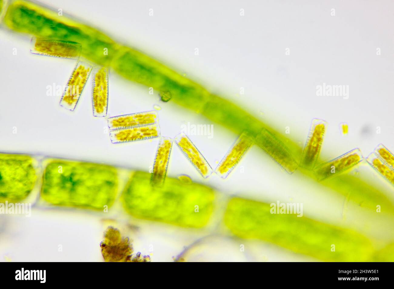 Microscopic view of a diatoms (Diatoma) and green algae filaments. Brightfield illumination. Stock Photohttps://www.alamy.com/image-license-details/?v=1https://www.alamy.com/microscopic-view-of-a-diatoms-diatoma-and-green-algae-filaments-brightfield-illumination-image449866649.html
RF2H3W5E1–Microscopic view of a diatoms (Diatoma) and green algae filaments. Brightfield illumination.
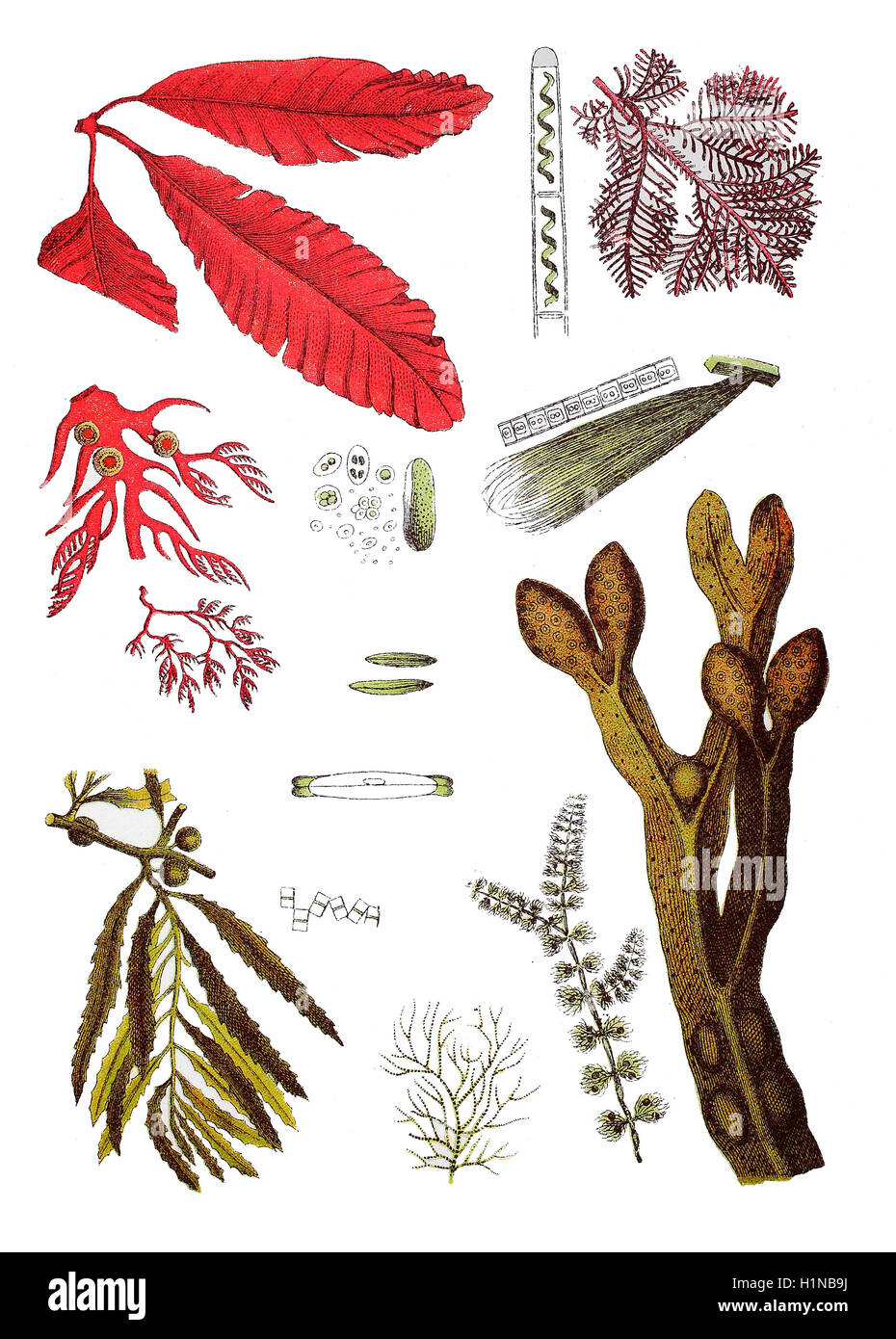 Navicula, Navicula viridis (3. von top center), Alga, Diatoma flocculosum (4. von top center), Alga, Frustulia (2. von bottem center), Spirogyra, Spirogyra quinina (top center), Alga, Protococcus viridis (2.von top center), Alga, Conferva linum (2. von top right), Bladder wrack, Fucus vesiculosus (right bottem), Sargassum, (Sargassum bacciferum), Alga, Batrachospermum moniliforme (2. und 3. bottem von left), Seaweed, Gelidium corneum (right top), Seaweedg, Plocamium coccineum (2. und 3. von top left), Delesseria, Delesseria sanguinea (top left) Stock Photohttps://www.alamy.com/image-license-details/?v=1https://www.alamy.com/stock-photo-navicula-navicula-viridis-3-von-top-center-alga-diatoma-flocculosum-121557118.html
RFH1NB9J–Navicula, Navicula viridis (3. von top center), Alga, Diatoma flocculosum (4. von top center), Alga, Frustulia (2. von bottem center), Spirogyra, Spirogyra quinina (top center), Alga, Protococcus viridis (2.von top center), Alga, Conferva linum (2. von top right), Bladder wrack, Fucus vesiculosus (right bottem), Sargassum, (Sargassum bacciferum), Alga, Batrachospermum moniliforme (2. und 3. bottem von left), Seaweed, Gelidium corneum (right top), Seaweedg, Plocamium coccineum (2. und 3. von top left), Delesseria, Delesseria sanguinea (top left)
 Diatoms, Yorkshire, UK, photomicrograph Stock Photohttps://www.alamy.com/image-license-details/?v=1https://www.alamy.com/diatoms-yorkshire-uk-photomicrograph-image403311797.html
RM2EC4C8N–Diatoms, Yorkshire, UK, photomicrograph
 A guide to Belfast and the counties of Down & Antrim . Near the Ormeau Bridge fine sections havebeen exposed in the Annadale and other brickworks, whilethe eskers at Lisburn are the remains of englacial depositscontemporaneous with the gravels and sands of the valleyabove noted. Pr.-egkr.—Raised Beaches of N.E. of Ireland. Proc. koyal IrishAcademy, 3rd series, vol. iv. No. i (1897). - Kstuarine Clays of the N.E. of Ireland. Ibid., vol. li, No. 2 (1892). 92 Guide to Belfast. The alluvial deposits of the river Bann yield diatoma-ceous earth in large quantities at Toome and at Portglenone.The ki Stock Photohttps://www.alamy.com/image-license-details/?v=1https://www.alamy.com/a-guide-to-belfast-and-the-counties-of-down-antrim-near-the-ormeau-bridge-fine-sections-havebeen-exposed-in-the-annadale-and-other-brickworks-whilethe-eskers-at-lisburn-are-the-remains-of-englacial-depositscontemporaneous-with-the-gravels-and-sands-of-the-valleyabove-noted-pr-egkrraised-beaches-of-ne-of-ireland-proc-koyal-irishacademy-3rd-series-vol-iv-no-i-1897-kstuarine-clays-of-the-ne-of-ireland-ibid-vol-li-no-2-1892-92-guide-to-belfast-the-alluvial-deposits-of-the-river-bann-yield-diatoma-ceous-earth-in-large-quantities-at-toome-and-at-portglenonethe-ki-image342708011.html
RM2AWFKFR–A guide to Belfast and the counties of Down & Antrim . Near the Ormeau Bridge fine sections havebeen exposed in the Annadale and other brickworks, whilethe eskers at Lisburn are the remains of englacial depositscontemporaneous with the gravels and sands of the valleyabove noted. Pr.-egkr.—Raised Beaches of N.E. of Ireland. Proc. koyal IrishAcademy, 3rd series, vol. iv. No. i (1897). - Kstuarine Clays of the N.E. of Ireland. Ibid., vol. li, No. 2 (1892). 92 Guide to Belfast. The alluvial deposits of the river Bann yield diatoma-ceous earth in large quantities at Toome and at Portglenone.The ki
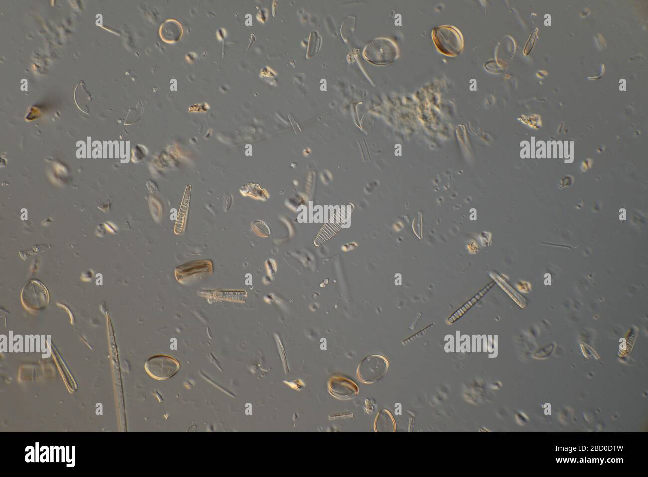 Freshwater diatoms Newton Beck, S Yorks. UK Stock Photohttps://www.alamy.com/image-license-details/?v=1https://www.alamy.com/freshwater-diatoms-newton-beck-s-yorks-uk-image352208777.html
RM2BD0DTW–Freshwater diatoms Newton Beck, S Yorks. UK
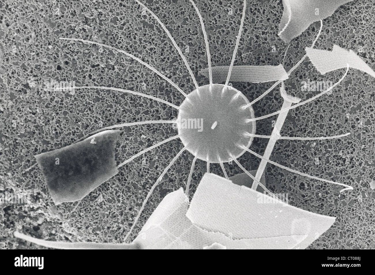 DIATOM, SEM Stock Photohttps://www.alamy.com/image-license-details/?v=1https://www.alamy.com/stock-photo-diatom-sem-49178994.html
RMCT088J–DIATOM, SEM
 . Diatomées marines de la côte occidentale d'Afrique. Diatomées delà Côte Occid^ d'Afrique PL. I. Birikés Frères. Castm DUeudujer Torlmotel ad nal del'. Please note that these images are extracted from scanned page images that may have been digitally enhanced for readability - coloration and appearance of these illustrations may not perfectly resemble the original work.. Leuduger-Fortmorel, Georges, 1803-1902. Saint-Brieuc, F. Guyon Stock Photohttps://www.alamy.com/image-license-details/?v=1https://www.alamy.com/diatomes-marines-de-la-cte-occidentale-dafrique-diatomes-del-cte-occid-dafrique-pl-i-biriks-frres-castm-dueudujer-torlmotel-ad-nal-del-please-note-that-these-images-are-extracted-from-scanned-page-images-that-may-have-been-digitally-enhanced-for-readability-coloration-and-appearance-of-these-illustrations-may-not-perfectly-resemble-the-original-work-leuduger-fortmorel-georges-1803-1902-saint-brieuc-f-guyon-image215893563.html
RMPF6PCB–. Diatomées marines de la côte occidentale d'Afrique. Diatomées delà Côte Occid^ d'Afrique PL. I. Birikés Frères. Castm DUeudujer Torlmotel ad nal del'. Please note that these images are extracted from scanned page images that may have been digitally enhanced for readability - coloration and appearance of these illustrations may not perfectly resemble the original work.. Leuduger-Fortmorel, Georges, 1803-1902. Saint-Brieuc, F. Guyon
 . Fig. 197. Meridion circulare (Grev.) Ag. A Schalen-, B Gürtelansieht (S00|l); C Kette (400|l). (A, IS nach Van Heurck; C nach Smith.) b. v. 12. a. Fragilarioideae-Fragilarieae-Diatominae. Schalenansicht rund, langelliptisch, bisquitförmig, kreuzförmig. Schalen ohne Kiel, mit Transversalrippen, von mehr oder minder tief ins Innere vorspringenden Septen her- rührend. Ohne Raphe. Pseudoraphe deutlich oder fehlend. Gürtelansicht rechteckig. Schalen mit Transversalsepten (Rippen' . A. Ohne gegabelte Sagittalrippe. a. Schale ohne Centralknoten und -Auge 109. Diatoma. b. Schale mit Centralknoten 11 Stock Photohttps://www.alamy.com/image-license-details/?v=1https://www.alamy.com/fig-197-meridion-circulare-grev-ag-a-schalen-b-grtelansieht-s00l-c-kette-400l-a-is-nach-van-heurck-c-nach-smith-b-v-12-a-fragilarioideae-fragilarieae-diatominae-schalenansicht-rund-langelliptisch-bisquitfrmig-kreuzfrmig-schalen-ohne-kiel-mit-transversalrippen-von-mehr-oder-minder-tief-ins-innere-vorspringenden-septen-her-rhrend-ohne-raphe-pseudoraphe-deutlich-oder-fehlend-grtelansicht-rechteckig-schalen-mit-transversalsepten-rippen-a-ohne-gegabelte-sagittalrippe-a-schale-ohne-centralknoten-und-auge-109-diatoma-b-schale-mit-centralknoten-11-image180021135.html
RMMCTJP7–. Fig. 197. Meridion circulare (Grev.) Ag. A Schalen-, B Gürtelansieht (S00|l); C Kette (400|l). (A, IS nach Van Heurck; C nach Smith.) b. v. 12. a. Fragilarioideae-Fragilarieae-Diatominae. Schalenansicht rund, langelliptisch, bisquitförmig, kreuzförmig. Schalen ohne Kiel, mit Transversalrippen, von mehr oder minder tief ins Innere vorspringenden Septen her- rührend. Ohne Raphe. Pseudoraphe deutlich oder fehlend. Gürtelansicht rechteckig. Schalen mit Transversalsepten (Rippen' . A. Ohne gegabelte Sagittalrippe. a. Schale ohne Centralknoten und -Auge 109. Diatoma. b. Schale mit Centralknoten 11
 Diatoma vulgare (Diatoma vulgare), with phase-contrast MRI Stock Photohttps://www.alamy.com/image-license-details/?v=1https://www.alamy.com/diatoma-vulgare-diatoma-vulgare-with-phase-contrast-mri-image9924535.html
RMA09FYM–Diatoma vulgare (Diatoma vulgare), with phase-contrast MRI
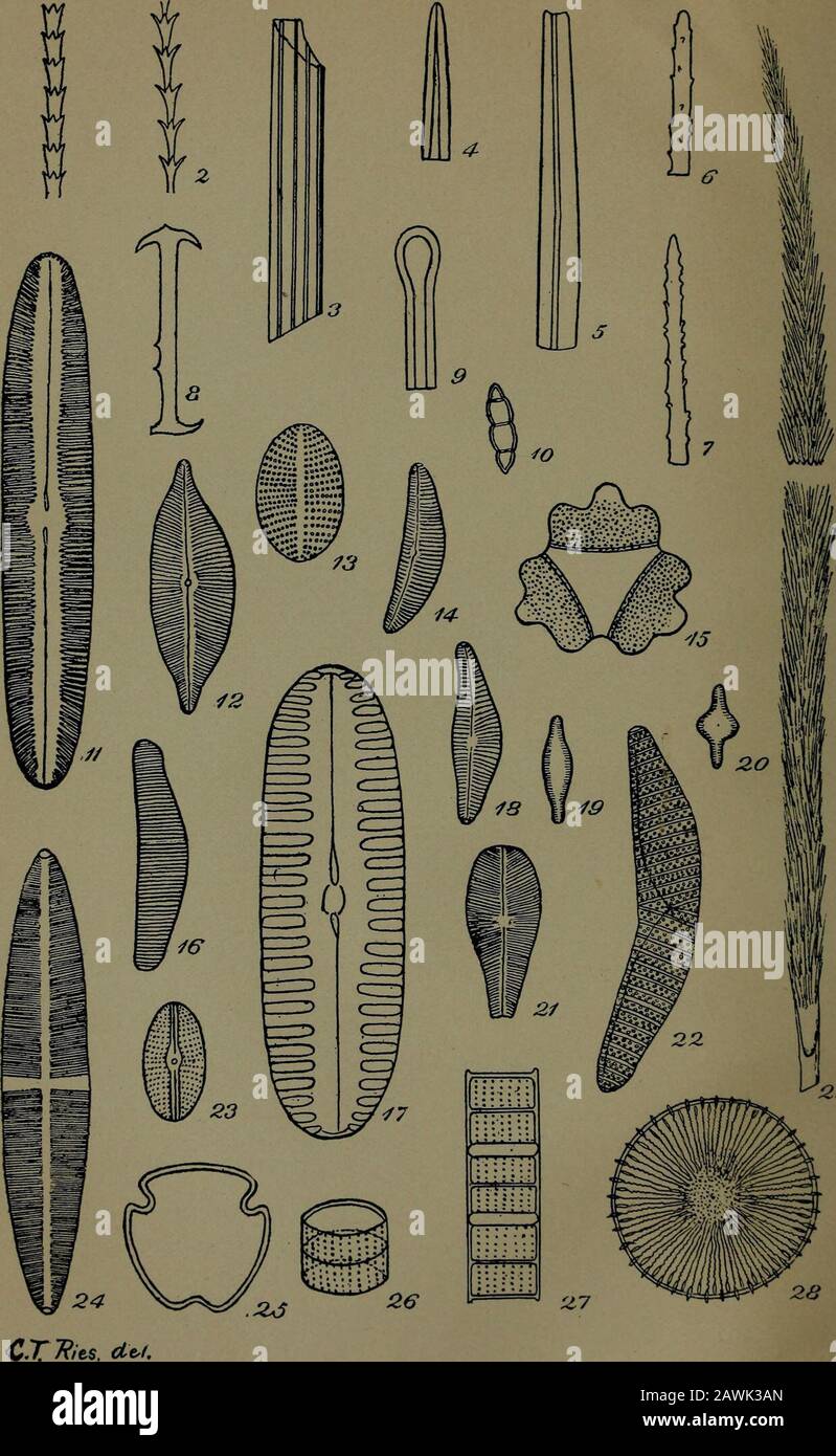 Annual report of the Regents . C. TRiG^, de). Micro-organisms from the clays of New York. Plate 15 To face page 601. iZ9 Micro-organisms from the clays of New York. CLAYS OF IS^EW YORK 601 (Magnified 500 diameters) Pig. 1, 2 Jointed liair. Wyandance, L. I. Fig. 3 Ridged tube from stoneware clay. Glencove, L. I. Fig. 4, 5 Spicules from cretaceous clay at Grlencove, L. I. Fig. 6-8 Spicules from Lloyds neck, L. I. Fig. 9 Spicule fragment? Farmingdale, L. I. Fig. 10 Diatoma hyemale. Glencove, L. I. Fig. 11 Navicula viridis Kutz. Lloyds neck, L. I. Fig. 12 Cymhella cuspidata Kutz. Lloyds neck, L. L Stock Photohttps://www.alamy.com/image-license-details/?v=1https://www.alamy.com/annual-report-of-the-regents-c-trig-de-micro-organisms-from-the-clays-of-new-york-plate-15-to-face-page-601-iz9-micro-organisms-from-the-clays-of-new-york-clays-of-isew-york-601-magnified-500-diameters-pig-1-2-jointed-liair-wyandance-l-i-fig-3-ridged-tube-from-stoneware-clay-glencove-l-i-fig-4-5-spicules-from-cretaceous-clay-at-grlencove-l-i-fig-6-8-spicules-from-lloyds-neck-l-i-fig-9-spicule-fragment-farmingdale-l-i-fig-10-diatoma-hyemale-glencove-l-i-fig-11-navicula-viridis-kutz-lloyds-neck-l-i-fig-12-cymhella-cuspidata-kutz-lloyds-neck-l-l-image342783133.html
RM2AWK3AN–Annual report of the Regents . C. TRiG^, de). Micro-organisms from the clays of New York. Plate 15 To face page 601. iZ9 Micro-organisms from the clays of New York. CLAYS OF IS^EW YORK 601 (Magnified 500 diameters) Pig. 1, 2 Jointed liair. Wyandance, L. I. Fig. 3 Ridged tube from stoneware clay. Glencove, L. I. Fig. 4, 5 Spicules from cretaceous clay at Grlencove, L. I. Fig. 6-8 Spicules from Lloyds neck, L. I. Fig. 9 Spicule fragment? Farmingdale, L. I. Fig. 10 Diatoma hyemale. Glencove, L. I. Fig. 11 Navicula viridis Kutz. Lloyds neck, L. I. Fig. 12 Cymhella cuspidata Kutz. Lloyds neck, L. L
 Freshwater diatoms Newton Beck, S Yorks. UK Stock Photohttps://www.alamy.com/image-license-details/?v=1https://www.alamy.com/freshwater-diatoms-newton-beck-s-yorks-uk-image352208936.html
RM2BD0E2G–Freshwater diatoms Newton Beck, S Yorks. UK
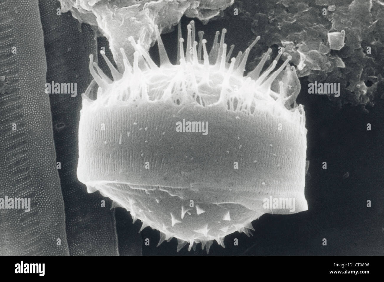 DIATOM, SEM Stock Photohttps://www.alamy.com/image-license-details/?v=1https://www.alamy.com/stock-photo-diatom-sem-49179010.html
RMCT0896–DIATOM, SEM
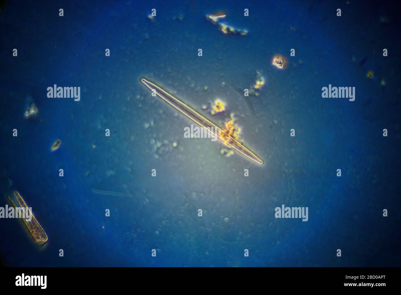 Freshwater diatoms, Synedra sp. Stock Photohttps://www.alamy.com/image-license-details/?v=1https://www.alamy.com/freshwater-diatoms-synedra-sp-image352206368.html
RM2BD0APT–Freshwater diatoms, Synedra sp.
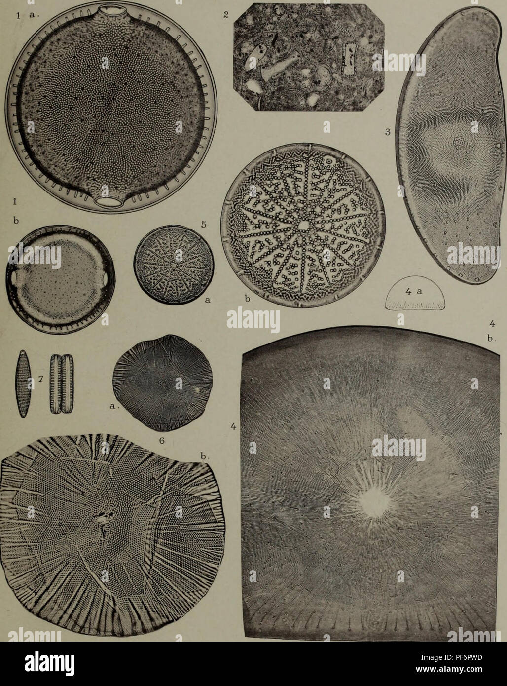 . Diatomées; espèces nouvelles marines, fossiles ou pélagiques. Diatomées PI.XV PlioCotypie Thevoz & C" à Genève e&pèceô rLovLv-ef&o 1891. # 1 |». Please note that these images are extracted from scanned page images that may have been digitally enhanced for readability - coloration and appearance of these illustrations may not perfectly resemble the original work.. Brun, Jacques, 1826-1908. Leipzig, Theodor Oswald Weigle Stock Photohttps://www.alamy.com/image-license-details/?v=1https://www.alamy.com/diatomes-espces-nouvelles-marines-fossiles-ou-plagiques-diatomes-pixv-pliocotypie-thevoz-amp-cquot-genve-eamppce-rlovlv-efampo-1891-1-please-note-that-these-images-are-extracted-from-scanned-page-images-that-may-have-been-digitally-enhanced-for-readability-coloration-and-appearance-of-these-illustrations-may-not-perfectly-resemble-the-original-work-brun-jacques-1826-1908-leipzig-theodor-oswald-weigle-image215893929.html
RMPF6PWD–. Diatomées; espèces nouvelles marines, fossiles ou pélagiques. Diatomées PI.XV PlioCotypie Thevoz & C" à Genève e&pèceô rLovLv-ef&o 1891. # 1 |». Please note that these images are extracted from scanned page images that may have been digitally enhanced for readability - coloration and appearance of these illustrations may not perfectly resemble the original work.. Brun, Jacques, 1826-1908. Leipzig, Theodor Oswald Weigle
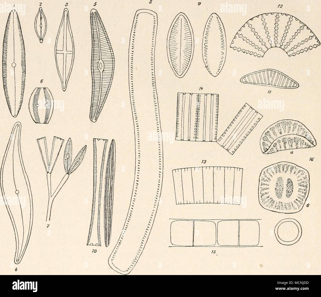 . Fig. 2. Kieselalgen, Bacillariaceen. i. Pätiiutaria — 2. A'avicula — 3. Staiironeis — 4. Pleuro- sigma — 5. Cymbella — 6. Aviphora — 7. Gompkonema — 8. Nitschia — 9. Surirella — 10. Synedra — 11. Epithemia — 12. Meridian — 13. Fragillaria — 14. Diatoma — 15. Melosira — 16. Campylodiscus (a von der Seite, b von oben). Stark vergrössert. Bei Amphora (Nr. 6) bildet die Streifung zwei eigentümliche Bänder, während andere Teile des Kieselpanzers ungestreift bleiben. Bei der Gattung Gomphonenia sitzen die einzelnen Zellen auf Gallert- stielen, welche ein vielfach verzweigtes Bäumchen darstellen (N Stock Photohttps://www.alamy.com/image-license-details/?v=1https://www.alamy.com/fig-2-kieselalgen-bacillariaceen-i-ptiiutaria-2-aavicula-3-staiironeis-4-pleuro-sigma-5-cymbella-6-aviphora-7-gompkonema-8-nitschia-9-surirella-10-synedra-11-epithemia-12-meridian-13-fragillaria-14-diatoma-15-melosira-16-campylodiscus-a-von-der-seite-b-von-oben-stark-vergrssert-bei-amphora-nr-6-bildet-die-streifung-zwei-eigentmliche-bnder-whrend-andere-teile-des-kieselpanzers-ungestreift-bleiben-bei-der-gattung-gomphonenia-sitzen-die-einzelnen-zellen-auf-gallert-stielen-welche-ein-vielfach-verzweigtes-bumchen-darstellen-n-image179955033.html
RMMCNJDD–. Fig. 2. Kieselalgen, Bacillariaceen. i. Pätiiutaria — 2. A'avicula — 3. Staiironeis — 4. Pleuro- sigma — 5. Cymbella — 6. Aviphora — 7. Gompkonema — 8. Nitschia — 9. Surirella — 10. Synedra — 11. Epithemia — 12. Meridian — 13. Fragillaria — 14. Diatoma — 15. Melosira — 16. Campylodiscus (a von der Seite, b von oben). Stark vergrössert. Bei Amphora (Nr. 6) bildet die Streifung zwei eigentümliche Bänder, während andere Teile des Kieselpanzers ungestreift bleiben. Bei der Gattung Gomphonenia sitzen die einzelnen Zellen auf Gallert- stielen, welche ein vielfach verzweigtes Bäumchen darstellen (N
 Diatoma vulgare (Diatoma vulgare), in shining-through light Stock Photohttps://www.alamy.com/image-license-details/?v=1https://www.alamy.com/diatoma-vulgare-diatoma-vulgare-in-shining-through-light-image8114641.html
RMAGG2N2–Diatoma vulgare (Diatoma vulgare), in shining-through light
 . Algæ. Vol. I. Myxophyceæ, Peridinieæ, Bacillarieæ, Chlorophyceæ, together with a brief summary of the occurrence and distribution of freshwat4er Algæ . cells arejoined in an irregularly zigzag manner at the corners of the valves (Tabel-laria, some species of Diatoma, Triceratium, arid Bidduiphia). In allthese cases the attachment is by mucus secreted by the cells themselves.The cells of the ribbon-shaped and thread-like colonies are joined eitherby a layer of mucus or by mucous strands between the closely applied valve-faces, and the cells of the zigzag colonies are united by small mucouscus Stock Photohttps://www.alamy.com/image-license-details/?v=1https://www.alamy.com/alg-vol-i-myxophyce-peridinie-bacillarie-chlorophyce-together-with-a-brief-summary-of-the-occurrence-and-distribution-of-freshwat4er-alg-cells-arejoined-in-an-irregularly-zigzag-manner-at-the-corners-of-the-valves-tabel-laria-some-species-of-diatoma-triceratium-arid-bidduiphia-in-allthese-cases-the-attachment-is-by-mucus-secreted-by-the-cells-themselvesthe-cells-of-the-ribbon-shaped-and-thread-like-colonies-are-joined-eitherby-a-layer-of-mucus-or-by-mucous-strands-between-the-closely-applied-valve-faces-and-the-cells-of-the-zigzag-colonies-are-united-by-small-mucouscus-image372070371.html
RM2CH97FF–. Algæ. Vol. I. Myxophyceæ, Peridinieæ, Bacillarieæ, Chlorophyceæ, together with a brief summary of the occurrence and distribution of freshwat4er Algæ . cells arejoined in an irregularly zigzag manner at the corners of the valves (Tabel-laria, some species of Diatoma, Triceratium, arid Bidduiphia). In allthese cases the attachment is by mucus secreted by the cells themselves.The cells of the ribbon-shaped and thread-like colonies are joined eitherby a layer of mucus or by mucous strands between the closely applied valve-faces, and the cells of the zigzag colonies are united by small mucouscus
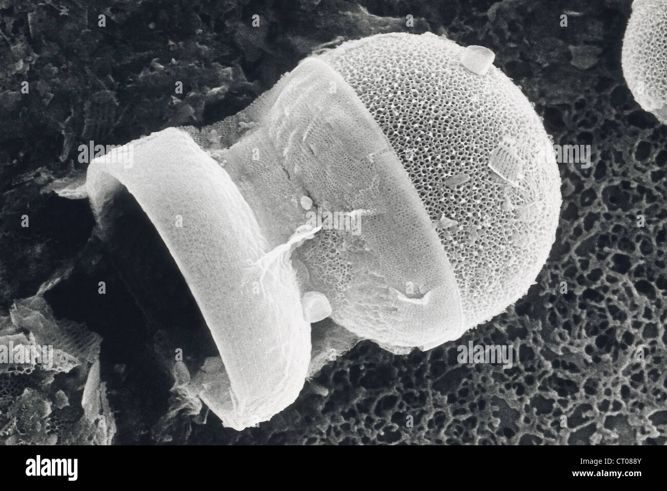 DIATOM, SEM Stock Photohttps://www.alamy.com/image-license-details/?v=1https://www.alamy.com/stock-photo-diatom-sem-49179003.html
RMCT088Y–DIATOM, SEM
 Freshwater diatoms Newton Beck, S Yorks. UK Stock Photohttps://www.alamy.com/image-license-details/?v=1https://www.alamy.com/freshwater-diatoms-newton-beck-s-yorks-uk-image352206684.html
RM2BD0B64–Freshwater diatoms Newton Beck, S Yorks. UK
 . Diatomées marines de France et des districts maritimes voisins. Diatoms. Peragallo. - Diatomées de France. n. wwiL. â Ml.TM^I, I. Please note that these images are extracted from scanned page images that may have been digitally enhanced for readability - coloration and appearance of these illustrations may not perfectly resemble the original work.. Péragallo, H. (Hippolyte), b. 1851; Péragallo, M. (Maurice), b. 1853. Grez-sur-Loing, M. J. Tempère Stock Photohttps://www.alamy.com/image-license-details/?v=1https://www.alamy.com/diatomes-marines-de-france-et-des-districts-maritimes-voisins-diatoms-peragallo-diatomes-de-france-n-wwil-mltmi-i-please-note-that-these-images-are-extracted-from-scanned-page-images-that-may-have-been-digitally-enhanced-for-readability-coloration-and-appearance-of-these-illustrations-may-not-perfectly-resemble-the-original-work-pragallo-h-hippolyte-b-1851-pragallo-m-maurice-b-1853-grez-sur-loing-m-j-tempre-image215893376.html
RMPF6P5M–. Diatomées marines de France et des districts maritimes voisins. Diatoms. Peragallo. - Diatomées de France. n. wwiL. â Ml.TM^I, I. Please note that these images are extracted from scanned page images that may have been digitally enhanced for readability - coloration and appearance of these illustrations may not perfectly resemble the original work.. Péragallo, H. (Hippolyte), b. 1851; Péragallo, M. (Maurice), b. 1853. Grez-sur-Loing, M. J. Tempère
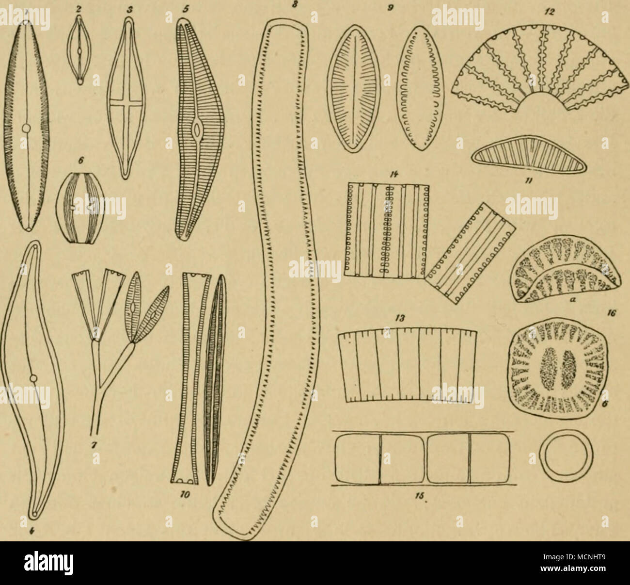 . Kig. 2. Kiesclalgcn, Bacillariaceen. i. Püinularia — 2. Navicula — 3. Siauroncis — 4. Pleuro- sigma — 5. Cymbella — 6. Amphora — 7. Gomphonema — 8. Nitschia — 9. Surirella — 10. Synedra — 11. Epithemia — 12. Meridian — 13. Fragillaria — 14. Diatoma — 15. Mclosira — 16. Campylodiscus (a von der Seite, b von oben). Stark vergrössert. Bei Amphora (Nr. 6) bildet die Streifung zwei eigentümliche Bänder, während andere Teile des Kicselpanzers ungestreift bleiben. Bei der Gattung Gomphouema sitzen die einzelnen Zellen auf Gallert- stielen, welche ein vielfach verzweigtes Bäumchen darstellen (Nr. 7) Stock Photohttps://www.alamy.com/image-license-details/?v=1https://www.alamy.com/kig-2-kiesclalgcn-bacillariaceen-i-pinularia-2-navicula-3-siauroncis-4-pleuro-sigma-5-cymbella-6-amphora-7-gomphonema-8-nitschia-9-surirella-10-synedra-11-epithemia-12-meridian-13-fragillaria-14-diatoma-15-mclosira-16-campylodiscus-a-von-der-seite-b-von-oben-stark-vergrssert-bei-amphora-nr-6-bildet-die-streifung-zwei-eigentmliche-bnder-whrend-andere-teile-des-kicselpanzers-ungestreift-bleiben-bei-der-gattung-gomphouema-sitzen-die-einzelnen-zellen-auf-gallert-stielen-welche-ein-vielfach-verzweigtes-bumchen-darstellen-nr-7-image179954553.html
RMMCNHT9–. Kig. 2. Kiesclalgcn, Bacillariaceen. i. Püinularia — 2. Navicula — 3. Siauroncis — 4. Pleuro- sigma — 5. Cymbella — 6. Amphora — 7. Gomphonema — 8. Nitschia — 9. Surirella — 10. Synedra — 11. Epithemia — 12. Meridian — 13. Fragillaria — 14. Diatoma — 15. Mclosira — 16. Campylodiscus (a von der Seite, b von oben). Stark vergrössert. Bei Amphora (Nr. 6) bildet die Streifung zwei eigentümliche Bänder, während andere Teile des Kicselpanzers ungestreift bleiben. Bei der Gattung Gomphouema sitzen die einzelnen Zellen auf Gallert- stielen, welche ein vielfach verzweigtes Bäumchen darstellen (Nr. 7)
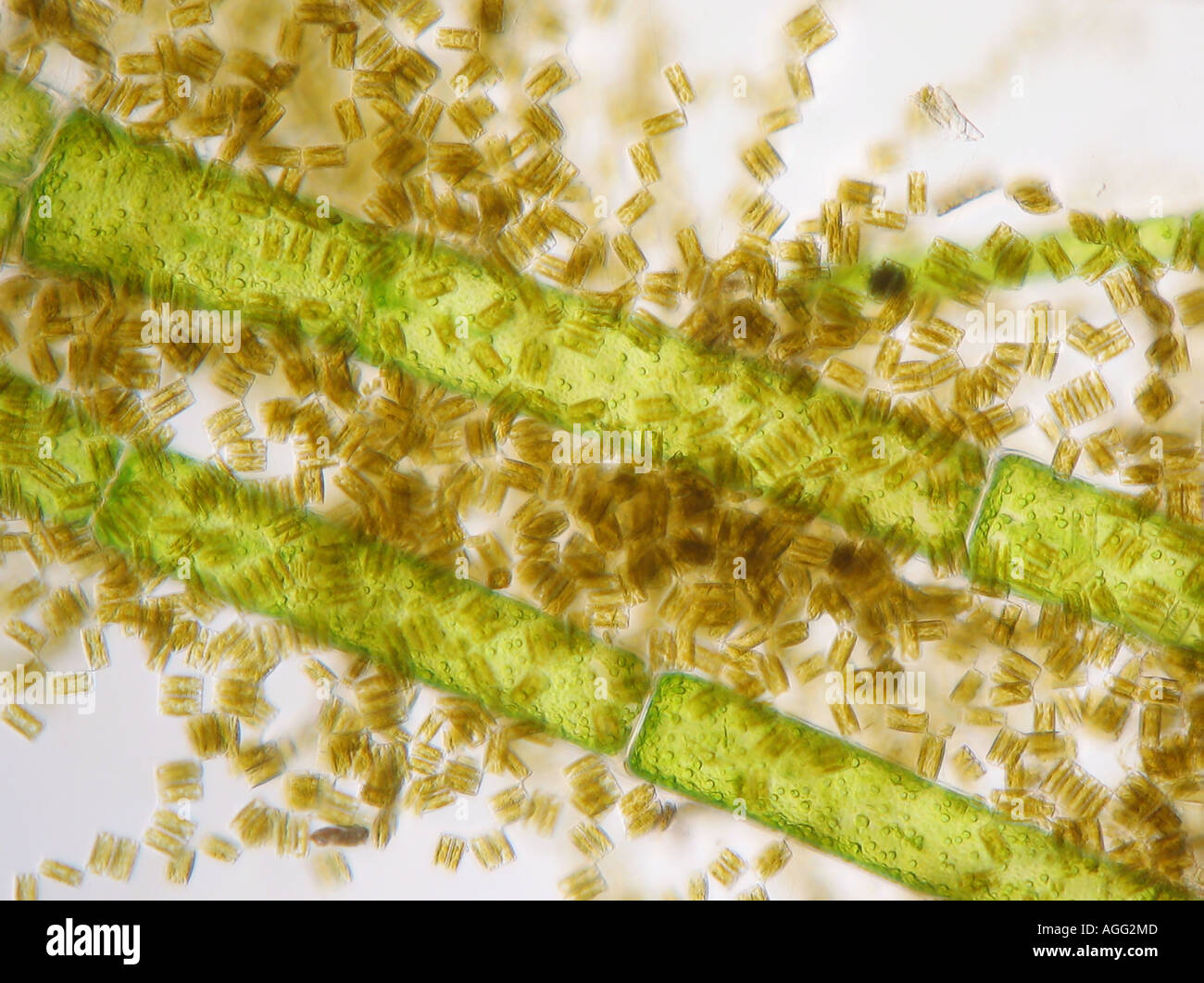 Diatoma vulgare (Diatoma vulgare), at Cladophora glomerata, in shining-through light Stock Photohttps://www.alamy.com/image-license-details/?v=1https://www.alamy.com/diatoma-vulgare-diatoma-vulgare-at-cladophora-glomerata-in-shining-image8114636.html
RMAGG2MD–Diatoma vulgare (Diatoma vulgare), at Cladophora glomerata, in shining-through light
 . Algæ. Vol. I. Myxophyceæ, Peridinieæ, Bacillarieæ, Chlorophyceæ, together with a brief summary of the occurrence and distribution of freshwat4er Algæ . lls arejoined in an irregularly zigzag manner at the corners of the valves (Tabel-laria, some species of Diatoma, Triceratium, arid Biddulphia). In allthese cases the attachment is by mucus secreted by the cells themselves.The cells of the ribbon-shaped and thread-like colonies are joined eitherby a layer of mucus or by mucous strands between the closely applied valve-faces, and the cells of the zigzag colonies are united by small mucouscushi Stock Photohttps://www.alamy.com/image-license-details/?v=1https://www.alamy.com/alg-vol-i-myxophyce-peridinie-bacillarie-chlorophyce-together-with-a-brief-summary-of-the-occurrence-and-distribution-of-freshwat4er-alg-lls-arejoined-in-an-irregularly-zigzag-manner-at-the-corners-of-the-valves-tabel-laria-some-species-of-diatoma-triceratium-arid-biddulphia-in-allthese-cases-the-attachment-is-by-mucus-secreted-by-the-cells-themselvesthe-cells-of-the-ribbon-shaped-and-thread-like-colonies-are-joined-eitherby-a-layer-of-mucus-or-by-mucous-strands-between-the-closely-applied-valve-faces-and-the-cells-of-the-zigzag-colonies-are-united-by-small-mucouscushi-image369755619.html
RM2CDFR1R–. Algæ. Vol. I. Myxophyceæ, Peridinieæ, Bacillarieæ, Chlorophyceæ, together with a brief summary of the occurrence and distribution of freshwat4er Algæ . lls arejoined in an irregularly zigzag manner at the corners of the valves (Tabel-laria, some species of Diatoma, Triceratium, arid Biddulphia). In allthese cases the attachment is by mucus secreted by the cells themselves.The cells of the ribbon-shaped and thread-like colonies are joined eitherby a layer of mucus or by mucous strands between the closely applied valve-faces, and the cells of the zigzag colonies are united by small mucouscushi
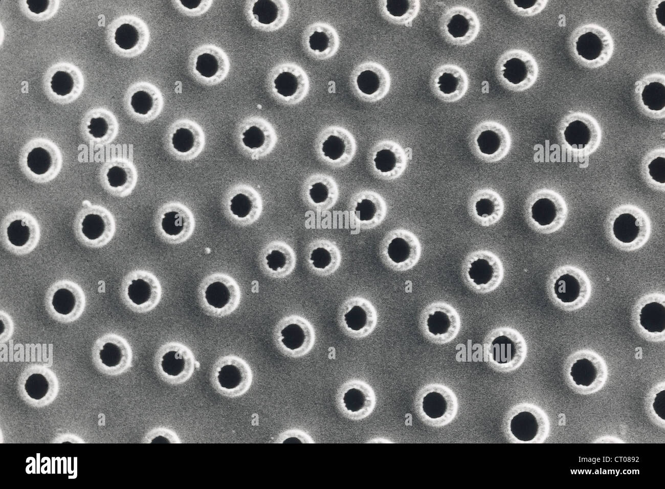 DIATOM, SEM Stock Photohttps://www.alamy.com/image-license-details/?v=1https://www.alamy.com/stock-photo-diatom-sem-49179006.html
RMCT0892–DIATOM, SEM
 Freshwater diatoms Newton Beck, S Yorks. UK Stock Photohttps://www.alamy.com/image-license-details/?v=1https://www.alamy.com/freshwater-diatoms-newton-beck-s-yorks-uk-image352206508.html
RM2BD0AYT–Freshwater diatoms Newton Beck, S Yorks. UK
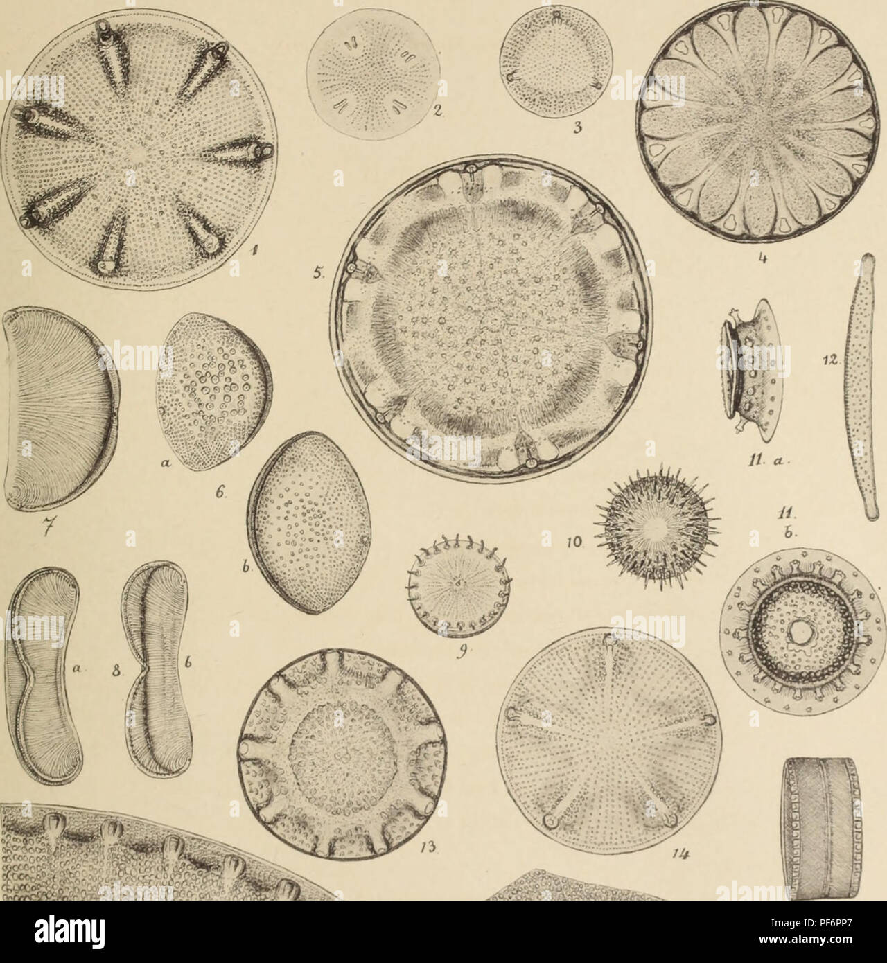 . Diatomées fossiles du Japon. Espèces marines & nouvelles des calcaires argileux de Sendaï & Yedo. Paleobotany. Diatomées Fossiles PI. IV.. '"^fal ' â â. Please note that these images are extracted from scanned page images that may have been digitally enhanced for readability - coloration and appearance of these illustrations may not perfectly resemble the original work.. Brun, Jacques, 1826-1908; Tempère, Joannes Albert, 1847-1926. Leipzig, Theodor Oswald Wigel, Leipzig Stock Photohttps://www.alamy.com/image-license-details/?v=1https://www.alamy.com/diatomes-fossiles-du-japon-espces-marines-amp-nouvelles-des-calcaires-argileux-de-senda-amp-yedo-paleobotany-diatomes-fossiles-pi-iv-quotfal-please-note-that-these-images-are-extracted-from-scanned-page-images-that-may-have-been-digitally-enhanced-for-readability-coloration-and-appearance-of-these-illustrations-may-not-perfectly-resemble-the-original-work-brun-jacques-1826-1908-tempre-joannes-albert-1847-1926-leipzig-theodor-oswald-wigel-leipzig-image215893839.html
RMPF6PP7–. Diatomées fossiles du Japon. Espèces marines & nouvelles des calcaires argileux de Sendaï & Yedo. Paleobotany. Diatomées Fossiles PI. IV.. '"^fal ' â â. Please note that these images are extracted from scanned page images that may have been digitally enhanced for readability - coloration and appearance of these illustrations may not perfectly resemble the original work.. Brun, Jacques, 1826-1908; Tempère, Joannes Albert, 1847-1926. Leipzig, Theodor Oswald Wigel, Leipzig
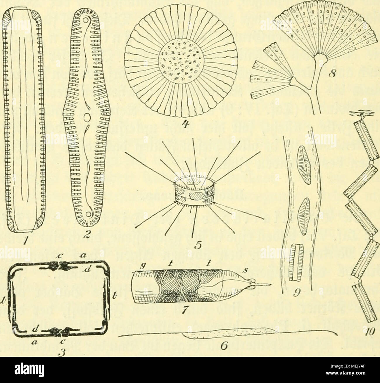 . Die Algen, Moose und Farnpflanzen. . <raf. vi. 1—3. Pinnularia viridis: 1. Don ber ©firtet&anbfeite, 2. bon ber ©dmtenfeite, 3.£utei1d)uitt: a Gdjate, b ©firtelMnber, c3fJopIic, dfiatnntctn. 4. Planktoniella Sol. 5. Stephanodiscus faec. 6. Rhizosolenia setigera. 7. Wembrcmhälfte »Ott Rhizosolenia styliformis: s Gdjale, t Shrtfdjenbätibet, g ©üttelbcnb. 8. Licmophora flabellata. 9. Cymbella (Encyonema) caespitoaa. 10. Diatoma vulgare. Seiften, Kammern unb Sßorcn in ber Membran ruft eine äufcerft äterlidje geidjnung berfetben l)croor (Xaf. VI: 1—3). Xer ftarre S5au bcr Membranen bebingt Stock Photohttps://www.alamy.com/image-license-details/?v=1https://www.alamy.com/die-algen-moose-und-farnpflanzen-ltraf-vi-13-pinnularia-viridis-1-don-ber-firtetampanbfeite-2-bon-ber-dmtenfeite-3utei1duitt-a-gdjate-b-firtelmnber-c3fjopiic-dfiatnntctn-4-planktoniella-sol-5-stephanodiscus-faec-6-rhizosolenia-setigera-7-wembrcmhlfte-ott-rhizosolenia-styliformis-s-gdjale-t-shrtfdjenbtibet-g-ttelbcnb-8-licmophora-flabellata-9-cymbella-encyonema-caespitoaa-10-diatoma-vulgare-seiften-kammern-unb-sorcn-in-ber-membran-ruft-eine-ufcerft-terlidje-geidjnung-berfetben-lcroor-xaf-vi-13-xer-ftarre-s5au-bcr-membranen-bebingt-image181125302.html
RMMEJY4P–. Die Algen, Moose und Farnpflanzen. . <raf. vi. 1—3. Pinnularia viridis: 1. Don ber ©firtet&anbfeite, 2. bon ber ©dmtenfeite, 3.£utei1d)uitt: a Gdjate, b ©firtelMnber, c3fJopIic, dfiatnntctn. 4. Planktoniella Sol. 5. Stephanodiscus faec. 6. Rhizosolenia setigera. 7. Wembrcmhälfte »Ott Rhizosolenia styliformis: s Gdjale, t Shrtfdjenbätibet, g ©üttelbcnb. 8. Licmophora flabellata. 9. Cymbella (Encyonema) caespitoaa. 10. Diatoma vulgare. Seiften, Kammern unb Sßorcn in ber Membran ruft eine äufcerft äterlidje geidjnung berfetben l)croor (Xaf. VI: 1—3). Xer ftarre S5au bcr Membranen bebingt
 . Algæ. Vol. I. Myxophyceæ, Peridinieæ, Bacillarieæ, Chlorophyceæ, together with a brief summary of the occurrence and distribution of freshwat4er Algæ . Fig. 72. Parts of the valves of various diatoms showing the pores through which mucus issecreted. A, Diatoma grande W. Sm. B and C, Grammatophora serpentina Kiitz., girdleand valve views respectively. D, Syncdra Ulna (Nitzsch) Ehrenb. var. splendens (Kiitz.)V. Heurck. E, Fragilaria virescens Ealfs. F, Tabellaria fenestrata (Lyngb.) Kiitz. p, pore.All x 2200. (After 0. Miiller.) In this diatom, and in the plankton-species of Asterionella, Voig Stock Photohttps://www.alamy.com/image-license-details/?v=1https://www.alamy.com/alg-vol-i-myxophyce-peridinie-bacillarie-chlorophyce-together-with-a-brief-summary-of-the-occurrence-and-distribution-of-freshwat4er-alg-fig-72-parts-of-the-valves-of-various-diatoms-showing-the-pores-through-which-mucus-issecreted-a-diatoma-grande-w-sm-b-and-c-grammatophora-serpentina-kiitz-girdleand-valve-views-respectively-d-syncdra-ulna-nitzsch-ehrenb-var-splendens-kiitzv-heurck-e-fragilaria-virescens-ealfs-f-tabellaria-fenestrata-lyngb-kiitz-p-poreall-x-2200-after-0-miiller-in-this-diatom-and-in-the-plankton-species-of-asterionella-voig-image372068622.html
RM2CH9592–. Algæ. Vol. I. Myxophyceæ, Peridinieæ, Bacillarieæ, Chlorophyceæ, together with a brief summary of the occurrence and distribution of freshwat4er Algæ . Fig. 72. Parts of the valves of various diatoms showing the pores through which mucus issecreted. A, Diatoma grande W. Sm. B and C, Grammatophora serpentina Kiitz., girdleand valve views respectively. D, Syncdra Ulna (Nitzsch) Ehrenb. var. splendens (Kiitz.)V. Heurck. E, Fragilaria virescens Ealfs. F, Tabellaria fenestrata (Lyngb.) Kiitz. p, pore.All x 2200. (After 0. Miiller.) In this diatom, and in the plankton-species of Asterionella, Voig
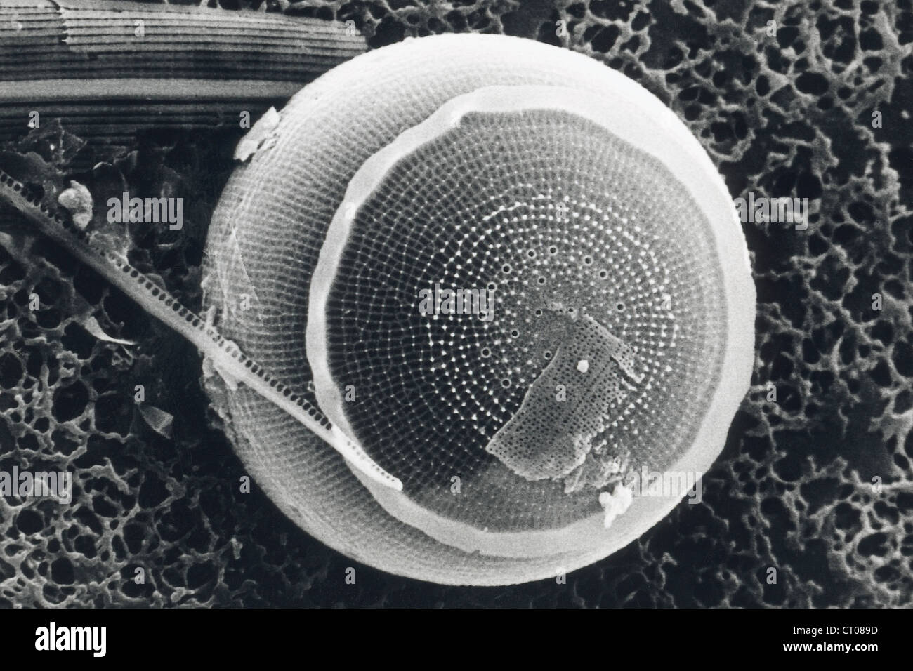 DIATOM, SEM Stock Photohttps://www.alamy.com/image-license-details/?v=1https://www.alamy.com/stock-photo-diatom-sem-49179017.html
RMCT089D–DIATOM, SEM
 Freshwater diatoms Newton Beck, S Yorks. UK Stock Photohttps://www.alamy.com/image-license-details/?v=1https://www.alamy.com/freshwater-diatoms-newton-beck-s-yorks-uk-image352206810.html
RM2BD0BAJ–Freshwater diatoms Newton Beck, S Yorks. UK
 . Diatomées marines de France et des districts maritimes voisins. Diatoms. Peragallo. - Diatomées de France. l'i. i.. i - â 'â '. Please note that these images are extracted from scanned page images that may have been digitally enhanced for readability - coloration and appearance of these illustrations may not perfectly resemble the original work.. Péragallo, H. (Hippolyte), b. 1851; Péragallo, M. (Maurice), b. 1853. Grez-sur-Loing, M. J. Tempère Stock Photohttps://www.alamy.com/image-license-details/?v=1https://www.alamy.com/diatomes-marines-de-france-et-des-districts-maritimes-voisins-diatoms-peragallo-diatomes-de-france-li-i-i-please-note-that-these-images-are-extracted-from-scanned-page-images-that-may-have-been-digitally-enhanced-for-readability-coloration-and-appearance-of-these-illustrations-may-not-perfectly-resemble-the-original-work-pragallo-h-hippolyte-b-1851-pragallo-m-maurice-b-1853-grez-sur-loing-m-j-tempre-image215893389.html
RMPF6P65–. Diatomées marines de France et des districts maritimes voisins. Diatoms. Peragallo. - Diatomées de France. l'i. i.. i - â 'â '. Please note that these images are extracted from scanned page images that may have been digitally enhanced for readability - coloration and appearance of these illustrations may not perfectly resemble the original work.. Péragallo, H. (Hippolyte), b. 1851; Péragallo, M. (Maurice), b. 1853. Grez-sur-Loing, M. J. Tempère
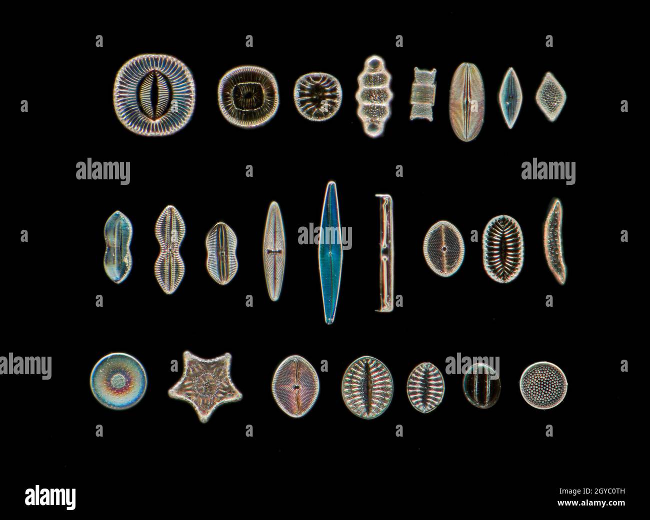 Diatom biodiversity, darkfield photomicrograph Stock Photohttps://www.alamy.com/image-license-details/?v=1https://www.alamy.com/diatom-biodiversity-darkfield-photomicrograph-image447119025.html
RM2GYC0TH–Diatom biodiversity, darkfield photomicrograph
![. Dictionnaire des sciences naturelles [electronic resource] : dans lequel on traite méthodiquement des différens êtres de la nature, considérés soit en eux-mêmes, d'après l'état actuel de nos connoissances, soit relativement à l'utilité qu'en peuvent retirer la médecine, l'agriculture, le commerce et les artes. Suivi d'une biographie des plus célèbres naturalistes . . . DIAJ'OMA Vlllgains. /I,>r,/ /^''â¢/â r/ax.r.A ,-/h ./,//m- ,/n>.r,r/.r. i.DIATOMA S wcutzii. /,/,â/ ô./)/I/O M A aiiiha./,//,</ i.FRAlrlLAIUA uni|)iiiio(ala . /,///,/. Stock Photo . Dictionnaire des sciences naturelles [electronic resource] : dans lequel on traite méthodiquement des différens êtres de la nature, considérés soit en eux-mêmes, d'après l'état actuel de nos connoissances, soit relativement à l'utilité qu'en peuvent retirer la médecine, l'agriculture, le commerce et les artes. Suivi d'une biographie des plus célèbres naturalistes . . . DIAJ'OMA Vlllgains. /I,>r,/ /^''â¢/â r/ax.r.A ,-/h ./,//m- ,/n>.r,r/.r. i.DIATOMA S wcutzii. /,/,â/ ô./)/I/O M A aiiiha./,//,</ i.FRAlrlLAIUA uni|)iiiio(ala . /,///,/. Stock Photo](https://c8.alamy.com/comp/MEM8FH/dictionnaire-des-sciences-naturelles-electronic-resource-dans-lequel-on-traite-mthodiquement-des-diffrens-tres-de-la-nature-considrs-soit-en-eux-mmes-daprs-ltat-actuel-de-nos-connoissances-soit-relativement-lutilit-quen-peuvent-retirer-la-mdecine-lagriculture-le-commerce-et-les-artes-suivi-dune-biographie-des-plus-clbres-naturalistes-diajoma-vlllgains-igtr-raxra-h-m-ngtrrr-idiatoma-s-wcutzii-io-m-a-aiiihalt-ifralrllaiua-uniiiiioala-MEM8FH.jpg) . Dictionnaire des sciences naturelles [electronic resource] : dans lequel on traite méthodiquement des différens êtres de la nature, considérés soit en eux-mêmes, d'après l'état actuel de nos connoissances, soit relativement à l'utilité qu'en peuvent retirer la médecine, l'agriculture, le commerce et les artes. Suivi d'une biographie des plus célèbres naturalistes . . . DIAJ'OMA Vlllgains. /I,>r,/ /^''â¢/â r/ax.r.A ,-/h ./,//m- ,/n>.r,r/.r. i.DIATOMA S wcutzii. /,/,â/ ô./)/I/O M A aiiiha./,//,</ i.FRAlrlLAIUA uni|)iiiio(ala . /,///,/. Stock Photohttps://www.alamy.com/image-license-details/?v=1https://www.alamy.com/dictionnaire-des-sciences-naturelles-electronic-resource-dans-lequel-on-traite-mthodiquement-des-diffrens-tres-de-la-nature-considrs-soit-en-eux-mmes-daprs-ltat-actuel-de-nos-connoissances-soit-relativement-lutilit-quen-peuvent-retirer-la-mdecine-lagriculture-le-commerce-et-les-artes-suivi-dune-biographie-des-plus-clbres-naturalistes-diajoma-vlllgains-igtr-raxra-h-m-ngtrrr-idiatoma-s-wcutzii-io-m-a-aiiihalt-ifralrllaiua-uniiiiioala-image181154613.html
. Dictionnaire des sciences naturelles [electronic resource] : dans lequel on traite méthodiquement des différens êtres de la nature, considérés soit en eux-mêmes, d'après l'état actuel de nos connoissances, soit relativement à l'utilité qu'en peuvent retirer la médecine, l'agriculture, le commerce et les artes. Suivi d'une biographie des plus célèbres naturalistes . . . DIAJ'OMA Vlllgains. /I,>r,/ /^''â¢/â r/ax.r.A ,-/h ./,//m- ,/n>.r,r/.r. i.DIATOMA S wcutzii. /,/,â/ ô./)/I/O M A aiiiha./,//,</ i.FRAlrlLAIUA uni|)iiiio(ala . /,///,/. Stock Photohttps://www.alamy.com/image-license-details/?v=1https://www.alamy.com/dictionnaire-des-sciences-naturelles-electronic-resource-dans-lequel-on-traite-mthodiquement-des-diffrens-tres-de-la-nature-considrs-soit-en-eux-mmes-daprs-ltat-actuel-de-nos-connoissances-soit-relativement-lutilit-quen-peuvent-retirer-la-mdecine-lagriculture-le-commerce-et-les-artes-suivi-dune-biographie-des-plus-clbres-naturalistes-diajoma-vlllgains-igtr-raxra-h-m-ngtrrr-idiatoma-s-wcutzii-io-m-a-aiiihalt-ifralrllaiua-uniiiiioala-image181154613.htmlRMMEM8FH–. Dictionnaire des sciences naturelles [electronic resource] : dans lequel on traite méthodiquement des différens êtres de la nature, considérés soit en eux-mêmes, d'après l'état actuel de nos connoissances, soit relativement à l'utilité qu'en peuvent retirer la médecine, l'agriculture, le commerce et les artes. Suivi d'une biographie des plus célèbres naturalistes . . . DIAJ'OMA Vlllgains. /I,>r,/ /^''â¢/â r/ax.r.A ,-/h ./,//m- ,/n>.r,r/.r. i.DIATOMA S wcutzii. /,/,â/ ô./)/I/O M A aiiiha./,//,</ i.FRAlrlLAIUA uni|)iiiio(ala . /,///,/.
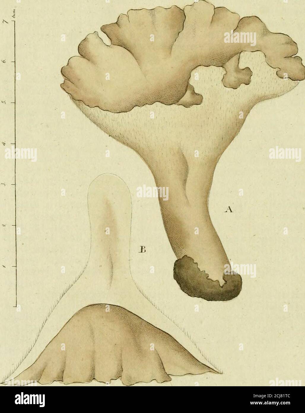 . Svensk botanik . ch den gröna massan, som förut liktett band genomstrukit tråden, samlar sig i vinklarne afleder ne. Af de här upptagne arter lefva de två i färskt vatten,neml. D Swortzii, som blifvit funnen både i Halland, Vest-manland och Vermland, och D. tennis som finnes i en bäckvid Fogelsang nära Lund. Deremot finnes D. fasciculalai Ii al vet på Ceramier, hvilka den som små k» ist fillika nå-lar betäcker. A * * *. Tab. Fig. x. Ett knippe af Diatoma Swartzii i nat. stor-lek. — 2. En tråd före copulationen. — 3. Två trådar co-pulerade samt upplöste, båda mycket förstorade. 4- Diatoma ten Stock Photohttps://www.alamy.com/image-license-details/?v=1https://www.alamy.com/svensk-botanik-ch-den-grna-massan-som-frut-liktett-band-genomstrukit-trden-samlar-sig-i-vinklarne-afleder-ne-af-de-hr-upptagne-arter-lefva-de-tv-i-frskt-vattenneml-d-swortzii-som-blifvit-funnen-bde-i-halland-vest-manland-och-vermland-och-d-tennis-som-finnes-i-en-bckvid-fogelsang-nra-lund-deremot-finnes-d-fasciculalai-ii-al-vet-p-ceramier-hvilka-den-som-sm-k-ist-fillika-n-lar-betcker-a-tab-fig-x-ett-knippe-af-diatoma-swartzii-i-nat-stor-lek-2-en-trd-fre-copulationen-3-tv-trdar-co-pulerade-samt-upplste-bda-mycket-frstorade-4-diatoma-ten-image372658620.html
RM2CJ81TC–. Svensk botanik . ch den gröna massan, som förut liktett band genomstrukit tråden, samlar sig i vinklarne afleder ne. Af de här upptagne arter lefva de två i färskt vatten,neml. D Swortzii, som blifvit funnen både i Halland, Vest-manland och Vermland, och D. tennis som finnes i en bäckvid Fogelsang nära Lund. Deremot finnes D. fasciculalai Ii al vet på Ceramier, hvilka den som små k» ist fillika nå-lar betäcker. A * * *. Tab. Fig. x. Ett knippe af Diatoma Swartzii i nat. stor-lek. — 2. En tråd före copulationen. — 3. Två trådar co-pulerade samt upplöste, båda mycket förstorade. 4- Diatoma ten
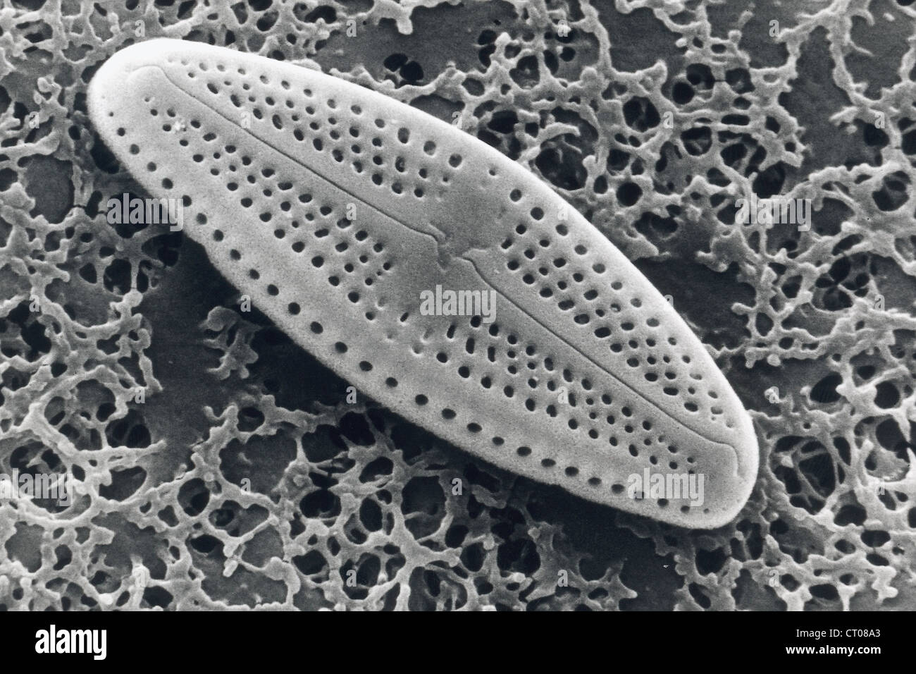 DIATOM, SEM Stock Photohttps://www.alamy.com/image-license-details/?v=1https://www.alamy.com/stock-photo-diatom-sem-49179035.html
RMCT08A3–DIATOM, SEM
 . Diatomées marines de France et des districts maritimes voisins. Diatoms. Peraqallo. - Diatomées de France. l'I I.WII.. I.' M ''..â ^n 1V.|. Please note that these images are extracted from scanned page images that may have been digitally enhanced for readability - coloration and appearance of these illustrations may not perfectly resemble the original work.. Péragallo, H. (Hippolyte), b. 1851; Péragallo, M. (Maurice), b. 1853. Grez-sur-Loing, M. J. Tempère Stock Photohttps://www.alamy.com/image-license-details/?v=1https://www.alamy.com/diatomes-marines-de-france-et-des-districts-maritimes-voisins-diatoms-peraqallo-diatomes-de-france-li-iwii-i-m-n-1v-please-note-that-these-images-are-extracted-from-scanned-page-images-that-may-have-been-digitally-enhanced-for-readability-coloration-and-appearance-of-these-illustrations-may-not-perfectly-resemble-the-original-work-pragallo-h-hippolyte-b-1851-pragallo-m-maurice-b-1853-grez-sur-loing-m-j-tempre-image215893296.html
RMPF6P2T–. Diatomées marines de France et des districts maritimes voisins. Diatoms. Peraqallo. - Diatomées de France. l'I I.WII.. I.' M ''..â ^n 1V.|. Please note that these images are extracted from scanned page images that may have been digitally enhanced for readability - coloration and appearance of these illustrations may not perfectly resemble the original work.. Péragallo, H. (Hippolyte), b. 1851; Péragallo, M. (Maurice), b. 1853. Grez-sur-Loing, M. J. Tempère
![. Dictionnaire des sciences naturelles [electronic resource] : dans lequel on traite méthodiquement des différens êtres de la nature, considérés soit en eux-mêmes, d'après l'état actuel de nos connoissances, soit relativement à l'utilité qu'en peuvent retirer la médecine, l'agriculture, le commerce et les artes. Suivi d'une biographie des plus célèbres naturalistes . . 7itr/<i',i ^à l.tff/ ,/,re.i f^ J,yrA.r Bon/ r/ Ayruffiir . .l/,uvâr,/ .r,i,//. iDLiTOMA TlIgai'is. /Iân, M-Z.c/a.i-j-.^i'/h.A/.ùÃiy/w.n.r. â i.DIATOMA Swai-tzii. /,//â/ Z.DIUOMA aiiriia./,//., i.FIiAd/lA/i/A un Stock Photo . Dictionnaire des sciences naturelles [electronic resource] : dans lequel on traite méthodiquement des différens êtres de la nature, considérés soit en eux-mêmes, d'après l'état actuel de nos connoissances, soit relativement à l'utilité qu'en peuvent retirer la médecine, l'agriculture, le commerce et les artes. Suivi d'une biographie des plus célèbres naturalistes . . 7itr/<i',i ^à l.tff/ ,/,re.i f^ J,yrA.r Bon/ r/ Ayruffiir . .l/,uvâr,/ .r,i,//. iDLiTOMA TlIgai'is. /Iân, M-Z.c/a.i-j-.^i'/h.A/.ùÃiy/w.n.r. â i.DIATOMA Swai-tzii. /,//â/ Z.DIUOMA aiiriia./,//., i.FIiAd/lA/i/A un Stock Photo](https://c8.alamy.com/comp/MEM8FB/dictionnaire-des-sciences-naturelles-electronic-resource-dans-lequel-on-traite-mthodiquement-des-diffrens-tres-de-la-nature-considrs-soit-en-eux-mmes-daprs-ltat-actuel-de-nos-connoissances-soit-relativement-lutilit-quen-peuvent-retirer-la-mdecine-lagriculture-le-commerce-et-les-artes-suivi-dune-biographie-des-plus-clbres-naturalistes-7itrltii-ltff-rei-f-jyrar-bon-r-ayruffiir-luvr-ri-idlitoma-tligaiis-in-m-zcai-j-ihaiywnr-idiatoma-swai-tzii-zdiuoma-aiiriia-ifiiadlaia-un-MEM8FB.jpg) . Dictionnaire des sciences naturelles [electronic resource] : dans lequel on traite méthodiquement des différens êtres de la nature, considérés soit en eux-mêmes, d'après l'état actuel de nos connoissances, soit relativement à l'utilité qu'en peuvent retirer la médecine, l'agriculture, le commerce et les artes. Suivi d'une biographie des plus célèbres naturalistes . . 7itr/<i',i ^à l.tff/ ,/,re.i f^ J,yrA.r Bon/ r/ Ayruffiir . .l/,uvâr,/ .r,i,//. iDLiTOMA TlIgai'is. /Iân, M-Z.c/a.i-j-.^i'/h.A/.ùÃiy/w.n.r. â i.DIATOMA Swai-tzii. /,//â/ Z.DIUOMA aiiriia./,//., i.FIiAd/lA/i/A un Stock Photohttps://www.alamy.com/image-license-details/?v=1https://www.alamy.com/dictionnaire-des-sciences-naturelles-electronic-resource-dans-lequel-on-traite-mthodiquement-des-diffrens-tres-de-la-nature-considrs-soit-en-eux-mmes-daprs-ltat-actuel-de-nos-connoissances-soit-relativement-lutilit-quen-peuvent-retirer-la-mdecine-lagriculture-le-commerce-et-les-artes-suivi-dune-biographie-des-plus-clbres-naturalistes-7itrltii-ltff-rei-f-jyrar-bon-r-ayruffiir-luvr-ri-idlitoma-tligaiis-in-m-zcai-j-ihaiywnr-idiatoma-swai-tzii-zdiuoma-aiiriia-ifiiadlaia-un-image181154607.html
. Dictionnaire des sciences naturelles [electronic resource] : dans lequel on traite méthodiquement des différens êtres de la nature, considérés soit en eux-mêmes, d'après l'état actuel de nos connoissances, soit relativement à l'utilité qu'en peuvent retirer la médecine, l'agriculture, le commerce et les artes. Suivi d'une biographie des plus célèbres naturalistes . . 7itr/<i',i ^à l.tff/ ,/,re.i f^ J,yrA.r Bon/ r/ Ayruffiir . .l/,uvâr,/ .r,i,//. iDLiTOMA TlIgai'is. /Iân, M-Z.c/a.i-j-.^i'/h.A/.ùÃiy/w.n.r. â i.DIATOMA Swai-tzii. /,//â/ Z.DIUOMA aiiriia./,//., i.FIiAd/lA/i/A un Stock Photohttps://www.alamy.com/image-license-details/?v=1https://www.alamy.com/dictionnaire-des-sciences-naturelles-electronic-resource-dans-lequel-on-traite-mthodiquement-des-diffrens-tres-de-la-nature-considrs-soit-en-eux-mmes-daprs-ltat-actuel-de-nos-connoissances-soit-relativement-lutilit-quen-peuvent-retirer-la-mdecine-lagriculture-le-commerce-et-les-artes-suivi-dune-biographie-des-plus-clbres-naturalistes-7itrltii-ltff-rei-f-jyrar-bon-r-ayruffiir-luvr-ri-idlitoma-tligaiis-in-m-zcai-j-ihaiywnr-idiatoma-swai-tzii-zdiuoma-aiiriia-ifiiadlaia-un-image181154607.htmlRMMEM8FB–. Dictionnaire des sciences naturelles [electronic resource] : dans lequel on traite méthodiquement des différens êtres de la nature, considérés soit en eux-mêmes, d'après l'état actuel de nos connoissances, soit relativement à l'utilité qu'en peuvent retirer la médecine, l'agriculture, le commerce et les artes. Suivi d'une biographie des plus célèbres naturalistes . . 7itr/<i',i ^à l.tff/ ,/,re.i f^ J,yrA.r Bon/ r/ Ayruffiir . .l/,uvâr,/ .r,i,//. iDLiTOMA TlIgai'is. /Iân, M-Z.c/a.i-j-.^i'/h.A/.ùÃiy/w.n.r. â i.DIATOMA Swai-tzii. /,//â/ Z.DIUOMA aiiriia./,//., i.FIiAd/lA/i/A un
 . An annotated key to the identification of commonly occurring and dominant genera of algae observed in the phytoplankton of the United States. Freshwater algae -- United States Identification; Freshwater algae -- United States; Freshwater phytoplankton -- United States Identification; Freshwater phytoplankton -- United States. DESCRIPTION OF THE GENERA 71. Figure 29.-Photomicrograph of Diatoma.. Please note that these images are extracted from scanned page images that may have been digitally enhanced for readability - coloration and appearance of these illustrations may not perfectly resemble Stock Photohttps://www.alamy.com/image-license-details/?v=1https://www.alamy.com/an-annotated-key-to-the-identification-of-commonly-occurring-and-dominant-genera-of-algae-observed-in-the-phytoplankton-of-the-united-states-freshwater-algae-united-states-identification-freshwater-algae-united-states-freshwater-phytoplankton-united-states-identification-freshwater-phytoplankton-united-states-description-of-the-genera-71-figure-29-photomicrograph-of-diatoma-please-note-that-these-images-are-extracted-from-scanned-page-images-that-may-have-been-digitally-enhanced-for-readability-coloration-and-appearance-of-these-illustrations-may-not-perfectly-resemble-image236393254.html
RMRMGJ06–. An annotated key to the identification of commonly occurring and dominant genera of algae observed in the phytoplankton of the United States. Freshwater algae -- United States Identification; Freshwater algae -- United States; Freshwater phytoplankton -- United States Identification; Freshwater phytoplankton -- United States. DESCRIPTION OF THE GENERA 71. Figure 29.-Photomicrograph of Diatoma.. Please note that these images are extracted from scanned page images that may have been digitally enhanced for readability - coloration and appearance of these illustrations may not perfectly resemble
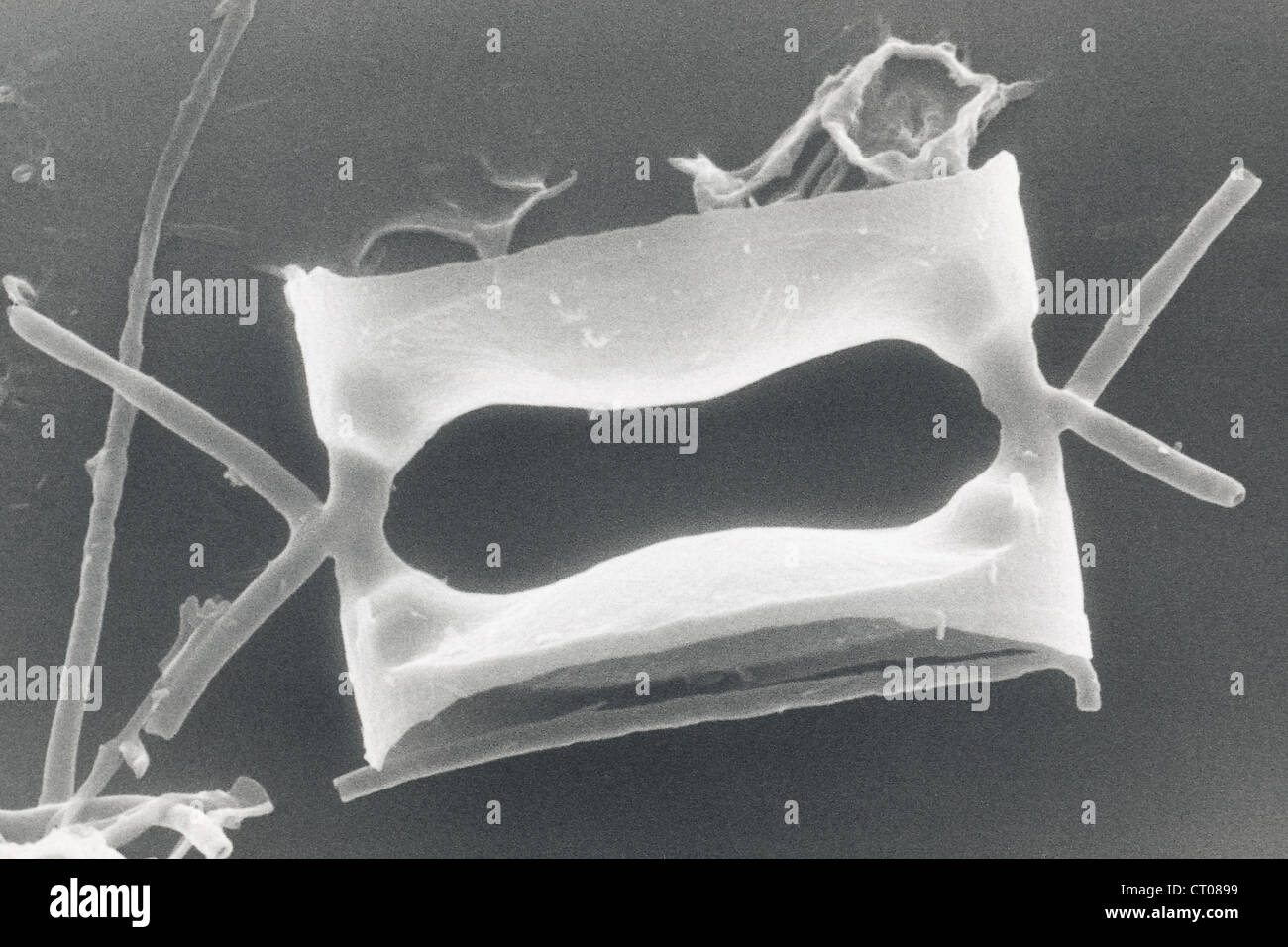 DIATOM, SEM Stock Photohttps://www.alamy.com/image-license-details/?v=1https://www.alamy.com/stock-photo-diatom-sem-49179013.html
RMCT0899–DIATOM, SEM
 . Diatomées marines de France et des districts maritimes voisins. Diatoms. Peragallo. - Diatomées de Ffance. PI. cxix. (. M l'Kh%i.MI-'i â !â. Please note that these images are extracted from scanned page images that may have been digitally enhanced for readability - coloration and appearance of these illustrations may not perfectly resemble the original work.. Péragallo, H. (Hippolyte), b. 1851; Péragallo, M. (Maurice), b. 1853. Grez-sur-Loing, M. J. Tempère Stock Photohttps://www.alamy.com/image-license-details/?v=1https://www.alamy.com/diatomes-marines-de-france-et-des-districts-maritimes-voisins-diatoms-peragallo-diatomes-de-ffance-pi-cxix-m-lkhimi-i-!-please-note-that-these-images-are-extracted-from-scanned-page-images-that-may-have-been-digitally-enhanced-for-readability-coloration-and-appearance-of-these-illustrations-may-not-perfectly-resemble-the-original-work-pragallo-h-hippolyte-b-1851-pragallo-m-maurice-b-1853-grez-sur-loing-m-j-tempre-image215893178.html
RMPF6NXJ–. Diatomées marines de France et des districts maritimes voisins. Diatoms. Peragallo. - Diatomées de Ffance. PI. cxix. (. M l'Kh%i.MI-'i â !â. Please note that these images are extracted from scanned page images that may have been digitally enhanced for readability - coloration and appearance of these illustrations may not perfectly resemble the original work.. Péragallo, H. (Hippolyte), b. 1851; Péragallo, M. (Maurice), b. 1853. Grez-sur-Loing, M. J. Tempère
![. Diatomaceæ of North America [microform] : illustrated with twenty-three hundred figures from the author's drawings on one hundred and twelve plates. Diatoms; Algae; Diatomées; Algues. p(7^^,,ii>s^. â m ^J3;V/. Please note that these images are extracted from scanned page images that may have been digitally enhanced for readability - coloration and appearance of these illustrations may not perfectly resemble the original work.. Wolle, Francis, 1817-1893. Bethlehem, Pa. : Comenius Press Stock Photo . Diatomaceæ of North America [microform] : illustrated with twenty-three hundred figures from the author's drawings on one hundred and twelve plates. Diatoms; Algae; Diatomées; Algues. p(7^^,,ii>s^. â m ^J3;V/. Please note that these images are extracted from scanned page images that may have been digitally enhanced for readability - coloration and appearance of these illustrations may not perfectly resemble the original work.. Wolle, Francis, 1817-1893. Bethlehem, Pa. : Comenius Press Stock Photo](https://c8.alamy.com/comp/RJ1974/diatomace-of-north-america-microform-illustrated-with-twenty-three-hundred-figures-from-the-authors-drawings-on-one-hundred-and-twelve-plates-diatoms-algae-diatomes-algues-p7iigts-m-j3v-please-note-that-these-images-are-extracted-from-scanned-page-images-that-may-have-been-digitally-enhanced-for-readability-coloration-and-appearance-of-these-illustrations-may-not-perfectly-resemble-the-original-work-wolle-francis-1817-1893-bethlehem-pa-comenius-press-RJ1974.jpg) . Diatomaceæ of North America [microform] : illustrated with twenty-three hundred figures from the author's drawings on one hundred and twelve plates. Diatoms; Algae; Diatomées; Algues. p(7^^,,ii>s^. â m ^J3;V/. Please note that these images are extracted from scanned page images that may have been digitally enhanced for readability - coloration and appearance of these illustrations may not perfectly resemble the original work.. Wolle, Francis, 1817-1893. Bethlehem, Pa. : Comenius Press Stock Photohttps://www.alamy.com/image-license-details/?v=1https://www.alamy.com/diatomace-of-north-america-microform-illustrated-with-twenty-three-hundred-figures-from-the-authors-drawings-on-one-hundred-and-twelve-plates-diatoms-algae-diatomes-algues-p7iigts-m-j3v-please-note-that-these-images-are-extracted-from-scanned-page-images-that-may-have-been-digitally-enhanced-for-readability-coloration-and-appearance-of-these-illustrations-may-not-perfectly-resemble-the-original-work-wolle-francis-1817-1893-bethlehem-pa-comenius-press-image234827800.html
. Diatomaceæ of North America [microform] : illustrated with twenty-three hundred figures from the author's drawings on one hundred and twelve plates. Diatoms; Algae; Diatomées; Algues. p(7^^,,ii>s^. â m ^J3;V/. Please note that these images are extracted from scanned page images that may have been digitally enhanced for readability - coloration and appearance of these illustrations may not perfectly resemble the original work.. Wolle, Francis, 1817-1893. Bethlehem, Pa. : Comenius Press Stock Photohttps://www.alamy.com/image-license-details/?v=1https://www.alamy.com/diatomace-of-north-america-microform-illustrated-with-twenty-three-hundred-figures-from-the-authors-drawings-on-one-hundred-and-twelve-plates-diatoms-algae-diatomes-algues-p7iigts-m-j3v-please-note-that-these-images-are-extracted-from-scanned-page-images-that-may-have-been-digitally-enhanced-for-readability-coloration-and-appearance-of-these-illustrations-may-not-perfectly-resemble-the-original-work-wolle-francis-1817-1893-bethlehem-pa-comenius-press-image234827800.htmlRMRJ1974–. Diatomaceæ of North America [microform] : illustrated with twenty-three hundred figures from the author's drawings on one hundred and twelve plates. Diatoms; Algae; Diatomées; Algues. p(7^^,,ii>s^. â m ^J3;V/. Please note that these images are extracted from scanned page images that may have been digitally enhanced for readability - coloration and appearance of these illustrations may not perfectly resemble the original work.. Wolle, Francis, 1817-1893. Bethlehem, Pa. : Comenius Press
 . Diatomées fossiles du Japon. Espèces marines & nouvelles des calcaires argileux de Sendaï & Yedo. Paleobotany. '"^fal ' â â. Del 3 Brun Pfiotolypio F Thovoi! à C". Please note that these images are extracted from scanned page images that may have been digitally enhanced for readability - coloration and appearance of these illustrations may not perfectly resemble the original work.. Brun, Jacques, 1826-1908; Tempère, Joannes Albert, 1847-1926. Leipzig, Theodor Oswald Wigel, Leipzig Stock Photohttps://www.alamy.com/image-license-details/?v=1https://www.alamy.com/diatomes-fossiles-du-japon-espces-marines-amp-nouvelles-des-calcaires-argileux-de-senda-amp-yedo-paleobotany-quotfal-del-3-brun-pfiotolypio-f-thovoi!-cquot-please-note-that-these-images-are-extracted-from-scanned-page-images-that-may-have-been-digitally-enhanced-for-readability-coloration-and-appearance-of-these-illustrations-may-not-perfectly-resemble-the-original-work-brun-jacques-1826-1908-tempre-joannes-albert-1847-1926-leipzig-theodor-oswald-wigel-leipzig-image215893814.html
RMPF6PNA–. Diatomées fossiles du Japon. Espèces marines & nouvelles des calcaires argileux de Sendaï & Yedo. Paleobotany. '"^fal ' â â. Del 3 Brun Pfiotolypio F Thovoi! à C". Please note that these images are extracted from scanned page images that may have been digitally enhanced for readability - coloration and appearance of these illustrations may not perfectly resemble the original work.. Brun, Jacques, 1826-1908; Tempère, Joannes Albert, 1847-1926. Leipzig, Theodor Oswald Wigel, Leipzig
![. Diatomaceæ of North America [microform] : illustrated with twenty-three hundred figures from the author's drawings on one hundred and twelve plates. Diatoms; Algae; Diatomées; Algues. ^U^L^'^Olc â ( j^ I'hiv lAXXiV. Please note that these images are extracted from scanned page images that may have been digitally enhanced for readability - coloration and appearance of these illustrations may not perfectly resemble the original work.. Wolle, Francis, 1817-1893. Bethlehem, Pa. : Comenius Press Stock Photo . Diatomaceæ of North America [microform] : illustrated with twenty-three hundred figures from the author's drawings on one hundred and twelve plates. Diatoms; Algae; Diatomées; Algues. ^U^L^'^Olc â ( j^ I'hiv lAXXiV. Please note that these images are extracted from scanned page images that may have been digitally enhanced for readability - coloration and appearance of these illustrations may not perfectly resemble the original work.. Wolle, Francis, 1817-1893. Bethlehem, Pa. : Comenius Press Stock Photo](https://c8.alamy.com/comp/RJ19JC/diatomace-of-north-america-microform-illustrated-with-twenty-three-hundred-figures-from-the-authors-drawings-on-one-hundred-and-twelve-plates-diatoms-algae-diatomes-algues-ulolc-j-ihiv-laxxiv-please-note-that-these-images-are-extracted-from-scanned-page-images-that-may-have-been-digitally-enhanced-for-readability-coloration-and-appearance-of-these-illustrations-may-not-perfectly-resemble-the-original-work-wolle-francis-1817-1893-bethlehem-pa-comenius-press-RJ19JC.jpg) . Diatomaceæ of North America [microform] : illustrated with twenty-three hundred figures from the author's drawings on one hundred and twelve plates. Diatoms; Algae; Diatomées; Algues. ^U^L^'^Olc â ( j^ I'hiv lAXXiV. Please note that these images are extracted from scanned page images that may have been digitally enhanced for readability - coloration and appearance of these illustrations may not perfectly resemble the original work.. Wolle, Francis, 1817-1893. Bethlehem, Pa. : Comenius Press Stock Photohttps://www.alamy.com/image-license-details/?v=1https://www.alamy.com/diatomace-of-north-america-microform-illustrated-with-twenty-three-hundred-figures-from-the-authors-drawings-on-one-hundred-and-twelve-plates-diatoms-algae-diatomes-algues-ulolc-j-ihiv-laxxiv-please-note-that-these-images-are-extracted-from-scanned-page-images-that-may-have-been-digitally-enhanced-for-readability-coloration-and-appearance-of-these-illustrations-may-not-perfectly-resemble-the-original-work-wolle-francis-1817-1893-bethlehem-pa-comenius-press-image234828116.html
. Diatomaceæ of North America [microform] : illustrated with twenty-three hundred figures from the author's drawings on one hundred and twelve plates. Diatoms; Algae; Diatomées; Algues. ^U^L^'^Olc â ( j^ I'hiv lAXXiV. Please note that these images are extracted from scanned page images that may have been digitally enhanced for readability - coloration and appearance of these illustrations may not perfectly resemble the original work.. Wolle, Francis, 1817-1893. Bethlehem, Pa. : Comenius Press Stock Photohttps://www.alamy.com/image-license-details/?v=1https://www.alamy.com/diatomace-of-north-america-microform-illustrated-with-twenty-three-hundred-figures-from-the-authors-drawings-on-one-hundred-and-twelve-plates-diatoms-algae-diatomes-algues-ulolc-j-ihiv-laxxiv-please-note-that-these-images-are-extracted-from-scanned-page-images-that-may-have-been-digitally-enhanced-for-readability-coloration-and-appearance-of-these-illustrations-may-not-perfectly-resemble-the-original-work-wolle-francis-1817-1893-bethlehem-pa-comenius-press-image234828116.htmlRMRJ19JC–. Diatomaceæ of North America [microform] : illustrated with twenty-three hundred figures from the author's drawings on one hundred and twelve plates. Diatoms; Algae; Diatomées; Algues. ^U^L^'^Olc â ( j^ I'hiv lAXXiV. Please note that these images are extracted from scanned page images that may have been digitally enhanced for readability - coloration and appearance of these illustrations may not perfectly resemble the original work.. Wolle, Francis, 1817-1893. Bethlehem, Pa. : Comenius Press
 . Diatomées marines de France et des districts maritimes voisins. Diatoms. Peragallo. - Diatomées de F^nce.. !.â M-i.,ïr;,|,li,- |.,, |.;,,.,, Vâl. i. F'I.. Please note that these images are extracted from scanned page images that may have been digitally enhanced for readability - coloration and appearance of these illustrations may not perfectly resemble the original work.. Péragallo, H. (Hippolyte), b. 1851; Péragallo, M. (Maurice), b. 1853. Grez-sur-Loing, M. J. Tempère Stock Photohttps://www.alamy.com/image-license-details/?v=1https://www.alamy.com/diatomes-marines-de-france-et-des-districts-maritimes-voisins-diatoms-peragallo-diatomes-de-fnce-!-m-irli-vl-i-fi-please-note-that-these-images-are-extracted-from-scanned-page-images-that-may-have-been-digitally-enhanced-for-readability-coloration-and-appearance-of-these-illustrations-may-not-perfectly-resemble-the-original-work-pragallo-h-hippolyte-b-1851-pragallo-m-maurice-b-1853-grez-sur-loing-m-j-tempre-image215893360.html
RMPF6P54–. Diatomées marines de France et des districts maritimes voisins. Diatoms. Peragallo. - Diatomées de F^nce.. !.â M-i.,ïr;,|,li,- |.,, |.;,,.,, Vâl. i. F'I.. Please note that these images are extracted from scanned page images that may have been digitally enhanced for readability - coloration and appearance of these illustrations may not perfectly resemble the original work.. Péragallo, H. (Hippolyte), b. 1851; Péragallo, M. (Maurice), b. 1853. Grez-sur-Loing, M. J. Tempère
![. Diatomaceæ of North America [microform] : illustrated with twenty-three hundred figures from the author's drawings on one hundred and twelve plates. Diatoms; Algae; Diatomées; Algues. â m ^J3;V/. <^7^f^ ^1 '. Please note that these images are extracted from scanned page images that may have been digitally enhanced for readability - coloration and appearance of these illustrations may not perfectly resemble the original work.. Wolle, Francis, 1817-1893. Bethlehem, Pa. : Comenius Press Stock Photo . Diatomaceæ of North America [microform] : illustrated with twenty-three hundred figures from the author's drawings on one hundred and twelve plates. Diatoms; Algae; Diatomées; Algues. â m ^J3;V/. <^7^f^ ^1 '. Please note that these images are extracted from scanned page images that may have been digitally enhanced for readability - coloration and appearance of these illustrations may not perfectly resemble the original work.. Wolle, Francis, 1817-1893. Bethlehem, Pa. : Comenius Press Stock Photo](https://c8.alamy.com/comp/RJ196D/diatomace-of-north-america-microform-illustrated-with-twenty-three-hundred-figures-from-the-authors-drawings-on-one-hundred-and-twelve-plates-diatoms-algae-diatomes-algues-m-j3v-lt7f-1-please-note-that-these-images-are-extracted-from-scanned-page-images-that-may-have-been-digitally-enhanced-for-readability-coloration-and-appearance-of-these-illustrations-may-not-perfectly-resemble-the-original-work-wolle-francis-1817-1893-bethlehem-pa-comenius-press-RJ196D.jpg) . Diatomaceæ of North America [microform] : illustrated with twenty-three hundred figures from the author's drawings on one hundred and twelve plates. Diatoms; Algae; Diatomées; Algues. â m ^J3;V/. <^7^f^ ^1 '. Please note that these images are extracted from scanned page images that may have been digitally enhanced for readability - coloration and appearance of these illustrations may not perfectly resemble the original work.. Wolle, Francis, 1817-1893. Bethlehem, Pa. : Comenius Press Stock Photohttps://www.alamy.com/image-license-details/?v=1https://www.alamy.com/diatomace-of-north-america-microform-illustrated-with-twenty-three-hundred-figures-from-the-authors-drawings-on-one-hundred-and-twelve-plates-diatoms-algae-diatomes-algues-m-j3v-lt7f-1-please-note-that-these-images-are-extracted-from-scanned-page-images-that-may-have-been-digitally-enhanced-for-readability-coloration-and-appearance-of-these-illustrations-may-not-perfectly-resemble-the-original-work-wolle-francis-1817-1893-bethlehem-pa-comenius-press-image234827781.html
. Diatomaceæ of North America [microform] : illustrated with twenty-three hundred figures from the author's drawings on one hundred and twelve plates. Diatoms; Algae; Diatomées; Algues. â m ^J3;V/. <^7^f^ ^1 '. Please note that these images are extracted from scanned page images that may have been digitally enhanced for readability - coloration and appearance of these illustrations may not perfectly resemble the original work.. Wolle, Francis, 1817-1893. Bethlehem, Pa. : Comenius Press Stock Photohttps://www.alamy.com/image-license-details/?v=1https://www.alamy.com/diatomace-of-north-america-microform-illustrated-with-twenty-three-hundred-figures-from-the-authors-drawings-on-one-hundred-and-twelve-plates-diatoms-algae-diatomes-algues-m-j3v-lt7f-1-please-note-that-these-images-are-extracted-from-scanned-page-images-that-may-have-been-digitally-enhanced-for-readability-coloration-and-appearance-of-these-illustrations-may-not-perfectly-resemble-the-original-work-wolle-francis-1817-1893-bethlehem-pa-comenius-press-image234827781.htmlRMRJ196D–. Diatomaceæ of North America [microform] : illustrated with twenty-three hundred figures from the author's drawings on one hundred and twelve plates. Diatoms; Algae; Diatomées; Algues. â m ^J3;V/. <^7^f^ ^1 '. Please note that these images are extracted from scanned page images that may have been digitally enhanced for readability - coloration and appearance of these illustrations may not perfectly resemble the original work.. Wolle, Francis, 1817-1893. Bethlehem, Pa. : Comenius Press
 . Diatomées marines de France et des districts maritimes voisins. Diatoms. Peragallo. â Diatomées de Ffanee. l'I. r.ii.. â â¢(.;..jiIh' l>i-é|far;ilt'ti. Please note that these images are extracted from scanned page images that may have been digitally enhanced for readability - coloration and appearance of these illustrations may not perfectly resemble the original work.. Péragallo, H. (Hippolyte), b. 1851; Péragallo, M. (Maurice), b. 1853. Grez-sur-Loing, M. J. Tempère Stock Photohttps://www.alamy.com/image-license-details/?v=1https://www.alamy.com/diatomes-marines-de-france-et-des-districts-maritimes-voisins-diatoms-peragallo-diatomes-de-ffanee-li-rii-jiih-lgti-fariltti-please-note-that-these-images-are-extracted-from-scanned-page-images-that-may-have-been-digitally-enhanced-for-readability-coloration-and-appearance-of-these-illustrations-may-not-perfectly-resemble-the-original-work-pragallo-h-hippolyte-b-1851-pragallo-m-maurice-b-1853-grez-sur-loing-m-j-tempre-image215893211.html
RMPF6NYR–. Diatomées marines de France et des districts maritimes voisins. Diatoms. Peragallo. â Diatomées de Ffanee. l'I. r.ii.. â â¢(.;..jiIh' l>i-é|far;ilt'ti. Please note that these images are extracted from scanned page images that may have been digitally enhanced for readability - coloration and appearance of these illustrations may not perfectly resemble the original work.. Péragallo, H. (Hippolyte), b. 1851; Péragallo, M. (Maurice), b. 1853. Grez-sur-Loing, M. J. Tempère
![. Diatomaceæ of North America [microform] : illustrated with twenty-three hundred figures from the author's drawings on one hundred and twelve plates. Diatoms; Algae; Diatomées; Algues. â hii. 1,XX 4. [. Please note that these images are extracted from scanned page images that may have been digitally enhanced for readability - coloration and appearance of these illustrations may not perfectly resemble the original work.. Wolle, Francis, 1817-1893. Bethlehem, Pa. : Comenius Press Stock Photo . Diatomaceæ of North America [microform] : illustrated with twenty-three hundred figures from the author's drawings on one hundred and twelve plates. Diatoms; Algae; Diatomées; Algues. â hii. 1,XX 4. [. Please note that these images are extracted from scanned page images that may have been digitally enhanced for readability - coloration and appearance of these illustrations may not perfectly resemble the original work.. Wolle, Francis, 1817-1893. Bethlehem, Pa. : Comenius Press Stock Photo](https://c8.alamy.com/comp/RJ1Y6M/diatomace-of-north-america-microform-illustrated-with-twenty-three-hundred-figures-from-the-authors-drawings-on-one-hundred-and-twelve-plates-diatoms-algae-diatomes-algues-hii-1xx-4-please-note-that-these-images-are-extracted-from-scanned-page-images-that-may-have-been-digitally-enhanced-for-readability-coloration-and-appearance-of-these-illustrations-may-not-perfectly-resemble-the-original-work-wolle-francis-1817-1893-bethlehem-pa-comenius-press-RJ1Y6M.jpg) . Diatomaceæ of North America [microform] : illustrated with twenty-three hundred figures from the author's drawings on one hundred and twelve plates. Diatoms; Algae; Diatomées; Algues. â hii. 1,XX 4. [. Please note that these images are extracted from scanned page images that may have been digitally enhanced for readability - coloration and appearance of these illustrations may not perfectly resemble the original work.. Wolle, Francis, 1817-1893. Bethlehem, Pa. : Comenius Press Stock Photohttps://www.alamy.com/image-license-details/?v=1https://www.alamy.com/diatomace-of-north-america-microform-illustrated-with-twenty-three-hundred-figures-from-the-authors-drawings-on-one-hundred-and-twelve-plates-diatoms-algae-diatomes-algues-hii-1xx-4-please-note-that-these-images-are-extracted-from-scanned-page-images-that-may-have-been-digitally-enhanced-for-readability-coloration-and-appearance-of-these-illustrations-may-not-perfectly-resemble-the-original-work-wolle-francis-1817-1893-bethlehem-pa-comenius-press-image234841900.html
. Diatomaceæ of North America [microform] : illustrated with twenty-three hundred figures from the author's drawings on one hundred and twelve plates. Diatoms; Algae; Diatomées; Algues. â hii. 1,XX 4. [. Please note that these images are extracted from scanned page images that may have been digitally enhanced for readability - coloration and appearance of these illustrations may not perfectly resemble the original work.. Wolle, Francis, 1817-1893. Bethlehem, Pa. : Comenius Press Stock Photohttps://www.alamy.com/image-license-details/?v=1https://www.alamy.com/diatomace-of-north-america-microform-illustrated-with-twenty-three-hundred-figures-from-the-authors-drawings-on-one-hundred-and-twelve-plates-diatoms-algae-diatomes-algues-hii-1xx-4-please-note-that-these-images-are-extracted-from-scanned-page-images-that-may-have-been-digitally-enhanced-for-readability-coloration-and-appearance-of-these-illustrations-may-not-perfectly-resemble-the-original-work-wolle-francis-1817-1893-bethlehem-pa-comenius-press-image234841900.htmlRMRJ1Y6M–. Diatomaceæ of North America [microform] : illustrated with twenty-three hundred figures from the author's drawings on one hundred and twelve plates. Diatoms; Algae; Diatomées; Algues. â hii. 1,XX 4. [. Please note that these images are extracted from scanned page images that may have been digitally enhanced for readability - coloration and appearance of these illustrations may not perfectly resemble the original work.. Wolle, Francis, 1817-1893. Bethlehem, Pa. : Comenius Press
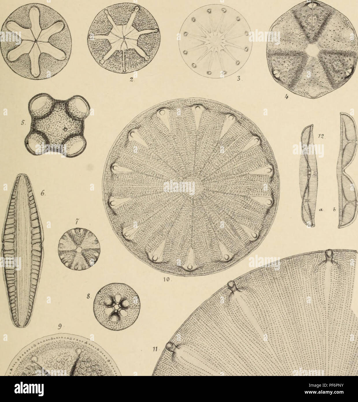 . Diatomées fossiles du Japon. Espèces marines & nouvelles des calcaires argileux de Sendaï & Yedo. Paleobotany. Diatomées Fossiles PI. III. *'''&. I .; "â 'â ' .- Biv.i.. Please note that these images are extracted from scanned page images that may have been digitally enhanced for readability - coloration and appearance of these illustrations may not perfectly resemble the original work.. Brun, Jacques, 1826-1908; Tempère, Joannes Albert, 1847-1926. Leipzig, Theodor Oswald Wigel, Leipzig Stock Photohttps://www.alamy.com/image-license-details/?v=1https://www.alamy.com/diatomes-fossiles-du-japon-espces-marines-amp-nouvelles-des-calcaires-argileux-de-senda-amp-yedo-paleobotany-diatomes-fossiles-pi-iii-amp-i-quot-bivi-please-note-that-these-images-are-extracted-from-scanned-page-images-that-may-have-been-digitally-enhanced-for-readability-coloration-and-appearance-of-these-illustrations-may-not-perfectly-resemble-the-original-work-brun-jacques-1826-1908-tempre-joannes-albert-1847-1926-leipzig-theodor-oswald-wigel-leipzig-image215893831.html
RMPF6PNY–. Diatomées fossiles du Japon. Espèces marines & nouvelles des calcaires argileux de Sendaï & Yedo. Paleobotany. Diatomées Fossiles PI. III. *'''&. I .; "â 'â ' .- Biv.i.. Please note that these images are extracted from scanned page images that may have been digitally enhanced for readability - coloration and appearance of these illustrations may not perfectly resemble the original work.. Brun, Jacques, 1826-1908; Tempère, Joannes Albert, 1847-1926. Leipzig, Theodor Oswald Wigel, Leipzig
![. Diatomaceæ of North America [microform] : illustrated with twenty-three hundred figures from the author's drawings on one hundred and twelve plates. Diatoms; Algae; Diatomées; Algues. i'l .â¢â â¢liXXW. I I 'I. Please note that these images are extracted from scanned page images that may have been digitally enhanced for readability - coloration and appearance of these illustrations may not perfectly resemble the original work.. Wolle, Francis, 1817-1893. Bethlehem, Pa. : Comenius Press Stock Photo . Diatomaceæ of North America [microform] : illustrated with twenty-three hundred figures from the author's drawings on one hundred and twelve plates. Diatoms; Algae; Diatomées; Algues. i'l .â¢â â¢liXXW. I I 'I. Please note that these images are extracted from scanned page images that may have been digitally enhanced for readability - coloration and appearance of these illustrations may not perfectly resemble the original work.. Wolle, Francis, 1817-1893. Bethlehem, Pa. : Comenius Press Stock Photo](https://c8.alamy.com/comp/RJ19GB/diatomace-of-north-america-microform-illustrated-with-twenty-three-hundred-figures-from-the-authors-drawings-on-one-hundred-and-twelve-plates-diatoms-algae-diatomes-algues-il-lixxw-i-i-i-please-note-that-these-images-are-extracted-from-scanned-page-images-that-may-have-been-digitally-enhanced-for-readability-coloration-and-appearance-of-these-illustrations-may-not-perfectly-resemble-the-original-work-wolle-francis-1817-1893-bethlehem-pa-comenius-press-RJ19GB.jpg) . Diatomaceæ of North America [microform] : illustrated with twenty-three hundred figures from the author's drawings on one hundred and twelve plates. Diatoms; Algae; Diatomées; Algues. i'l .â¢â â¢liXXW. I I 'I. Please note that these images are extracted from scanned page images that may have been digitally enhanced for readability - coloration and appearance of these illustrations may not perfectly resemble the original work.. Wolle, Francis, 1817-1893. Bethlehem, Pa. : Comenius Press Stock Photohttps://www.alamy.com/image-license-details/?v=1https://www.alamy.com/diatomace-of-north-america-microform-illustrated-with-twenty-three-hundred-figures-from-the-authors-drawings-on-one-hundred-and-twelve-plates-diatoms-algae-diatomes-algues-il-lixxw-i-i-i-please-note-that-these-images-are-extracted-from-scanned-page-images-that-may-have-been-digitally-enhanced-for-readability-coloration-and-appearance-of-these-illustrations-may-not-perfectly-resemble-the-original-work-wolle-francis-1817-1893-bethlehem-pa-comenius-press-image234828059.html
. Diatomaceæ of North America [microform] : illustrated with twenty-three hundred figures from the author's drawings on one hundred and twelve plates. Diatoms; Algae; Diatomées; Algues. i'l .â¢â â¢liXXW. I I 'I. Please note that these images are extracted from scanned page images that may have been digitally enhanced for readability - coloration and appearance of these illustrations may not perfectly resemble the original work.. Wolle, Francis, 1817-1893. Bethlehem, Pa. : Comenius Press Stock Photohttps://www.alamy.com/image-license-details/?v=1https://www.alamy.com/diatomace-of-north-america-microform-illustrated-with-twenty-three-hundred-figures-from-the-authors-drawings-on-one-hundred-and-twelve-plates-diatoms-algae-diatomes-algues-il-lixxw-i-i-i-please-note-that-these-images-are-extracted-from-scanned-page-images-that-may-have-been-digitally-enhanced-for-readability-coloration-and-appearance-of-these-illustrations-may-not-perfectly-resemble-the-original-work-wolle-francis-1817-1893-bethlehem-pa-comenius-press-image234828059.htmlRMRJ19GB–. Diatomaceæ of North America [microform] : illustrated with twenty-three hundred figures from the author's drawings on one hundred and twelve plates. Diatoms; Algae; Diatomées; Algues. i'l .â¢â â¢liXXW. I I 'I. Please note that these images are extracted from scanned page images that may have been digitally enhanced for readability - coloration and appearance of these illustrations may not perfectly resemble the original work.. Wolle, Francis, 1817-1893. Bethlehem, Pa. : Comenius Press
 . Diatomées marines de France et des districts maritimes voisins. Diatoms. Peragallo. â Diatomées de franee. Pi. LXXVII.. â ii M Le Micrographe Préparaleur Vul. l'I.. Please note that these images are extracted from scanned page images that may have been digitally enhanced for readability - coloration and appearance of these illustrations may not perfectly resemble the original work.. Péragallo, H. (Hippolyte), b. 1851; Péragallo, M. (Maurice), b. 1853. Grez-sur-Loing, M. J. Tempère Stock Photohttps://www.alamy.com/image-license-details/?v=1https://www.alamy.com/diatomes-marines-de-france-et-des-districts-maritimes-voisins-diatoms-peragallo-diatomes-de-franee-pi-lxxvii-ii-m-le-micrographe-prparaleur-vul-li-please-note-that-these-images-are-extracted-from-scanned-page-images-that-may-have-been-digitally-enhanced-for-readability-coloration-and-appearance-of-these-illustrations-may-not-perfectly-resemble-the-original-work-pragallo-h-hippolyte-b-1851-pragallo-m-maurice-b-1853-grez-sur-loing-m-j-tempre-image215893275.html
RMPF6P23–. Diatomées marines de France et des districts maritimes voisins. Diatoms. Peragallo. â Diatomées de franee. Pi. LXXVII.. â ii M Le Micrographe Préparaleur Vul. l'I.. Please note that these images are extracted from scanned page images that may have been digitally enhanced for readability - coloration and appearance of these illustrations may not perfectly resemble the original work.. Péragallo, H. (Hippolyte), b. 1851; Péragallo, M. (Maurice), b. 1853. Grez-sur-Loing, M. J. Tempère
![. Diatomaceæ of North America [microform] : illustrated with twenty-three hundred figures from the author's drawings on one hundred and twelve plates. Diatoms; Algae; Diatomées; Algues. .»â ⢠i,.x-. > ! i I. Please note that these images are extracted from scanned page images that may have been digitally enhanced for readability - coloration and appearance of these illustrations may not perfectly resemble the original work.. Wolle, Francis, 1817-1893. Bethlehem, Pa. : Comenius Press Stock Photo . Diatomaceæ of North America [microform] : illustrated with twenty-three hundred figures from the author's drawings on one hundred and twelve plates. Diatoms; Algae; Diatomées; Algues. .»â ⢠i,.x-. > ! i I. Please note that these images are extracted from scanned page images that may have been digitally enhanced for readability - coloration and appearance of these illustrations may not perfectly resemble the original work.. Wolle, Francis, 1817-1893. Bethlehem, Pa. : Comenius Press Stock Photo](https://c8.alamy.com/comp/RJ1XRW/diatomace-of-north-america-microform-illustrated-with-twenty-three-hundred-figures-from-the-authors-drawings-on-one-hundred-and-twelve-plates-diatoms-algae-diatomes-algues-ix-gt-!-i-i-please-note-that-these-images-are-extracted-from-scanned-page-images-that-may-have-been-digitally-enhanced-for-readability-coloration-and-appearance-of-these-illustrations-may-not-perfectly-resemble-the-original-work-wolle-francis-1817-1893-bethlehem-pa-comenius-press-RJ1XRW.jpg) . Diatomaceæ of North America [microform] : illustrated with twenty-three hundred figures from the author's drawings on one hundred and twelve plates. Diatoms; Algae; Diatomées; Algues. .»â ⢠i,.x-. > ! i I. Please note that these images are extracted from scanned page images that may have been digitally enhanced for readability - coloration and appearance of these illustrations may not perfectly resemble the original work.. Wolle, Francis, 1817-1893. Bethlehem, Pa. : Comenius Press Stock Photohttps://www.alamy.com/image-license-details/?v=1https://www.alamy.com/diatomace-of-north-america-microform-illustrated-with-twenty-three-hundred-figures-from-the-authors-drawings-on-one-hundred-and-twelve-plates-diatoms-algae-diatomes-algues-ix-gt-!-i-i-please-note-that-these-images-are-extracted-from-scanned-page-images-that-may-have-been-digitally-enhanced-for-readability-coloration-and-appearance-of-these-illustrations-may-not-perfectly-resemble-the-original-work-wolle-francis-1817-1893-bethlehem-pa-comenius-press-image234841597.html
. Diatomaceæ of North America [microform] : illustrated with twenty-three hundred figures from the author's drawings on one hundred and twelve plates. Diatoms; Algae; Diatomées; Algues. .»â ⢠i,.x-. > ! i I. Please note that these images are extracted from scanned page images that may have been digitally enhanced for readability - coloration and appearance of these illustrations may not perfectly resemble the original work.. Wolle, Francis, 1817-1893. Bethlehem, Pa. : Comenius Press Stock Photohttps://www.alamy.com/image-license-details/?v=1https://www.alamy.com/diatomace-of-north-america-microform-illustrated-with-twenty-three-hundred-figures-from-the-authors-drawings-on-one-hundred-and-twelve-plates-diatoms-algae-diatomes-algues-ix-gt-!-i-i-please-note-that-these-images-are-extracted-from-scanned-page-images-that-may-have-been-digitally-enhanced-for-readability-coloration-and-appearance-of-these-illustrations-may-not-perfectly-resemble-the-original-work-wolle-francis-1817-1893-bethlehem-pa-comenius-press-image234841597.htmlRMRJ1XRW–. Diatomaceæ of North America [microform] : illustrated with twenty-three hundred figures from the author's drawings on one hundred and twelve plates. Diatoms; Algae; Diatomées; Algues. .»â ⢠i,.x-. > ! i I. Please note that these images are extracted from scanned page images that may have been digitally enhanced for readability - coloration and appearance of these illustrations may not perfectly resemble the original work.. Wolle, Francis, 1817-1893. Bethlehem, Pa. : Comenius Press
 . Diatomées fossiles du Japon. Espèces marines & nouvelles des calcaires argileux de Sendaï & Yedo. Paleobotany. *'''&. I .; "â 'â ' .- Biv.i.. Phobti/ptc I TAe'iA â -â 8 ( ''.. Please note that these images are extracted from scanned page images that may have been digitally enhanced for readability - coloration and appearance of these illustrations may not perfectly resemble the original work.. Brun, Jacques, 1826-1908; Tempère, Joannes Albert, 1847-1926. Leipzig, Theodor Oswald Wigel, Leipzig Stock Photohttps://www.alamy.com/image-license-details/?v=1https://www.alamy.com/diatomes-fossiles-du-japon-espces-marines-amp-nouvelles-des-calcaires-argileux-de-senda-amp-yedo-paleobotany-amp-i-quot-bivi-phobtiptc-i-taeia-8-please-note-that-these-images-are-extracted-from-scanned-page-images-that-may-have-been-digitally-enhanced-for-readability-coloration-and-appearance-of-these-illustrations-may-not-perfectly-resemble-the-original-work-brun-jacques-1826-1908-tempre-joannes-albert-1847-1926-leipzig-theodor-oswald-wigel-leipzig-image215893820.html
RMPF6PNG–. Diatomées fossiles du Japon. Espèces marines & nouvelles des calcaires argileux de Sendaï & Yedo. Paleobotany. *'''&. I .; "â 'â ' .- Biv.i.. Phobti/ptc I TAe'iA â -â 8 ( ''.. Please note that these images are extracted from scanned page images that may have been digitally enhanced for readability - coloration and appearance of these illustrations may not perfectly resemble the original work.. Brun, Jacques, 1826-1908; Tempère, Joannes Albert, 1847-1926. Leipzig, Theodor Oswald Wigel, Leipzig
![. Diatomaceæ of North America [microform] : illustrated with twenty-three hundred figures from the author's drawings on one hundred and twelve plates. Diatoms; Algae; Diatomées; Algues. Plate :a,iv. I f ' hi M l if ! â pi :.a. Please note that these images are extracted from scanned page images that may have been digitally enhanced for readability - coloration and appearance of these illustrations may not perfectly resemble the original work.. Wolle, Francis, 1817-1893. Bethlehem, Pa. : Comenius Press Stock Photo . Diatomaceæ of North America [microform] : illustrated with twenty-three hundred figures from the author's drawings on one hundred and twelve plates. Diatoms; Algae; Diatomées; Algues. Plate :a,iv. I f ' hi M l if ! â pi :.a. Please note that these images are extracted from scanned page images that may have been digitally enhanced for readability - coloration and appearance of these illustrations may not perfectly resemble the original work.. Wolle, Francis, 1817-1893. Bethlehem, Pa. : Comenius Press Stock Photo](https://c8.alamy.com/comp/RJ1A18/diatomace-of-north-america-microform-illustrated-with-twenty-three-hundred-figures-from-the-authors-drawings-on-one-hundred-and-twelve-plates-diatoms-algae-diatomes-algues-plate-aiv-i-f-hi-m-l-if-!-pi-a-please-note-that-these-images-are-extracted-from-scanned-page-images-that-may-have-been-digitally-enhanced-for-readability-coloration-and-appearance-of-these-illustrations-may-not-perfectly-resemble-the-original-work-wolle-francis-1817-1893-bethlehem-pa-comenius-press-RJ1A18.jpg) . Diatomaceæ of North America [microform] : illustrated with twenty-three hundred figures from the author's drawings on one hundred and twelve plates. Diatoms; Algae; Diatomées; Algues. Plate :a,iv. I f ' hi M l if ! â pi :.a. Please note that these images are extracted from scanned page images that may have been digitally enhanced for readability - coloration and appearance of these illustrations may not perfectly resemble the original work.. Wolle, Francis, 1817-1893. Bethlehem, Pa. : Comenius Press Stock Photohttps://www.alamy.com/image-license-details/?v=1https://www.alamy.com/diatomace-of-north-america-microform-illustrated-with-twenty-three-hundred-figures-from-the-authors-drawings-on-one-hundred-and-twelve-plates-diatoms-algae-diatomes-algues-plate-aiv-i-f-hi-m-l-if-!-pi-a-please-note-that-these-images-are-extracted-from-scanned-page-images-that-may-have-been-digitally-enhanced-for-readability-coloration-and-appearance-of-these-illustrations-may-not-perfectly-resemble-the-original-work-wolle-francis-1817-1893-bethlehem-pa-comenius-press-image234828420.html
. Diatomaceæ of North America [microform] : illustrated with twenty-three hundred figures from the author's drawings on one hundred and twelve plates. Diatoms; Algae; Diatomées; Algues. Plate :a,iv. I f ' hi M l if ! â pi :.a. Please note that these images are extracted from scanned page images that may have been digitally enhanced for readability - coloration and appearance of these illustrations may not perfectly resemble the original work.. Wolle, Francis, 1817-1893. Bethlehem, Pa. : Comenius Press Stock Photohttps://www.alamy.com/image-license-details/?v=1https://www.alamy.com/diatomace-of-north-america-microform-illustrated-with-twenty-three-hundred-figures-from-the-authors-drawings-on-one-hundred-and-twelve-plates-diatoms-algae-diatomes-algues-plate-aiv-i-f-hi-m-l-if-!-pi-a-please-note-that-these-images-are-extracted-from-scanned-page-images-that-may-have-been-digitally-enhanced-for-readability-coloration-and-appearance-of-these-illustrations-may-not-perfectly-resemble-the-original-work-wolle-francis-1817-1893-bethlehem-pa-comenius-press-image234828420.htmlRMRJ1A18–. Diatomaceæ of North America [microform] : illustrated with twenty-three hundred figures from the author's drawings on one hundred and twelve plates. Diatoms; Algae; Diatomées; Algues. Plate :a,iv. I f ' hi M l if ! â pi :.a. Please note that these images are extracted from scanned page images that may have been digitally enhanced for readability - coloration and appearance of these illustrations may not perfectly resemble the original work.. Wolle, Francis, 1817-1893. Bethlehem, Pa. : Comenius Press
 . Diatomées marines de France et des districts maritimes voisins. Diatoms. j-inli illUiiiii.^ â â â , / â sïi^ ^tiiiiiii'' 4- â¢^T-!- * â * V amm. Please note that these images are extracted from scanned page images that may have been digitally enhanced for readability - coloration and appearance of these illustrations may not perfectly resemble the original work.. Péragallo, H. (Hippolyte), b. 1851; Péragallo, M. (Maurice), b. 1853. Grez-sur-Loing, M. J. Tempère Stock Photohttps://www.alamy.com/image-license-details/?v=1https://www.alamy.com/diatomes-marines-de-france-et-des-districts-maritimes-voisins-diatoms-j-inli-illuiiiii-si-tiiiiiii-4-t-!-v-amm-please-note-that-these-images-are-extracted-from-scanned-page-images-that-may-have-been-digitally-enhanced-for-readability-coloration-and-appearance-of-these-illustrations-may-not-perfectly-resemble-the-original-work-pragallo-h-hippolyte-b-1851-pragallo-m-maurice-b-1853-grez-sur-loing-m-j-tempre-image215893163.html
RMPF6NX3–. Diatomées marines de France et des districts maritimes voisins. Diatoms. j-inli illUiiiii.^ â â â , / â sïi^ ^tiiiiiii'' 4- â¢^T-!- * â * V amm. Please note that these images are extracted from scanned page images that may have been digitally enhanced for readability - coloration and appearance of these illustrations may not perfectly resemble the original work.. Péragallo, H. (Hippolyte), b. 1851; Péragallo, M. (Maurice), b. 1853. Grez-sur-Loing, M. J. Tempère
![. Diatomaceæ of North America [microform] : illustrated with twenty-three hundred figures from the author's drawings on one hundred and twelve plates. Diatoms; Algae; Diatomées; Algues. I'hii.'l.NXXM'l. .r<-^j^s^^m. J : '-^â â ^^f^' I. Please note that these images are extracted from scanned page images that may have been digitally enhanced for readability - coloration and appearance of these illustrations may not perfectly resemble the original work.. Wolle, Francis, 1817-1893. Bethlehem, Pa. : Comenius Press Stock Photo . Diatomaceæ of North America [microform] : illustrated with twenty-three hundred figures from the author's drawings on one hundred and twelve plates. Diatoms; Algae; Diatomées; Algues. I'hii.'l.NXXM'l. .r<-^j^s^^m. J : '-^â â ^^f^' I. Please note that these images are extracted from scanned page images that may have been digitally enhanced for readability - coloration and appearance of these illustrations may not perfectly resemble the original work.. Wolle, Francis, 1817-1893. Bethlehem, Pa. : Comenius Press Stock Photo](https://c8.alamy.com/comp/RJ19CK/diatomace-of-north-america-microform-illustrated-with-twenty-three-hundred-figures-from-the-authors-drawings-on-one-hundred-and-twelve-plates-diatoms-algae-diatomes-algues-ihiilnxxml-rlt-jsm-j-f-i-please-note-that-these-images-are-extracted-from-scanned-page-images-that-may-have-been-digitally-enhanced-for-readability-coloration-and-appearance-of-these-illustrations-may-not-perfectly-resemble-the-original-work-wolle-francis-1817-1893-bethlehem-pa-comenius-press-RJ19CK.jpg) . Diatomaceæ of North America [microform] : illustrated with twenty-three hundred figures from the author's drawings on one hundred and twelve plates. Diatoms; Algae; Diatomées; Algues. I'hii.'l.NXXM'l. .r<-^j^s^^m. J : '-^â â ^^f^' I. Please note that these images are extracted from scanned page images that may have been digitally enhanced for readability - coloration and appearance of these illustrations may not perfectly resemble the original work.. Wolle, Francis, 1817-1893. Bethlehem, Pa. : Comenius Press Stock Photohttps://www.alamy.com/image-license-details/?v=1https://www.alamy.com/diatomace-of-north-america-microform-illustrated-with-twenty-three-hundred-figures-from-the-authors-drawings-on-one-hundred-and-twelve-plates-diatoms-algae-diatomes-algues-ihiilnxxml-rlt-jsm-j-f-i-please-note-that-these-images-are-extracted-from-scanned-page-images-that-may-have-been-digitally-enhanced-for-readability-coloration-and-appearance-of-these-illustrations-may-not-perfectly-resemble-the-original-work-wolle-francis-1817-1893-bethlehem-pa-comenius-press-image234827955.html
. Diatomaceæ of North America [microform] : illustrated with twenty-three hundred figures from the author's drawings on one hundred and twelve plates. Diatoms; Algae; Diatomées; Algues. I'hii.'l.NXXM'l. .r<-^j^s^^m. J : '-^â â ^^f^' I. Please note that these images are extracted from scanned page images that may have been digitally enhanced for readability - coloration and appearance of these illustrations may not perfectly resemble the original work.. Wolle, Francis, 1817-1893. Bethlehem, Pa. : Comenius Press Stock Photohttps://www.alamy.com/image-license-details/?v=1https://www.alamy.com/diatomace-of-north-america-microform-illustrated-with-twenty-three-hundred-figures-from-the-authors-drawings-on-one-hundred-and-twelve-plates-diatoms-algae-diatomes-algues-ihiilnxxml-rlt-jsm-j-f-i-please-note-that-these-images-are-extracted-from-scanned-page-images-that-may-have-been-digitally-enhanced-for-readability-coloration-and-appearance-of-these-illustrations-may-not-perfectly-resemble-the-original-work-wolle-francis-1817-1893-bethlehem-pa-comenius-press-image234827955.htmlRMRJ19CK–. Diatomaceæ of North America [microform] : illustrated with twenty-three hundred figures from the author's drawings on one hundred and twelve plates. Diatoms; Algae; Diatomées; Algues. I'hii.'l.NXXM'l. .r<-^j^s^^m. J : '-^â â ^^f^' I. Please note that these images are extracted from scanned page images that may have been digitally enhanced for readability - coloration and appearance of these illustrations may not perfectly resemble the original work.. Wolle, Francis, 1817-1893. Bethlehem, Pa. : Comenius Press
 . Diatomées marines de France et des districts maritimes voisins. Diatoms. 1%^ Â¥*»â .«/' 'm 10 Le MîcrDgrapbe Préparateur Vol. PI.. Please note that these images are extracted from scanned page images that may have been digitally enhanced for readability - coloration and appearance of these illustrations may not perfectly resemble the original work.. Péragallo, H. (Hippolyte), b. 1851; Péragallo, M. (Maurice), b. 1853. Grez-sur-Loing, M. J. Tempère Stock Photohttps://www.alamy.com/image-license-details/?v=1https://www.alamy.com/diatomes-marines-de-france-et-des-districts-maritimes-voisins-diatoms-1-m-10-le-mcrdgrapbe-prparateur-vol-pi-please-note-that-these-images-are-extracted-from-scanned-page-images-that-may-have-been-digitally-enhanced-for-readability-coloration-and-appearance-of-these-illustrations-may-not-perfectly-resemble-the-original-work-pragallo-h-hippolyte-b-1851-pragallo-m-maurice-b-1853-grez-sur-loing-m-j-tempre-image215893156.html
RMPF6NWT–. Diatomées marines de France et des districts maritimes voisins. Diatoms. 1%^ Â¥*»â .«/' 'm 10 Le MîcrDgrapbe Préparateur Vol. PI.. Please note that these images are extracted from scanned page images that may have been digitally enhanced for readability - coloration and appearance of these illustrations may not perfectly resemble the original work.. Péragallo, H. (Hippolyte), b. 1851; Péragallo, M. (Maurice), b. 1853. Grez-sur-Loing, M. J. Tempère
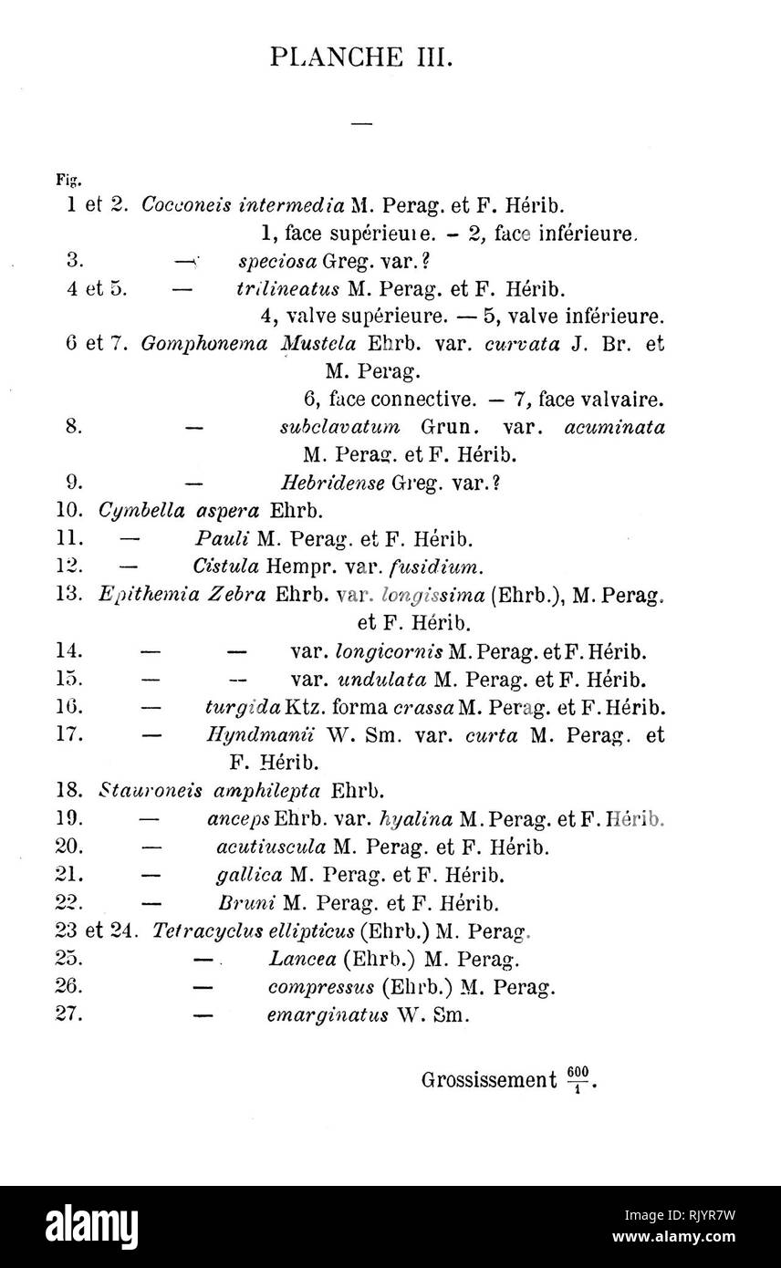 . Les diatomées d'Auvergne. Diatoms; Fragilariaceae; Paleontology. Dtafomees d Au verqne. JL'»s./â ââ «,/â :. ./.,.. Please note that these images are extracted from scanned page images that may have been digitally enhanced for readability - coloration and appearance of these illustrations may not perfectly resemble the original work.. Héribaud Joseph, brother, 1841-1918. Clermont-Ferrand : Pensionnat des Frères des écoles chrétiennes ; Paris : Librairie des sciences naturelles, P. Klincksieck Stock Photohttps://www.alamy.com/image-license-details/?v=1https://www.alamy.com/les-diatomes-dauvergne-diatoms-fragilariaceae-paleontology-dtafomees-d-au-verqne-jls-please-note-that-these-images-are-extracted-from-scanned-page-images-that-may-have-been-digitally-enhanced-for-readability-coloration-and-appearance-of-these-illustrations-may-not-perfectly-resemble-the-original-work-hribaud-joseph-brother-1841-1918-clermont-ferrand-pensionnat-des-frres-des-coles-chrtiennes-paris-librairie-des-sciences-naturelles-p-klincksieck-image235409549.html
RMRJYR7W–. Les diatomées d'Auvergne. Diatoms; Fragilariaceae; Paleontology. Dtafomees d Au verqne. JL'»s./â ââ «,/â :. ./.,.. Please note that these images are extracted from scanned page images that may have been digitally enhanced for readability - coloration and appearance of these illustrations may not perfectly resemble the original work.. Héribaud Joseph, brother, 1841-1918. Clermont-Ferrand : Pensionnat des Frères des écoles chrétiennes ; Paris : Librairie des sciences naturelles, P. Klincksieck
 . Diatomées marines de France et des districts maritimes voisins. Diatoms. Peragallo. â Diatomées de franee. n. (vv.. !.â¢â Mi.T .pi-.[|jlK' l'i-.-|. Please note that these images are extracted from scanned page images that may have been digitally enhanced for readability - coloration and appearance of these illustrations may not perfectly resemble the original work.. Péragallo, H. (Hippolyte), b. 1851; Péragallo, M. (Maurice), b. 1853. Grez-sur-Loing, M. J. Tempère Stock Photohttps://www.alamy.com/image-license-details/?v=1https://www.alamy.com/diatomes-marines-de-france-et-des-districts-maritimes-voisins-diatoms-peragallo-diatomes-de-franee-n-vv-!-mit-pi-jlk-li-please-note-that-these-images-are-extracted-from-scanned-page-images-that-may-have-been-digitally-enhanced-for-readability-coloration-and-appearance-of-these-illustrations-may-not-perfectly-resemble-the-original-work-pragallo-h-hippolyte-b-1851-pragallo-m-maurice-b-1853-grez-sur-loing-m-j-tempre-image215893215.html
RMPF6NYY–. Diatomées marines de France et des districts maritimes voisins. Diatoms. Peragallo. â Diatomées de franee. n. (vv.. !.â¢â Mi.T .pi-.[|jlK' l'i-.-|. Please note that these images are extracted from scanned page images that may have been digitally enhanced for readability - coloration and appearance of these illustrations may not perfectly resemble the original work.. Péragallo, H. (Hippolyte), b. 1851; Péragallo, M. (Maurice), b. 1853. Grez-sur-Loing, M. J. Tempère
![. Diatomaceæ of North America [microform] : illustrated with twenty-three hundred figures from the author's drawings on one hundred and twelve plates. Diatoms; Algae; Diatomées; Algues. Plat., xi.ir. n n â i â¢' ' V M'l. Please note that these images are extracted from scanned page images that may have been digitally enhanced for readability - coloration and appearance of these illustrations may not perfectly resemble the original work.. Wolle, Francis, 1817-1893. Bethlehem, Pa. : Comenius Press Stock Photo . Diatomaceæ of North America [microform] : illustrated with twenty-three hundred figures from the author's drawings on one hundred and twelve plates. Diatoms; Algae; Diatomées; Algues. Plat., xi.ir. n n â i â¢' ' V M'l. Please note that these images are extracted from scanned page images that may have been digitally enhanced for readability - coloration and appearance of these illustrations may not perfectly resemble the original work.. Wolle, Francis, 1817-1893. Bethlehem, Pa. : Comenius Press Stock Photo](https://c8.alamy.com/comp/RJ1A2Y/diatomace-of-north-america-microform-illustrated-with-twenty-three-hundred-figures-from-the-authors-drawings-on-one-hundred-and-twelve-plates-diatoms-algae-diatomes-algues-plat-xiir-n-n-i-v-ml-please-note-that-these-images-are-extracted-from-scanned-page-images-that-may-have-been-digitally-enhanced-for-readability-coloration-and-appearance-of-these-illustrations-may-not-perfectly-resemble-the-original-work-wolle-francis-1817-1893-bethlehem-pa-comenius-press-RJ1A2Y.jpg) . Diatomaceæ of North America [microform] : illustrated with twenty-three hundred figures from the author's drawings on one hundred and twelve plates. Diatoms; Algae; Diatomées; Algues. Plat., xi.ir. n n â i â¢' ' V M'l. Please note that these images are extracted from scanned page images that may have been digitally enhanced for readability - coloration and appearance of these illustrations may not perfectly resemble the original work.. Wolle, Francis, 1817-1893. Bethlehem, Pa. : Comenius Press Stock Photohttps://www.alamy.com/image-license-details/?v=1https://www.alamy.com/diatomace-of-north-america-microform-illustrated-with-twenty-three-hundred-figures-from-the-authors-drawings-on-one-hundred-and-twelve-plates-diatoms-algae-diatomes-algues-plat-xiir-n-n-i-v-ml-please-note-that-these-images-are-extracted-from-scanned-page-images-that-may-have-been-digitally-enhanced-for-readability-coloration-and-appearance-of-these-illustrations-may-not-perfectly-resemble-the-original-work-wolle-francis-1817-1893-bethlehem-pa-comenius-press-image234828467.html
. Diatomaceæ of North America [microform] : illustrated with twenty-three hundred figures from the author's drawings on one hundred and twelve plates. Diatoms; Algae; Diatomées; Algues. Plat., xi.ir. n n â i â¢' ' V M'l. Please note that these images are extracted from scanned page images that may have been digitally enhanced for readability - coloration and appearance of these illustrations may not perfectly resemble the original work.. Wolle, Francis, 1817-1893. Bethlehem, Pa. : Comenius Press Stock Photohttps://www.alamy.com/image-license-details/?v=1https://www.alamy.com/diatomace-of-north-america-microform-illustrated-with-twenty-three-hundred-figures-from-the-authors-drawings-on-one-hundred-and-twelve-plates-diatoms-algae-diatomes-algues-plat-xiir-n-n-i-v-ml-please-note-that-these-images-are-extracted-from-scanned-page-images-that-may-have-been-digitally-enhanced-for-readability-coloration-and-appearance-of-these-illustrations-may-not-perfectly-resemble-the-original-work-wolle-francis-1817-1893-bethlehem-pa-comenius-press-image234828467.htmlRMRJ1A2Y–. Diatomaceæ of North America [microform] : illustrated with twenty-three hundred figures from the author's drawings on one hundred and twelve plates. Diatoms; Algae; Diatomées; Algues. Plat., xi.ir. n n â i â¢' ' V M'l. Please note that these images are extracted from scanned page images that may have been digitally enhanced for readability - coloration and appearance of these illustrations may not perfectly resemble the original work.. Wolle, Francis, 1817-1893. Bethlehem, Pa. : Comenius Press
 . Diatomées marines de France et des districts maritimes voisins. Diatoms. Peragallo. â Diatomées de fvà nQe. V. Cl.. I.e MiiTofi-upht; Préparalcur. Please note that these images are extracted from scanned page images that may have been digitally enhanced for readability - coloration and appearance of these illustrations may not perfectly resemble the original work.. Péragallo, H. (Hippolyte), b. 1851; Péragallo, M. (Maurice), b. 1853. Grez-sur-Loing, M. J. Tempère Stock Photohttps://www.alamy.com/image-license-details/?v=1https://www.alamy.com/diatomes-marines-de-france-et-des-districts-maritimes-voisins-diatoms-peragallo-diatomes-de-fv-nqe-v-cl-ie-miitofi-upht-prparalcur-please-note-that-these-images-are-extracted-from-scanned-page-images-that-may-have-been-digitally-enhanced-for-readability-coloration-and-appearance-of-these-illustrations-may-not-perfectly-resemble-the-original-work-pragallo-h-hippolyte-b-1851-pragallo-m-maurice-b-1853-grez-sur-loing-m-j-tempre-image215893217.html
RMPF6P01–. Diatomées marines de France et des districts maritimes voisins. Diatoms. Peragallo. â Diatomées de fvà nQe. V. Cl.. I.e MiiTofi-upht; Préparalcur. Please note that these images are extracted from scanned page images that may have been digitally enhanced for readability - coloration and appearance of these illustrations may not perfectly resemble the original work.. Péragallo, H. (Hippolyte), b. 1851; Péragallo, M. (Maurice), b. 1853. Grez-sur-Loing, M. J. Tempère
 . Brigham Young University science bulletin. Biology -- Periodicals. Selected Genera: 1 = 25 algae ml Apr. June July Sept. Nov. Jan. Mar.. Diatoma Fig. .36. Seasonal dislriliulioii of selertpd iiaiiMiipl.inktoii at Stiiail Station Csite 6).. Please note that these images are extracted from scanned page images that may have been digitally enhanced for readability - coloration and appearance of these illustrations may not perfectly resemble the original work.. Brigham Young University. Provo, Utah : Brigham Young University Stock Photohttps://www.alamy.com/image-license-details/?v=1https://www.alamy.com/brigham-young-university-science-bulletin-biology-periodicals-selected-genera-1-=-25-algae-ml-apr-june-july-sept-nov-jan-mar-diatoma-fig-36-seasonal-dislriliulioii-of-selertpd-iiaiimiiplinktoii-at-stiiail-station-csite-6-please-note-that-these-images-are-extracted-from-scanned-page-images-that-may-have-been-digitally-enhanced-for-readability-coloration-and-appearance-of-these-illustrations-may-not-perfectly-resemble-the-original-work-brigham-young-university-provo-utah-brigham-young-university-image234269382.html
RMRH3TYJ–. Brigham Young University science bulletin. Biology -- Periodicals. Selected Genera: 1 = 25 algae ml Apr. June July Sept. Nov. Jan. Mar.. Diatoma Fig. .36. Seasonal dislriliulioii of selertpd iiaiiMiipl.inktoii at Stiiail Station Csite 6).. Please note that these images are extracted from scanned page images that may have been digitally enhanced for readability - coloration and appearance of these illustrations may not perfectly resemble the original work.. Brigham Young University. Provo, Utah : Brigham Young University
 . Diatomées marines de France et des districts maritimes voisins. Diatoms. Peragallo. Diatomées d» France. l'I l.MII. ..â .Ml. i«!;i-.|,l,.- l'TV|.nral.'. Please note that these images are extracted from scanned page images that may have been digitally enhanced for readability - coloration and appearance of these illustrations may not perfectly resemble the original work.. Péragallo, H. (Hippolyte), b. 1851; Péragallo, M. (Maurice), b. 1853. Grez-sur-Loing, M. J. Tempère Stock Photohttps://www.alamy.com/image-license-details/?v=1https://www.alamy.com/diatomes-marines-de-france-et-des-districts-maritimes-voisins-diatoms-peragallo-diatomes-d-france-li-lmii-ml-i!i-l-ltvnral-please-note-that-these-images-are-extracted-from-scanned-page-images-that-may-have-been-digitally-enhanced-for-readability-coloration-and-appearance-of-these-illustrations-may-not-perfectly-resemble-the-original-work-pragallo-h-hippolyte-b-1851-pragallo-m-maurice-b-1853-grez-sur-loing-m-j-tempre-image215893308.html
RMPF6P38–. Diatomées marines de France et des districts maritimes voisins. Diatoms. Peragallo. Diatomées d» France. l'I l.MII. ..â .Ml. i«!;i-.|,l,.- l'TV|.nral.'. Please note that these images are extracted from scanned page images that may have been digitally enhanced for readability - coloration and appearance of these illustrations may not perfectly resemble the original work.. Péragallo, H. (Hippolyte), b. 1851; Péragallo, M. (Maurice), b. 1853. Grez-sur-Loing, M. J. Tempère
 . Bilder-Atlas des Pflanzenreichs nach dem natürlichen System. Bilder-Atlas; Pflanzenreich; Botanik; Natürliches System; Pflanzen; Naturgeschichte; Systematik; Nomenklatur; Lehrmittel. 3. (Stäbcfyenalge. Diatoma floceulosum. ©tMdjenctlge. Frustalia. J£. •Mut- i V•Min / > ©cfjnrimmenber Seerentang. 9. ©em. $rofd)latcI)alge. Sargassum natans. Batrachospermum moniliforme. 7. ©emetner Slajentcmg. Fucus vesiculosus.. Please note that these images are extracted from scanned page images that may have been digitally enhanced for readability - coloration and appearance of these illustrations may n Stock Photohttps://www.alamy.com/image-license-details/?v=1https://www.alamy.com/bilder-atlas-des-pflanzenreichs-nach-dem-natrlichen-system-bilder-atlas-pflanzenreich-botanik-natrliches-system-pflanzen-naturgeschichte-systematik-nomenklatur-lehrmittel-3-stbcfyenalge-diatoma-floceulosum-tmdjenctlge-frustalia-j-mut-i-vmin-gt-cfjnrimmenber-seerentang-9-em-rofdlatcialge-sargassum-natans-batrachospermum-moniliforme-7-emetner-slajentcmg-fucus-vesiculosus-please-note-that-these-images-are-extracted-from-scanned-page-images-that-may-have-been-digitally-enhanced-for-readability-coloration-and-appearance-of-these-illustrations-may-n-image234705272.html
RMRHRMY4–. Bilder-Atlas des Pflanzenreichs nach dem natürlichen System. Bilder-Atlas; Pflanzenreich; Botanik; Natürliches System; Pflanzen; Naturgeschichte; Systematik; Nomenklatur; Lehrmittel. 3. (Stäbcfyenalge. Diatoma floceulosum. ©tMdjenctlge. Frustalia. J£. •Mut- i V•Min / > ©cfjnrimmenber Seerentang. 9. ©em. $rofd)latcI)alge. Sargassum natans. Batrachospermum moniliforme. 7. ©emetner Slajentcmg. Fucus vesiculosus.. Please note that these images are extracted from scanned page images that may have been digitally enhanced for readability - coloration and appearance of these illustrations may n
 . Diatomées marines de France et des districts maritimes voisins. Diatoms. Peragallo. Diatomées de France )â !. l.MV.. L.I' M (.T' ji'.iphi- Pp»-|iiirrikur. Please note that these images are extracted from scanned page images that may have been digitally enhanced for readability - coloration and appearance of these illustrations may not perfectly resemble the original work.. Péragallo, H. (Hippolyte), b. 1851; Péragallo, M. (Maurice), b. 1853. Grez-sur-Loing, M. J. Tempère Stock Photohttps://www.alamy.com/image-license-details/?v=1https://www.alamy.com/diatomes-marines-de-france-et-des-districts-maritimes-voisins-diatoms-peragallo-diatomes-de-france-!-lmv-li-m-t-jiiphi-pp-iiirrikur-please-note-that-these-images-are-extracted-from-scanned-page-images-that-may-have-been-digitally-enhanced-for-readability-coloration-and-appearance-of-these-illustrations-may-not-perfectly-resemble-the-original-work-pragallo-h-hippolyte-b-1851-pragallo-m-maurice-b-1853-grez-sur-loing-m-j-tempre-image215893305.html
RMPF6P35–. Diatomées marines de France et des districts maritimes voisins. Diatoms. Peragallo. Diatomées de France )â !. l.MV.. L.I' M (.T' ji'.iphi- Pp»-|iiirrikur. Please note that these images are extracted from scanned page images that may have been digitally enhanced for readability - coloration and appearance of these illustrations may not perfectly resemble the original work.. Péragallo, H. (Hippolyte), b. 1851; Péragallo, M. (Maurice), b. 1853. Grez-sur-Loing, M. J. Tempère
![. Diatomaceæ of North America [microform] : illustrated with twenty-three hundred figures from the author's drawings on one hundred and twelve plates. Diatoms; Algae; Diatomées; Algues. I'late XIA'l.. : f ] it tf i: 'â ^ ! ! P K^ I' 1. Please note that these images are extracted from scanned page images that may have been digitally enhanced for readability - coloration and appearance of these illustrations may not perfectly resemble the original work.. Wolle, Francis, 1817-1893. Bethlehem, Pa. : Comenius Press Stock Photo . Diatomaceæ of North America [microform] : illustrated with twenty-three hundred figures from the author's drawings on one hundred and twelve plates. Diatoms; Algae; Diatomées; Algues. I'late XIA'l.. : f ] it tf i: 'â ^ ! ! P K^ I' 1. Please note that these images are extracted from scanned page images that may have been digitally enhanced for readability - coloration and appearance of these illustrations may not perfectly resemble the original work.. Wolle, Francis, 1817-1893. Bethlehem, Pa. : Comenius Press Stock Photo](https://c8.alamy.com/comp/RJ19YA/diatomace-of-north-america-microform-illustrated-with-twenty-three-hundred-figures-from-the-authors-drawings-on-one-hundred-and-twelve-plates-diatoms-algae-diatomes-algues-ilate-xial-f-it-tf-i-!-!-p-k-i-1-please-note-that-these-images-are-extracted-from-scanned-page-images-that-may-have-been-digitally-enhanced-for-readability-coloration-and-appearance-of-these-illustrations-may-not-perfectly-resemble-the-original-work-wolle-francis-1817-1893-bethlehem-pa-comenius-press-RJ19YA.jpg) . Diatomaceæ of North America [microform] : illustrated with twenty-three hundred figures from the author's drawings on one hundred and twelve plates. Diatoms; Algae; Diatomées; Algues. I'late XIA'l.. : f ] it tf i: 'â ^ ! ! P K^ I' 1. Please note that these images are extracted from scanned page images that may have been digitally enhanced for readability - coloration and appearance of these illustrations may not perfectly resemble the original work.. Wolle, Francis, 1817-1893. Bethlehem, Pa. : Comenius Press Stock Photohttps://www.alamy.com/image-license-details/?v=1https://www.alamy.com/diatomace-of-north-america-microform-illustrated-with-twenty-three-hundred-figures-from-the-authors-drawings-on-one-hundred-and-twelve-plates-diatoms-algae-diatomes-algues-ilate-xial-f-it-tf-i-!-!-p-k-i-1-please-note-that-these-images-are-extracted-from-scanned-page-images-that-may-have-been-digitally-enhanced-for-readability-coloration-and-appearance-of-these-illustrations-may-not-perfectly-resemble-the-original-work-wolle-francis-1817-1893-bethlehem-pa-comenius-press-image234828366.html
. Diatomaceæ of North America [microform] : illustrated with twenty-three hundred figures from the author's drawings on one hundred and twelve plates. Diatoms; Algae; Diatomées; Algues. I'late XIA'l.. : f ] it tf i: 'â ^ ! ! P K^ I' 1. Please note that these images are extracted from scanned page images that may have been digitally enhanced for readability - coloration and appearance of these illustrations may not perfectly resemble the original work.. Wolle, Francis, 1817-1893. Bethlehem, Pa. : Comenius Press Stock Photohttps://www.alamy.com/image-license-details/?v=1https://www.alamy.com/diatomace-of-north-america-microform-illustrated-with-twenty-three-hundred-figures-from-the-authors-drawings-on-one-hundred-and-twelve-plates-diatoms-algae-diatomes-algues-ilate-xial-f-it-tf-i-!-!-p-k-i-1-please-note-that-these-images-are-extracted-from-scanned-page-images-that-may-have-been-digitally-enhanced-for-readability-coloration-and-appearance-of-these-illustrations-may-not-perfectly-resemble-the-original-work-wolle-francis-1817-1893-bethlehem-pa-comenius-press-image234828366.htmlRMRJ19YA–. Diatomaceæ of North America [microform] : illustrated with twenty-three hundred figures from the author's drawings on one hundred and twelve plates. Diatoms; Algae; Diatomées; Algues. I'late XIA'l.. : f ] it tf i: 'â ^ ! ! P K^ I' 1. Please note that these images are extracted from scanned page images that may have been digitally enhanced for readability - coloration and appearance of these illustrations may not perfectly resemble the original work.. Wolle, Francis, 1817-1893. Bethlehem, Pa. : Comenius Press
 . Diatomées; espèces nouvelles marines, fossiles ou pélagiques. PI .XXII f 1 II 1 â 'y I 1 } 1 1 I 1 Il 1 " j I lÃ' ^ ;! i II 1 J1 1. Please note that these images are extracted from scanned page images that may have been digitally enhanced for readability - coloration and appearance of these illustrations may not perfectly resemble the original work.. Brun, Jacques, 1826-1908. Leipzig, Theodor Oswald Weigle Stock Photohttps://www.alamy.com/image-license-details/?v=1https://www.alamy.com/diatomes-espces-nouvelles-marines-fossiles-ou-plagiques-pi-xxii-f-1-ii-1-y-i-1-1-1-i-1-il-1-quot-j-i-l-!-i-ii-1-j1-1-please-note-that-these-images-are-extracted-from-scanned-page-images-that-may-have-been-digitally-enhanced-for-readability-coloration-and-appearance-of-these-illustrations-may-not-perfectly-resemble-the-original-work-brun-jacques-1826-1908-leipzig-theodor-oswald-weigle-image215893903.html
RMPF6PTF–. Diatomées; espèces nouvelles marines, fossiles ou pélagiques. PI .XXII f 1 II 1 â 'y I 1 } 1 1 I 1 Il 1 " j I lÃ' ^ ;! i II 1 J1 1. Please note that these images are extracted from scanned page images that may have been digitally enhanced for readability - coloration and appearance of these illustrations may not perfectly resemble the original work.. Brun, Jacques, 1826-1908. Leipzig, Theodor Oswald Weigle
 . Les diatomées d'Auvergne. Diatoms; Fragilariaceae; Paleontology. Diatomées d'Auvergne 2 PI. V.. Thorotypic /. ThéVQZ à C" Uesi^. Please note that these images are extracted from scanned page images that may have been digitally enhanced for readability - coloration and appearance of these illustrations may not perfectly resemble the original work.. Héribaud Joseph, brother, 1841-1918. Clermont-Ferrand : Pensionnat des Frères des écoles chrétiennes ; Paris : Librairie des sciences naturelles, P. Klincksieck Stock Photohttps://www.alamy.com/image-license-details/?v=1https://www.alamy.com/les-diatomes-dauvergne-diatoms-fragilariaceae-paleontology-diatomes-dauvergne-2-pi-v-thorotypic-thvqz-cquot-uesi-please-note-that-these-images-are-extracted-from-scanned-page-images-that-may-have-been-digitally-enhanced-for-readability-coloration-and-appearance-of-these-illustrations-may-not-perfectly-resemble-the-original-work-hribaud-joseph-brother-1841-1918-clermont-ferrand-pensionnat-des-frres-des-coles-chrtiennes-paris-librairie-des-sciences-naturelles-p-klincksieck-image235409527.html
RMRJYR73–. Les diatomées d'Auvergne. Diatoms; Fragilariaceae; Paleontology. Diatomées d'Auvergne 2 PI. V.. Thorotypic /. ThéVQZ à C" Uesi^. Please note that these images are extracted from scanned page images that may have been digitally enhanced for readability - coloration and appearance of these illustrations may not perfectly resemble the original work.. Héribaud Joseph, brother, 1841-1918. Clermont-Ferrand : Pensionnat des Frères des écoles chrétiennes ; Paris : Librairie des sciences naturelles, P. Klincksieck
 . Diatomées marines de France et des districts maritimes voisins. Diatoms. ")«'Ooooooooooooooo â¢oaoooooooooooooo 00000000000000000 XjO 000000000 000000 }o 000 00000 ooocoooo oooooooooooooooot t/ooooooooooooooooo ^°'^°°oooooooooooo, >|'00000000000000000 jo000000000000000 0 000000000000000( ;ooooooo 000000000 jOOOOOOOOOOoOOoor ^o 00 000 000000co 00 00000000000000 OOOOOOOOOQOOOOO 'OOOOOOOOOOOOOOO )0000000000000000 -^OOOOOOOOOOOOOOO 000000000000000 r jwv7--Jiio'0 000 ooooooon nor- Wg^ooooocoooo''oooo° iOPA^-i^ ^oOoOOooOQOOoOOn -A 00 oooooooo 00000, 000000000 ,-rr?r?r?.?(. m Le Mic Stock Photohttps://www.alamy.com/image-license-details/?v=1https://www.alamy.com/diatomes-marines-de-france-et-des-districts-maritimes-voisins-diatoms-quotooooooooooooooo-oaoooooooooooooo-00000000000000000-xjo-000000000-000000-o-000-00000-ooocoooo-oooooooooooooooot-tooooooooooooooooo-oooooooooooo-gt00000000000000000-jo000000000000000-0-000000000000000-ooooooo-000000000-jooooooooooooooor-o-00-000-000000co-00-00000000000000-oooooooooqooooo-ooooooooooooooo-0000000000000000-ooooooooooooooo-000000000000000-r-jwv7-jiio0-000-ooooooon-nor-wgooooocoooooooo-iopa-i-ooooooooqooooon-a-00-oooooooo-00000-000000000-rrrr-m-le-mic-image215893226.html
RMPF6P0A–. Diatomées marines de France et des districts maritimes voisins. Diatoms. ")«'Ooooooooooooooo â¢oaoooooooooooooo 00000000000000000 XjO 000000000 000000 }o 000 00000 ooocoooo oooooooooooooooot t/ooooooooooooooooo ^°'^°°oooooooooooo, >|'00000000000000000 jo000000000000000 0 000000000000000( ;ooooooo 000000000 jOOOOOOOOOOoOOoor ^o 00 000 000000co 00 00000000000000 OOOOOOOOOQOOOOO 'OOOOOOOOOOOOOOO )0000000000000000 -^OOOOOOOOOOOOOOO 000000000000000 r jwv7--Jiio'0 000 ooooooon nor- Wg^ooooocoooo''oooo° iOPA^-i^ ^oOoOOooOQOOoOOn -A 00 oooooooo 00000, 000000000 ,-rr?r?r?.?(. m Le Mic
 . Bulletin of the Geological Society of America. Geology. 250 J. F. XEWSOM—CLASTIC DIKES -,^^S^^^^^^ :,i^-^. been observed along the coast in this region. This intrusion was ob- served by Dr Ralph Arnold, and the following notes and diagram (figure 18) are supplied by him. The sea-cliff is 100 feet high where cut by the dike. The upper 40 feet of the cliff is made up of Pleistocene sands, gravels, and clays, resting unconformably on the diatomaceous Miocene shales. The intrusion has an even thickness of 1 foot from the bottom to the top of the diatoma- ceous shales. The sides of the fissure in Stock Photohttps://www.alamy.com/image-license-details/?v=1https://www.alamy.com/bulletin-of-the-geological-society-of-america-geology-250-j-f-xewsomclastic-dikes-s-i-been-observed-along-the-coast-in-this-region-this-intrusion-was-ob-served-by-dr-ralph-arnold-and-the-following-notes-and-diagram-figure-18-are-supplied-by-him-the-sea-cliff-is-100-feet-high-where-cut-by-the-dike-the-upper-40-feet-of-the-cliff-is-made-up-of-pleistocene-sands-gravels-and-clays-resting-unconformably-on-the-diatomaceous-miocene-shales-the-intrusion-has-an-even-thickness-of-1-foot-from-the-bottom-to-the-top-of-the-diatoma-ceous-shales-the-sides-of-the-fissure-in-image233941062.html
RMRGGX5X–. Bulletin of the Geological Society of America. Geology. 250 J. F. XEWSOM—CLASTIC DIKES -,^^S^^^^^^ :,i^-^. been observed along the coast in this region. This intrusion was ob- served by Dr Ralph Arnold, and the following notes and diagram (figure 18) are supplied by him. The sea-cliff is 100 feet high where cut by the dike. The upper 40 feet of the cliff is made up of Pleistocene sands, gravels, and clays, resting unconformably on the diatomaceous Miocene shales. The intrusion has an even thickness of 1 foot from the bottom to the top of the diatoma- ceous shales. The sides of the fissure in
![. Diatomées marines de France et des districts maritimes voisins. Diatoms. Peragallo- Diatomées de fraRce li .XX'. Il â â ! M Heba.;all.> .Ifl Le MftTo;]raphe F'rpparâleur o. PI. Please note that these images are extracted from scanned page images that may have been digitally enhanced for readability - coloration and appearance of these illustrations may not perfectly resemble the original work.. Péragallo, H. (Hippolyte), b. 1851; Péragallo, M. (Maurice), b. 1853. Grez-sur-Loing, M. J. Tempère Stock Photo . Diatomées marines de France et des districts maritimes voisins. Diatoms. Peragallo- Diatomées de fraRce li .XX'. Il â â ! M Heba.;all.> .Ifl Le MftTo;]raphe F'rpparâleur o. PI. Please note that these images are extracted from scanned page images that may have been digitally enhanced for readability - coloration and appearance of these illustrations may not perfectly resemble the original work.. Péragallo, H. (Hippolyte), b. 1851; Péragallo, M. (Maurice), b. 1853. Grez-sur-Loing, M. J. Tempère Stock Photo](https://c8.alamy.com/comp/PF6P5B/diatomes-marines-de-france-et-des-districts-maritimes-voisins-diatoms-peragallo-diatomes-de-frarce-li-xx-il-!-m-hebaallgt-ifl-le-mftto-raphe-frpparleur-o-pi-please-note-that-these-images-are-extracted-from-scanned-page-images-that-may-have-been-digitally-enhanced-for-readability-coloration-and-appearance-of-these-illustrations-may-not-perfectly-resemble-the-original-work-pragallo-h-hippolyte-b-1851-pragallo-m-maurice-b-1853-grez-sur-loing-m-j-tempre-PF6P5B.jpg) . Diatomées marines de France et des districts maritimes voisins. Diatoms. Peragallo- Diatomées de fraRce li .XX'. Il â â ! M Heba.;all.> .Ifl Le MftTo;]raphe F'rpparâleur o. PI. Please note that these images are extracted from scanned page images that may have been digitally enhanced for readability - coloration and appearance of these illustrations may not perfectly resemble the original work.. Péragallo, H. (Hippolyte), b. 1851; Péragallo, M. (Maurice), b. 1853. Grez-sur-Loing, M. J. Tempère Stock Photohttps://www.alamy.com/image-license-details/?v=1https://www.alamy.com/diatomes-marines-de-france-et-des-districts-maritimes-voisins-diatoms-peragallo-diatomes-de-frarce-li-xx-il-!-m-hebaallgt-ifl-le-mftto-raphe-frpparleur-o-pi-please-note-that-these-images-are-extracted-from-scanned-page-images-that-may-have-been-digitally-enhanced-for-readability-coloration-and-appearance-of-these-illustrations-may-not-perfectly-resemble-the-original-work-pragallo-h-hippolyte-b-1851-pragallo-m-maurice-b-1853-grez-sur-loing-m-j-tempre-image215893367.html
. Diatomées marines de France et des districts maritimes voisins. Diatoms. Peragallo- Diatomées de fraRce li .XX'. Il â â ! M Heba.;all.> .Ifl Le MftTo;]raphe F'rpparâleur o. PI. Please note that these images are extracted from scanned page images that may have been digitally enhanced for readability - coloration and appearance of these illustrations may not perfectly resemble the original work.. Péragallo, H. (Hippolyte), b. 1851; Péragallo, M. (Maurice), b. 1853. Grez-sur-Loing, M. J. Tempère Stock Photohttps://www.alamy.com/image-license-details/?v=1https://www.alamy.com/diatomes-marines-de-france-et-des-districts-maritimes-voisins-diatoms-peragallo-diatomes-de-frarce-li-xx-il-!-m-hebaallgt-ifl-le-mftto-raphe-frpparleur-o-pi-please-note-that-these-images-are-extracted-from-scanned-page-images-that-may-have-been-digitally-enhanced-for-readability-coloration-and-appearance-of-these-illustrations-may-not-perfectly-resemble-the-original-work-pragallo-h-hippolyte-b-1851-pragallo-m-maurice-b-1853-grez-sur-loing-m-j-tempre-image215893367.htmlRMPF6P5B–. Diatomées marines de France et des districts maritimes voisins. Diatoms. Peragallo- Diatomées de fraRce li .XX'. Il â â ! M Heba.;all.> .Ifl Le MftTo;]raphe F'rpparâleur o. PI. Please note that these images are extracted from scanned page images that may have been digitally enhanced for readability - coloration and appearance of these illustrations may not perfectly resemble the original work.. Péragallo, H. (Hippolyte), b. 1851; Péragallo, M. (Maurice), b. 1853. Grez-sur-Loing, M. J. Tempère
![. Diatomaceæ of North America [microform] : illustrated with twenty-three hundred figures from the author's drawings on one hundred and twelve plates. Diatoms; Algae; Diatomées; Algues. 'l.'.l, ( A" â rt. vfi fVH^' 'v- '-i .â¢â¢ /. h. i i 1,. Please note that these images are extracted from scanned page images that may have been digitally enhanced for readability - coloration and appearance of these illustrations may not perfectly resemble the original work.. Wolle, Francis, 1817-1893. Bethlehem, Pa. : Comenius Press Stock Photo . Diatomaceæ of North America [microform] : illustrated with twenty-three hundred figures from the author's drawings on one hundred and twelve plates. Diatoms; Algae; Diatomées; Algues. 'l.'.l, ( A" â rt. vfi fVH^' 'v- '-i .â¢â¢ /. h. i i 1,. Please note that these images are extracted from scanned page images that may have been digitally enhanced for readability - coloration and appearance of these illustrations may not perfectly resemble the original work.. Wolle, Francis, 1817-1893. Bethlehem, Pa. : Comenius Press Stock Photo](https://c8.alamy.com/comp/RJ194D/diatomace-of-north-america-microform-illustrated-with-twenty-three-hundred-figures-from-the-authors-drawings-on-one-hundred-and-twelve-plates-diatoms-algae-diatomes-algues-ll-aquot-rt-vfi-fvh-v-i-h-i-i-1-please-note-that-these-images-are-extracted-from-scanned-page-images-that-may-have-been-digitally-enhanced-for-readability-coloration-and-appearance-of-these-illustrations-may-not-perfectly-resemble-the-original-work-wolle-francis-1817-1893-bethlehem-pa-comenius-press-RJ194D.jpg) . Diatomaceæ of North America [microform] : illustrated with twenty-three hundred figures from the author's drawings on one hundred and twelve plates. Diatoms; Algae; Diatomées; Algues. 'l.'.l, ( A" â rt. vfi fVH^' 'v- '-i .â¢â¢ /. h. i i 1,. Please note that these images are extracted from scanned page images that may have been digitally enhanced for readability - coloration and appearance of these illustrations may not perfectly resemble the original work.. Wolle, Francis, 1817-1893. Bethlehem, Pa. : Comenius Press Stock Photohttps://www.alamy.com/image-license-details/?v=1https://www.alamy.com/diatomace-of-north-america-microform-illustrated-with-twenty-three-hundred-figures-from-the-authors-drawings-on-one-hundred-and-twelve-plates-diatoms-algae-diatomes-algues-ll-aquot-rt-vfi-fvh-v-i-h-i-i-1-please-note-that-these-images-are-extracted-from-scanned-page-images-that-may-have-been-digitally-enhanced-for-readability-coloration-and-appearance-of-these-illustrations-may-not-perfectly-resemble-the-original-work-wolle-francis-1817-1893-bethlehem-pa-comenius-press-image234827725.html
. Diatomaceæ of North America [microform] : illustrated with twenty-three hundred figures from the author's drawings on one hundred and twelve plates. Diatoms; Algae; Diatomées; Algues. 'l.'.l, ( A" â rt. vfi fVH^' 'v- '-i .â¢â¢ /. h. i i 1,. Please note that these images are extracted from scanned page images that may have been digitally enhanced for readability - coloration and appearance of these illustrations may not perfectly resemble the original work.. Wolle, Francis, 1817-1893. Bethlehem, Pa. : Comenius Press Stock Photohttps://www.alamy.com/image-license-details/?v=1https://www.alamy.com/diatomace-of-north-america-microform-illustrated-with-twenty-three-hundred-figures-from-the-authors-drawings-on-one-hundred-and-twelve-plates-diatoms-algae-diatomes-algues-ll-aquot-rt-vfi-fvh-v-i-h-i-i-1-please-note-that-these-images-are-extracted-from-scanned-page-images-that-may-have-been-digitally-enhanced-for-readability-coloration-and-appearance-of-these-illustrations-may-not-perfectly-resemble-the-original-work-wolle-francis-1817-1893-bethlehem-pa-comenius-press-image234827725.htmlRMRJ194D–. Diatomaceæ of North America [microform] : illustrated with twenty-three hundred figures from the author's drawings on one hundred and twelve plates. Diatoms; Algae; Diatomées; Algues. 'l.'.l, ( A" â rt. vfi fVH^' 'v- '-i .â¢â¢ /. h. i i 1,. Please note that these images are extracted from scanned page images that may have been digitally enhanced for readability - coloration and appearance of these illustrations may not perfectly resemble the original work.. Wolle, Francis, 1817-1893. Bethlehem, Pa. : Comenius Press
 . Diatomées marines de France et des districts maritimes voisins. Diatoms. Peragallo. - Diatomées de fpance I â : ii.. I..- Ml.r..-i:i|ilii- IT. I .â n..l.'Ui. Please note that these images are extracted from scanned page images that may have been digitally enhanced for readability - coloration and appearance of these illustrations may not perfectly resemble the original work.. Péragallo, H. (Hippolyte), b. 1851; Péragallo, M. (Maurice), b. 1853. Grez-sur-Loing, M. J. Tempère Stock Photohttps://www.alamy.com/image-license-details/?v=1https://www.alamy.com/diatomes-marines-de-france-et-des-districts-maritimes-voisins-diatoms-peragallo-diatomes-de-fpance-i-ii-i-mlr-iiilii-it-i-nlui-please-note-that-these-images-are-extracted-from-scanned-page-images-that-may-have-been-digitally-enhanced-for-readability-coloration-and-appearance-of-these-illustrations-may-not-perfectly-resemble-the-original-work-pragallo-h-hippolyte-b-1851-pragallo-m-maurice-b-1853-grez-sur-loing-m-j-tempre-image215893315.html
RMPF6P3F–. Diatomées marines de France et des districts maritimes voisins. Diatoms. Peragallo. - Diatomées de fpance I â : ii.. I..- Ml.r..-i:i|ilii- IT. I .â n..l.'Ui. Please note that these images are extracted from scanned page images that may have been digitally enhanced for readability - coloration and appearance of these illustrations may not perfectly resemble the original work.. Péragallo, H. (Hippolyte), b. 1851; Péragallo, M. (Maurice), b. 1853. Grez-sur-Loing, M. J. Tempère
![. Diatomaceæ of North America [microform] : illustrated with twenty-three hundred figures from the author's drawings on one hundred and twelve plates. Diatoms; Algae; Diatomées; Algues. I'lair lA il. H.. " v53 II ^^^ 3' immu mrr aa^w/ Bia as ! â If i;. Please note that these images are extracted from scanned page images that may have been digitally enhanced for readability - coloration and appearance of these illustrations may not perfectly resemble the original work.. Wolle, Francis, 1817-1893. Bethlehem, Pa. : Comenius Press Stock Photo . Diatomaceæ of North America [microform] : illustrated with twenty-three hundred figures from the author's drawings on one hundred and twelve plates. Diatoms; Algae; Diatomées; Algues. I'lair lA il. H.. " v53 II ^^^ 3' immu mrr aa^w/ Bia as ! â If i;. Please note that these images are extracted from scanned page images that may have been digitally enhanced for readability - coloration and appearance of these illustrations may not perfectly resemble the original work.. Wolle, Francis, 1817-1893. Bethlehem, Pa. : Comenius Press Stock Photo](https://c8.alamy.com/comp/RJ200J/diatomace-of-north-america-microform-illustrated-with-twenty-three-hundred-figures-from-the-authors-drawings-on-one-hundred-and-twelve-plates-diatoms-algae-diatomes-algues-ilair-la-il-h-quot-v53-ii-3-immu-mrr-aaw-bia-as-!-if-i-please-note-that-these-images-are-extracted-from-scanned-page-images-that-may-have-been-digitally-enhanced-for-readability-coloration-and-appearance-of-these-illustrations-may-not-perfectly-resemble-the-original-work-wolle-francis-1817-1893-bethlehem-pa-comenius-press-RJ200J.jpg) . Diatomaceæ of North America [microform] : illustrated with twenty-three hundred figures from the author's drawings on one hundred and twelve plates. Diatoms; Algae; Diatomées; Algues. I'lair lA il. H.. " v53 II ^^^ 3' immu mrr aa^w/ Bia as ! â If i;. Please note that these images are extracted from scanned page images that may have been digitally enhanced for readability - coloration and appearance of these illustrations may not perfectly resemble the original work.. Wolle, Francis, 1817-1893. Bethlehem, Pa. : Comenius Press Stock Photohttps://www.alamy.com/image-license-details/?v=1https://www.alamy.com/diatomace-of-north-america-microform-illustrated-with-twenty-three-hundred-figures-from-the-authors-drawings-on-one-hundred-and-twelve-plates-diatoms-algae-diatomes-algues-ilair-la-il-h-quot-v53-ii-3-immu-mrr-aaw-bia-as-!-if-i-please-note-that-these-images-are-extracted-from-scanned-page-images-that-may-have-been-digitally-enhanced-for-readability-coloration-and-appearance-of-these-illustrations-may-not-perfectly-resemble-the-original-work-wolle-francis-1817-1893-bethlehem-pa-comenius-press-image234842514.html
. Diatomaceæ of North America [microform] : illustrated with twenty-three hundred figures from the author's drawings on one hundred and twelve plates. Diatoms; Algae; Diatomées; Algues. I'lair lA il. H.. " v53 II ^^^ 3' immu mrr aa^w/ Bia as ! â If i;. Please note that these images are extracted from scanned page images that may have been digitally enhanced for readability - coloration and appearance of these illustrations may not perfectly resemble the original work.. Wolle, Francis, 1817-1893. Bethlehem, Pa. : Comenius Press Stock Photohttps://www.alamy.com/image-license-details/?v=1https://www.alamy.com/diatomace-of-north-america-microform-illustrated-with-twenty-three-hundred-figures-from-the-authors-drawings-on-one-hundred-and-twelve-plates-diatoms-algae-diatomes-algues-ilair-la-il-h-quot-v53-ii-3-immu-mrr-aaw-bia-as-!-if-i-please-note-that-these-images-are-extracted-from-scanned-page-images-that-may-have-been-digitally-enhanced-for-readability-coloration-and-appearance-of-these-illustrations-may-not-perfectly-resemble-the-original-work-wolle-francis-1817-1893-bethlehem-pa-comenius-press-image234842514.htmlRMRJ200J–. Diatomaceæ of North America [microform] : illustrated with twenty-three hundred figures from the author's drawings on one hundred and twelve plates. Diatoms; Algae; Diatomées; Algues. I'lair lA il. H.. " v53 II ^^^ 3' immu mrr aa^w/ Bia as ! â If i;. Please note that these images are extracted from scanned page images that may have been digitally enhanced for readability - coloration and appearance of these illustrations may not perfectly resemble the original work.. Wolle, Francis, 1817-1893. Bethlehem, Pa. : Comenius Press
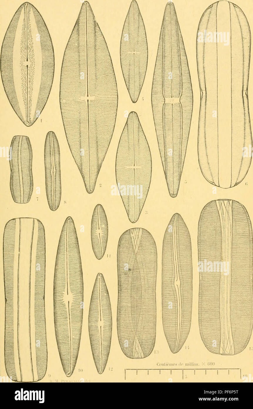 . Diatomées marines de France et des districts maritimes voisins. Diatoms. Peragallo. - Diatomées de France. l'I WMII,. â Mi.'f..;:iM|,li,- l'!r|,:ii;.l.-n VmL m l'I. Please note that these images are extracted from scanned page images that may have been digitally enhanced for readability - coloration and appearance of these illustrations may not perfectly resemble the original work.. Péragallo, H. (Hippolyte), b. 1851; Péragallo, M. (Maurice), b. 1853. Grez-sur-Loing, M. J. Tempère Stock Photohttps://www.alamy.com/image-license-details/?v=1https://www.alamy.com/diatomes-marines-de-france-et-des-districts-maritimes-voisins-diatoms-peragallo-diatomes-de-france-li-wmii-mifimli-l!riil-n-vml-m-li-please-note-that-these-images-are-extracted-from-scanned-page-images-that-may-have-been-digitally-enhanced-for-readability-coloration-and-appearance-of-these-illustrations-may-not-perfectly-resemble-the-original-work-pragallo-h-hippolyte-b-1851-pragallo-m-maurice-b-1853-grez-sur-loing-m-j-tempre-image215893380.html
RMPF6P5T–. Diatomées marines de France et des districts maritimes voisins. Diatoms. Peragallo. - Diatomées de France. l'I WMII,. â Mi.'f..;:iM|,li,- l'!r|,:ii;.l.-n VmL m l'I. Please note that these images are extracted from scanned page images that may have been digitally enhanced for readability - coloration and appearance of these illustrations may not perfectly resemble the original work.. Péragallo, H. (Hippolyte), b. 1851; Péragallo, M. (Maurice), b. 1853. Grez-sur-Loing, M. J. Tempère
![. Diatomaceæ of North America [microform] : illustrated with twenty-three hundred figures from the author's drawings on one hundred and twelve plates. Diatoms; Algae; Diatomées; Algues. I'l..t, l.XXXIX. M'lllll. â 'oliin. I'. 14, , r. 17. i^. t(K> . At., luie as. ti. Please note that these images are extracted from scanned page images that may have been digitally enhanced for readability - coloration and appearance of these illustrations may not perfectly resemble the original work.. Wolle, Francis, 1817-1893. Bethlehem, Pa. : Comenius Press Stock Photo . Diatomaceæ of North America [microform] : illustrated with twenty-three hundred figures from the author's drawings on one hundred and twelve plates. Diatoms; Algae; Diatomées; Algues. I'l..t, l.XXXIX. M'lllll. â 'oliin. I'. 14, , r. 17. i^. t(K> . At., luie as. ti. Please note that these images are extracted from scanned page images that may have been digitally enhanced for readability - coloration and appearance of these illustrations may not perfectly resemble the original work.. Wolle, Francis, 1817-1893. Bethlehem, Pa. : Comenius Press Stock Photo](https://c8.alamy.com/comp/RJ19C6/diatomace-of-north-america-microform-illustrated-with-twenty-three-hundred-figures-from-the-authors-drawings-on-one-hundred-and-twelve-plates-diatoms-algae-diatomes-algues-ilt-lxxxix-mlllll-oliin-i-14-r-17-i-tkgt-at-luie-as-ti-please-note-that-these-images-are-extracted-from-scanned-page-images-that-may-have-been-digitally-enhanced-for-readability-coloration-and-appearance-of-these-illustrations-may-not-perfectly-resemble-the-original-work-wolle-francis-1817-1893-bethlehem-pa-comenius-press-RJ19C6.jpg) . Diatomaceæ of North America [microform] : illustrated with twenty-three hundred figures from the author's drawings on one hundred and twelve plates. Diatoms; Algae; Diatomées; Algues. I'l..t, l.XXXIX. M'lllll. â 'oliin. I'. 14, , r. 17. i^. t(K> . At., luie as. ti. Please note that these images are extracted from scanned page images that may have been digitally enhanced for readability - coloration and appearance of these illustrations may not perfectly resemble the original work.. Wolle, Francis, 1817-1893. Bethlehem, Pa. : Comenius Press Stock Photohttps://www.alamy.com/image-license-details/?v=1https://www.alamy.com/diatomace-of-north-america-microform-illustrated-with-twenty-three-hundred-figures-from-the-authors-drawings-on-one-hundred-and-twelve-plates-diatoms-algae-diatomes-algues-ilt-lxxxix-mlllll-oliin-i-14-r-17-i-tkgt-at-luie-as-ti-please-note-that-these-images-are-extracted-from-scanned-page-images-that-may-have-been-digitally-enhanced-for-readability-coloration-and-appearance-of-these-illustrations-may-not-perfectly-resemble-the-original-work-wolle-francis-1817-1893-bethlehem-pa-comenius-press-image234827942.html
. Diatomaceæ of North America [microform] : illustrated with twenty-three hundred figures from the author's drawings on one hundred and twelve plates. Diatoms; Algae; Diatomées; Algues. I'l..t, l.XXXIX. M'lllll. â 'oliin. I'. 14, , r. 17. i^. t(K> . At., luie as. ti. Please note that these images are extracted from scanned page images that may have been digitally enhanced for readability - coloration and appearance of these illustrations may not perfectly resemble the original work.. Wolle, Francis, 1817-1893. Bethlehem, Pa. : Comenius Press Stock Photohttps://www.alamy.com/image-license-details/?v=1https://www.alamy.com/diatomace-of-north-america-microform-illustrated-with-twenty-three-hundred-figures-from-the-authors-drawings-on-one-hundred-and-twelve-plates-diatoms-algae-diatomes-algues-ilt-lxxxix-mlllll-oliin-i-14-r-17-i-tkgt-at-luie-as-ti-please-note-that-these-images-are-extracted-from-scanned-page-images-that-may-have-been-digitally-enhanced-for-readability-coloration-and-appearance-of-these-illustrations-may-not-perfectly-resemble-the-original-work-wolle-francis-1817-1893-bethlehem-pa-comenius-press-image234827942.htmlRMRJ19C6–. Diatomaceæ of North America [microform] : illustrated with twenty-three hundred figures from the author's drawings on one hundred and twelve plates. Diatoms; Algae; Diatomées; Algues. I'l..t, l.XXXIX. M'lllll. â 'oliin. I'. 14, , r. 17. i^. t(K> . At., luie as. ti. Please note that these images are extracted from scanned page images that may have been digitally enhanced for readability - coloration and appearance of these illustrations may not perfectly resemble the original work.. Wolle, Francis, 1817-1893. Bethlehem, Pa. : Comenius Press
 . Diatomées marines de France et des districts maritimes voisins. Diatoms. Peragallo. â Diatomées de France l'i. xciv.. I.'- M â¢â¢â â iik'f;l|'hi' Prf|i3r.ili'ii. Please note that these images are extracted from scanned page images that may have been digitally enhanced for readability - coloration and appearance of these illustrations may not perfectly resemble the original work.. Péragallo, H. (Hippolyte), b. 1851; Péragallo, M. (Maurice), b. 1853. Grez-sur-Loing, M. J. Tempère Stock Photohttps://www.alamy.com/image-license-details/?v=1https://www.alamy.com/diatomes-marines-de-france-et-des-districts-maritimes-voisins-diatoms-peragallo-diatomes-de-france-li-xciv-i-m-iikflhi-prfi3riliii-please-note-that-these-images-are-extracted-from-scanned-page-images-that-may-have-been-digitally-enhanced-for-readability-coloration-and-appearance-of-these-illustrations-may-not-perfectly-resemble-the-original-work-pragallo-h-hippolyte-b-1851-pragallo-m-maurice-b-1853-grez-sur-loing-m-j-tempre-image215893236.html
RMPF6P0M–. Diatomées marines de France et des districts maritimes voisins. Diatoms. Peragallo. â Diatomées de France l'i. xciv.. I.'- M â¢â¢â â iik'f;l|'hi' Prf|i3r.ili'ii. Please note that these images are extracted from scanned page images that may have been digitally enhanced for readability - coloration and appearance of these illustrations may not perfectly resemble the original work.. Péragallo, H. (Hippolyte), b. 1851; Péragallo, M. (Maurice), b. 1853. Grez-sur-Loing, M. J. Tempère
 . First forms of vegetation. Botany; Cryptogams. FiG. 24.—DiATOMA BiDDULPHiANA—magnified. pearance, and more richly and elaborately carved than the costliest bracelet on the arm of a queen. Some resemble miniature flags or fans, adorned with the most exquisite figures; some graceful boats, frost- ed and granulated, in which a tiny animalcule might float over a dew-drop; and some little trees (Fig. 25), covered with variegated leaves, arranged in fan- like clusters, as though intended for microscopic Fig 25.—exilaeia tlabellata. models of 3. grove of fan- palms. In short, they form circles, tri Stock Photohttps://www.alamy.com/image-license-details/?v=1https://www.alamy.com/first-forms-of-vegetation-botany-cryptogams-fig-24diatoma-biddulphianamagnified-pearance-and-more-richly-and-elaborately-carved-than-the-costliest-bracelet-on-the-arm-of-a-queen-some-resemble-miniature-flags-or-fans-adorned-with-the-most-exquisite-figures-some-graceful-boats-frost-ed-and-granulated-in-which-a-tiny-animalcule-might-float-over-a-dew-drop-and-some-little-trees-fig-25-covered-with-variegated-leaves-arranged-in-fan-like-clusters-as-though-intended-for-microscopic-fig-25exilaeia-tlabellata-models-of-3-grove-of-fan-palms-in-short-they-form-circles-tri-image232290321.html
RMRDWMJW–. First forms of vegetation. Botany; Cryptogams. FiG. 24.—DiATOMA BiDDULPHiANA—magnified. pearance, and more richly and elaborately carved than the costliest bracelet on the arm of a queen. Some resemble miniature flags or fans, adorned with the most exquisite figures; some graceful boats, frost- ed and granulated, in which a tiny animalcule might float over a dew-drop; and some little trees (Fig. 25), covered with variegated leaves, arranged in fan- like clusters, as though intended for microscopic Fig 25.—exilaeia tlabellata. models of 3. grove of fan- palms. In short, they form circles, tri
 . Diatomées marines de France et des districts maritimes voisins. Diatoms. Peragallo. - Diatomées de france. l'i. .. [ H. Perailm-lo. ilc'l, I â¢â¢ â II- â â |.l,.- l'K. Please note that these images are extracted from scanned page images that may have been digitally enhanced for readability - coloration and appearance of these illustrations may not perfectly resemble the original work.. Péragallo, H. (Hippolyte), b. 1851; Péragallo, M. (Maurice), b. 1853. Grez-sur-Loing, M. J. Tempère Stock Photohttps://www.alamy.com/image-license-details/?v=1https://www.alamy.com/diatomes-marines-de-france-et-des-districts-maritimes-voisins-diatoms-peragallo-diatomes-de-france-li-h-perailm-lo-ilcl-i-ii-l-lk-please-note-that-these-images-are-extracted-from-scanned-page-images-that-may-have-been-digitally-enhanced-for-readability-coloration-and-appearance-of-these-illustrations-may-not-perfectly-resemble-the-original-work-pragallo-h-hippolyte-b-1851-pragallo-m-maurice-b-1853-grez-sur-loing-m-j-tempre-image215893393.html
RMPF6P69–. Diatomées marines de France et des districts maritimes voisins. Diatoms. Peragallo. - Diatomées de france. l'i. .. [ H. Perailm-lo. ilc'l, I â¢â¢ â II- â â |.l,.- l'K. Please note that these images are extracted from scanned page images that may have been digitally enhanced for readability - coloration and appearance of these illustrations may not perfectly resemble the original work.. Péragallo, H. (Hippolyte), b. 1851; Péragallo, M. (Maurice), b. 1853. Grez-sur-Loing, M. J. Tempère
 . Contributions to the fauna of the New York Croton water : microscopial observations during the years 1870-'71. Water-supply; Water. from nature by the author's own hand, and engraved on stone by Mr. F. Rlxinger, of New York. Also, seven wood-cuts, made by Mr. Bernstein, have been placed in the body of this pamphlet. Of the orders and families having representatives in this water, there have been only one or two drawings made of each order. Of the frequently found alg^e, fractures and single al- gse, {Conferva, Spirogyra, Desmidium, Diatoma, &c.)I have had no drawings made. The crustacean Stock Photohttps://www.alamy.com/image-license-details/?v=1https://www.alamy.com/contributions-to-the-fauna-of-the-new-york-croton-water-microscopial-observations-during-the-years-1870-71-water-supply-water-from-nature-by-the-authors-own-hand-and-engraved-on-stone-by-mr-f-rlxinger-of-new-york-also-seven-wood-cuts-made-by-mr-bernstein-have-been-placed-in-the-body-of-this-pamphlet-of-the-orders-and-families-having-representatives-in-this-water-there-have-been-only-one-or-two-drawings-made-of-each-order-of-the-frequently-found-alge-fractures-and-single-al-gse-conferva-spirogyra-desmidium-diatoma-ampci-have-had-no-drawings-made-the-crustacean-image232550459.html
RMRE9GDF–. Contributions to the fauna of the New York Croton water : microscopial observations during the years 1870-'71. Water-supply; Water. from nature by the author's own hand, and engraved on stone by Mr. F. Rlxinger, of New York. Also, seven wood-cuts, made by Mr. Bernstein, have been placed in the body of this pamphlet. Of the orders and families having representatives in this water, there have been only one or two drawings made of each order. Of the frequently found alg^e, fractures and single al- gse, {Conferva, Spirogyra, Desmidium, Diatoma, &c.)I have had no drawings made. The crustacean
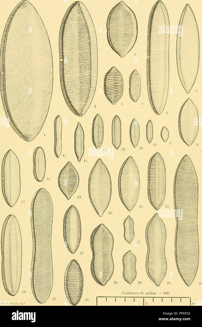 . Diatomées marines de France et des districts maritimes voisins. Diatoms. Peraqallo. - Diatomées de France. IM. Ll.. l'mAii.vr.i.c.l.-l !,<â Mil l'iiL'rnphe Pn-par.il.i. Please note that these images are extracted from scanned page images that may have been digitally enhanced for readability - coloration and appearance of these illustrations may not perfectly resemble the original work.. Péragallo, H. (Hippolyte), b. 1851; Péragallo, M. (Maurice), b. 1853. Grez-sur-Loing, M. J. Tempère Stock Photohttps://www.alamy.com/image-license-details/?v=1https://www.alamy.com/diatomes-marines-de-france-et-des-districts-maritimes-voisins-diatoms-peraqallo-diatomes-de-france-im-ll-lmaiivricl-l-!lt-mil-liilrnphe-pn-parili-please-note-that-these-images-are-extracted-from-scanned-page-images-that-may-have-been-digitally-enhanced-for-readability-coloration-and-appearance-of-these-illustrations-may-not-perfectly-resemble-the-original-work-pragallo-h-hippolyte-b-1851-pragallo-m-maurice-b-1853-grez-sur-loing-m-j-tempre-image215893298.html
RMPF6P2X–. Diatomées marines de France et des districts maritimes voisins. Diatoms. Peraqallo. - Diatomées de France. IM. Ll.. l'mAii.vr.i.c.l.-l !,<â Mil l'iiL'rnphe Pn-par.il.i. Please note that these images are extracted from scanned page images that may have been digitally enhanced for readability - coloration and appearance of these illustrations may not perfectly resemble the original work.. Péragallo, H. (Hippolyte), b. 1851; Péragallo, M. (Maurice), b. 1853. Grez-sur-Loing, M. J. Tempère
![. Diatomaceæ of North America [microform] : illustrated with twenty-three hundred figures from the author's drawings on one hundred and twelve plates. Diatoms; Algae; Diatomées; Algues. v^ â ''^>I^ ^o. 23 WEST MAIN STREET WEBSTER, N.Y. 14SS0 (716)S72-4S03 â¢^^. Please note that these images are extracted from scanned page images that may have been digitally enhanced for readability - coloration and appearance of these illustrations may not perfectly resemble the original work.. Wolle, Francis, 1817-1893. Bethlehem, Pa. : Comenius Press Stock Photo . Diatomaceæ of North America [microform] : illustrated with twenty-three hundred figures from the author's drawings on one hundred and twelve plates. Diatoms; Algae; Diatomées; Algues. v^ â ''^>I^ ^o. 23 WEST MAIN STREET WEBSTER, N.Y. 14SS0 (716)S72-4S03 â¢^^. Please note that these images are extracted from scanned page images that may have been digitally enhanced for readability - coloration and appearance of these illustrations may not perfectly resemble the original work.. Wolle, Francis, 1817-1893. Bethlehem, Pa. : Comenius Press Stock Photo](https://c8.alamy.com/comp/RJ1YGE/diatomace-of-north-america-microform-illustrated-with-twenty-three-hundred-figures-from-the-authors-drawings-on-one-hundred-and-twelve-plates-diatoms-algae-diatomes-algues-v-gti-o-23-west-main-street-webster-ny-14ss0-716s72-4s03-please-note-that-these-images-are-extracted-from-scanned-page-images-that-may-have-been-digitally-enhanced-for-readability-coloration-and-appearance-of-these-illustrations-may-not-perfectly-resemble-the-original-work-wolle-francis-1817-1893-bethlehem-pa-comenius-press-RJ1YGE.jpg) . Diatomaceæ of North America [microform] : illustrated with twenty-three hundred figures from the author's drawings on one hundred and twelve plates. Diatoms; Algae; Diatomées; Algues. v^ â ''^>I^ ^o. 23 WEST MAIN STREET WEBSTER, N.Y. 14SS0 (716)S72-4S03 â¢^^. Please note that these images are extracted from scanned page images that may have been digitally enhanced for readability - coloration and appearance of these illustrations may not perfectly resemble the original work.. Wolle, Francis, 1817-1893. Bethlehem, Pa. : Comenius Press Stock Photohttps://www.alamy.com/image-license-details/?v=1https://www.alamy.com/diatomace-of-north-america-microform-illustrated-with-twenty-three-hundred-figures-from-the-authors-drawings-on-one-hundred-and-twelve-plates-diatoms-algae-diatomes-algues-v-gti-o-23-west-main-street-webster-ny-14ss0-716s72-4s03-please-note-that-these-images-are-extracted-from-scanned-page-images-that-may-have-been-digitally-enhanced-for-readability-coloration-and-appearance-of-these-illustrations-may-not-perfectly-resemble-the-original-work-wolle-francis-1817-1893-bethlehem-pa-comenius-press-image234842174.html
. Diatomaceæ of North America [microform] : illustrated with twenty-three hundred figures from the author's drawings on one hundred and twelve plates. Diatoms; Algae; Diatomées; Algues. v^ â ''^>I^ ^o. 23 WEST MAIN STREET WEBSTER, N.Y. 14SS0 (716)S72-4S03 â¢^^. Please note that these images are extracted from scanned page images that may have been digitally enhanced for readability - coloration and appearance of these illustrations may not perfectly resemble the original work.. Wolle, Francis, 1817-1893. Bethlehem, Pa. : Comenius Press Stock Photohttps://www.alamy.com/image-license-details/?v=1https://www.alamy.com/diatomace-of-north-america-microform-illustrated-with-twenty-three-hundred-figures-from-the-authors-drawings-on-one-hundred-and-twelve-plates-diatoms-algae-diatomes-algues-v-gti-o-23-west-main-street-webster-ny-14ss0-716s72-4s03-please-note-that-these-images-are-extracted-from-scanned-page-images-that-may-have-been-digitally-enhanced-for-readability-coloration-and-appearance-of-these-illustrations-may-not-perfectly-resemble-the-original-work-wolle-francis-1817-1893-bethlehem-pa-comenius-press-image234842174.htmlRMRJ1YGE–. Diatomaceæ of North America [microform] : illustrated with twenty-three hundred figures from the author's drawings on one hundred and twelve plates. Diatoms; Algae; Diatomées; Algues. v^ â ''^>I^ ^o. 23 WEST MAIN STREET WEBSTER, N.Y. 14SS0 (716)S72-4S03 â¢^^. Please note that these images are extracted from scanned page images that may have been digitally enhanced for readability - coloration and appearance of these illustrations may not perfectly resemble the original work.. Wolle, Francis, 1817-1893. Bethlehem, Pa. : Comenius Press