Quick filters:
Diatoms in Stock Photos and Images
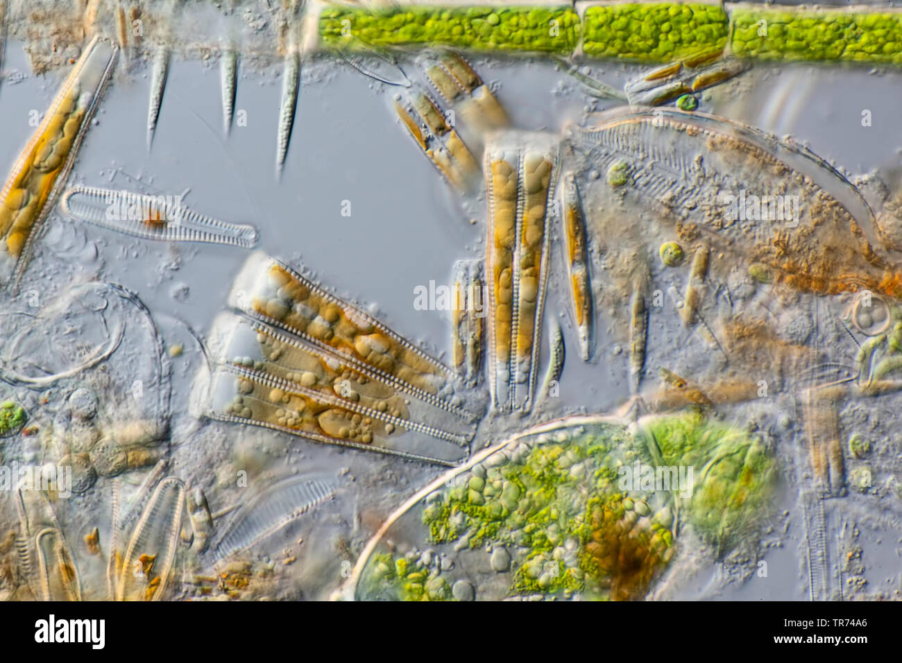 diatom (Diatomeae), living diatoms from West Iceland, in differential interference contrast, x 80, Iceland Stock Photohttps://www.alamy.com/image-license-details/?v=1https://www.alamy.com/diatom-diatomeae-living-diatoms-from-west-iceland-in-differential-interference-contrast-x-80-iceland-image255239326.html
RMTR74A6–diatom (Diatomeae), living diatoms from West Iceland, in differential interference contrast, x 80, Iceland
 Thermal water of the Svartsengi geothermal power plant, colored blue by diatoms, in the moss-covered lava field Illahraun near Grindavik on the Reykjanes peninsula. in the background the volcano Thordarfell. in the evening light. Stock Photohttps://www.alamy.com/image-license-details/?v=1https://www.alamy.com/thermal-water-of-the-svartsengi-geothermal-power-plant-colored-blue-by-diatoms-in-the-moss-covered-lava-field-illahraun-near-grindavik-on-the-reykjanes-peninsula-in-the-background-the-volcano-thordarfell-in-the-evening-light-image357611129.html
RM2BNPGJ1–Thermal water of the Svartsengi geothermal power plant, colored blue by diatoms, in the moss-covered lava field Illahraun near Grindavik on the Reykjanes peninsula. in the background the volcano Thordarfell. in the evening light.
 Diatoms in the water under the microskop Stock Photohttps://www.alamy.com/image-license-details/?v=1https://www.alamy.com/diatoms-in-the-water-under-the-microskop-image245269242.html
RMT70YBP–Diatoms in the water under the microskop
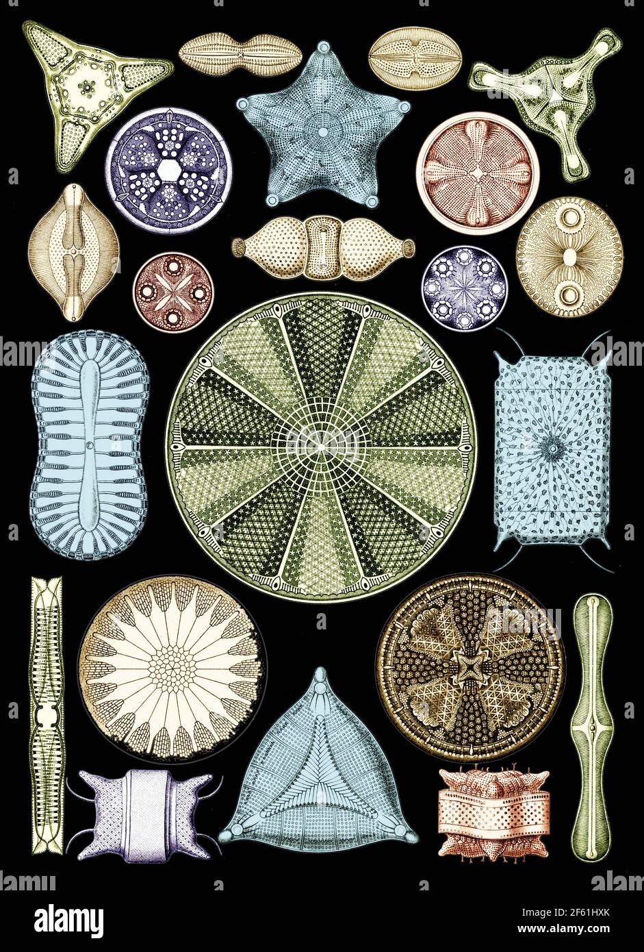 Ernst Haeckel, Diatoms, Microalgae Stock Photohttps://www.alamy.com/image-license-details/?v=1https://www.alamy.com/ernst-haeckel-diatoms-microalgae-image416772795.html
RM2F61HXK–Ernst Haeckel, Diatoms, Microalgae
 Diatoms in a gerden pond, darkground illumination Stock Photohttps://www.alamy.com/image-license-details/?v=1https://www.alamy.com/diatoms-in-a-gerden-pond-darkground-illumination-image351084666.html
RM2BB5822–Diatoms in a gerden pond, darkground illumination
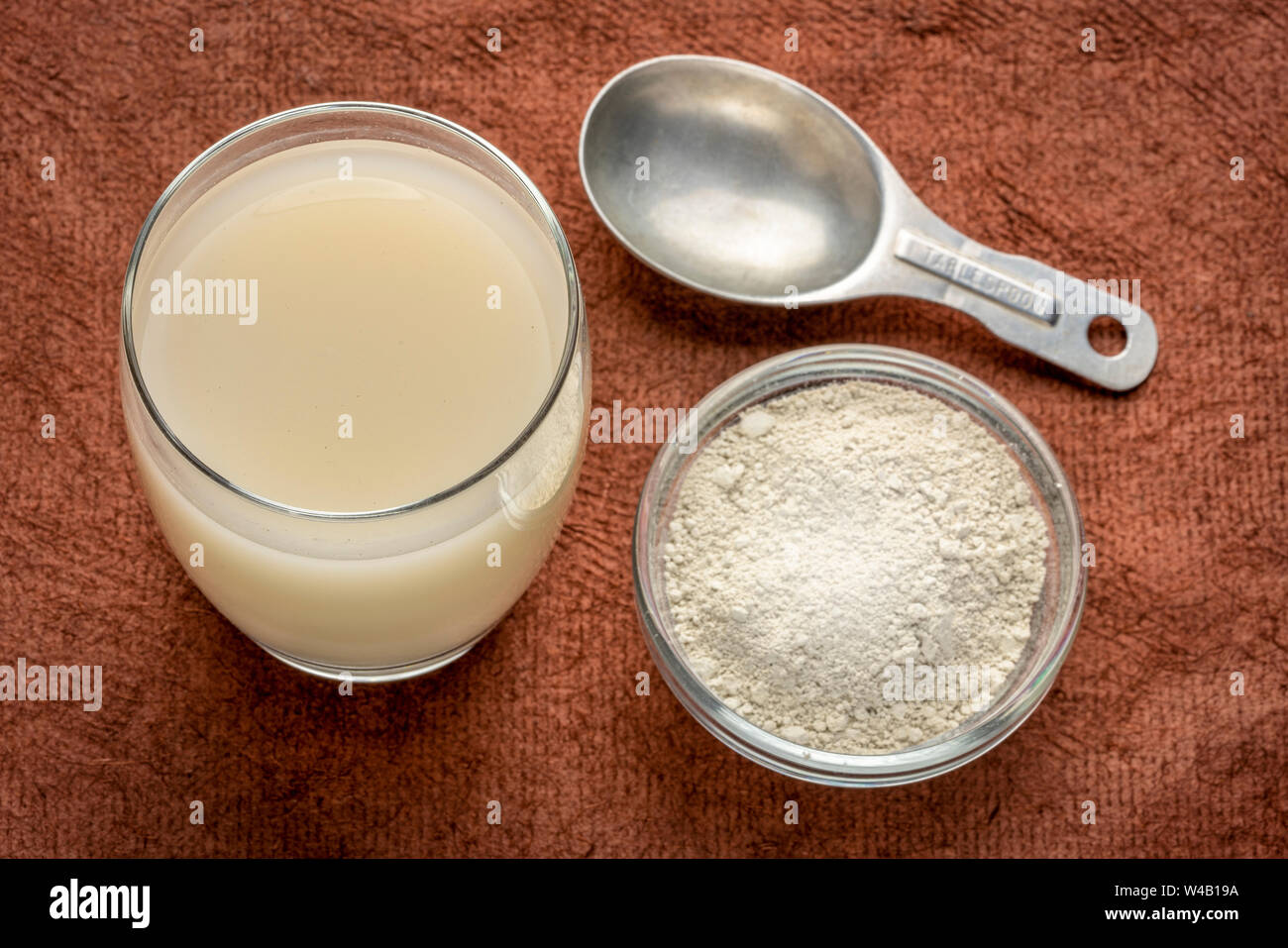 food grade diatomaceous earth supplement - powder and in a glass of water with measuring scoop Stock Photohttps://www.alamy.com/image-license-details/?v=1https://www.alamy.com/food-grade-diatomaceous-earth-supplement-powder-and-in-a-glass-of-water-with-measuring-scoop-image260856662.html
RFW4B19A–food grade diatomaceous earth supplement - powder and in a glass of water with measuring scoop
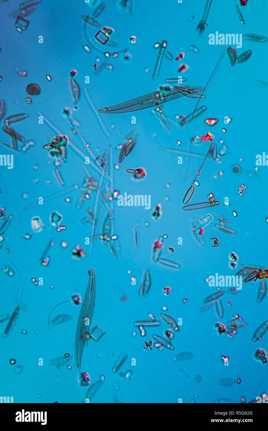 diatoms in the water Stock Photohttps://www.alamy.com/image-license-details/?v=1https://www.alamy.com/diatoms-in-the-water-image227166424.html
RFR5G92G–diatoms in the water
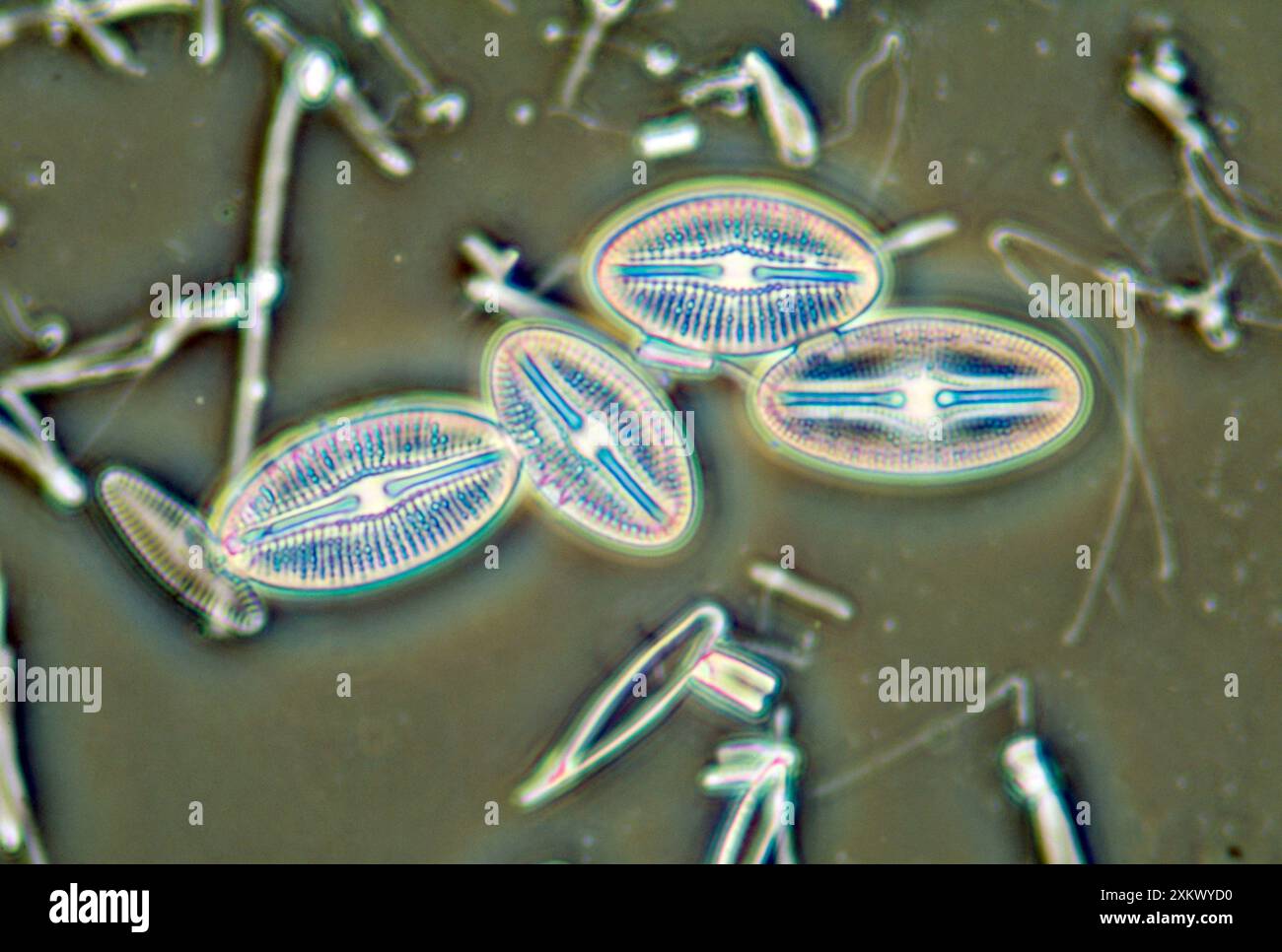 Diatoms - from marine plankton sample Stock Photohttps://www.alamy.com/image-license-details/?v=1https://www.alamy.com/diatoms-from-marine-plankton-sample-image614611676.html
RM2XKWYD0–Diatoms - from marine plankton sample
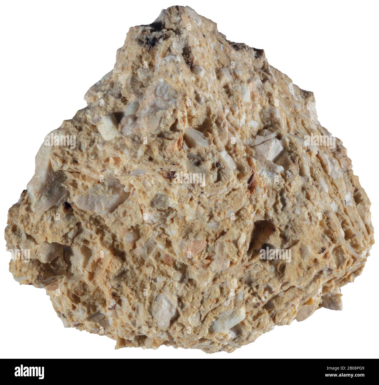 Tripolite, Carbonate, Italy A light-colored porous rock composed of the shells of diatoms. Stock Photohttps://www.alamy.com/image-license-details/?v=1https://www.alamy.com/tripolite-carbonate-italy-a-light-colored-porous-rock-composed-of-the-shells-of-diatoms-image344400681.html
RM2B08PG9–Tripolite, Carbonate, Italy A light-colored porous rock composed of the shells of diatoms.
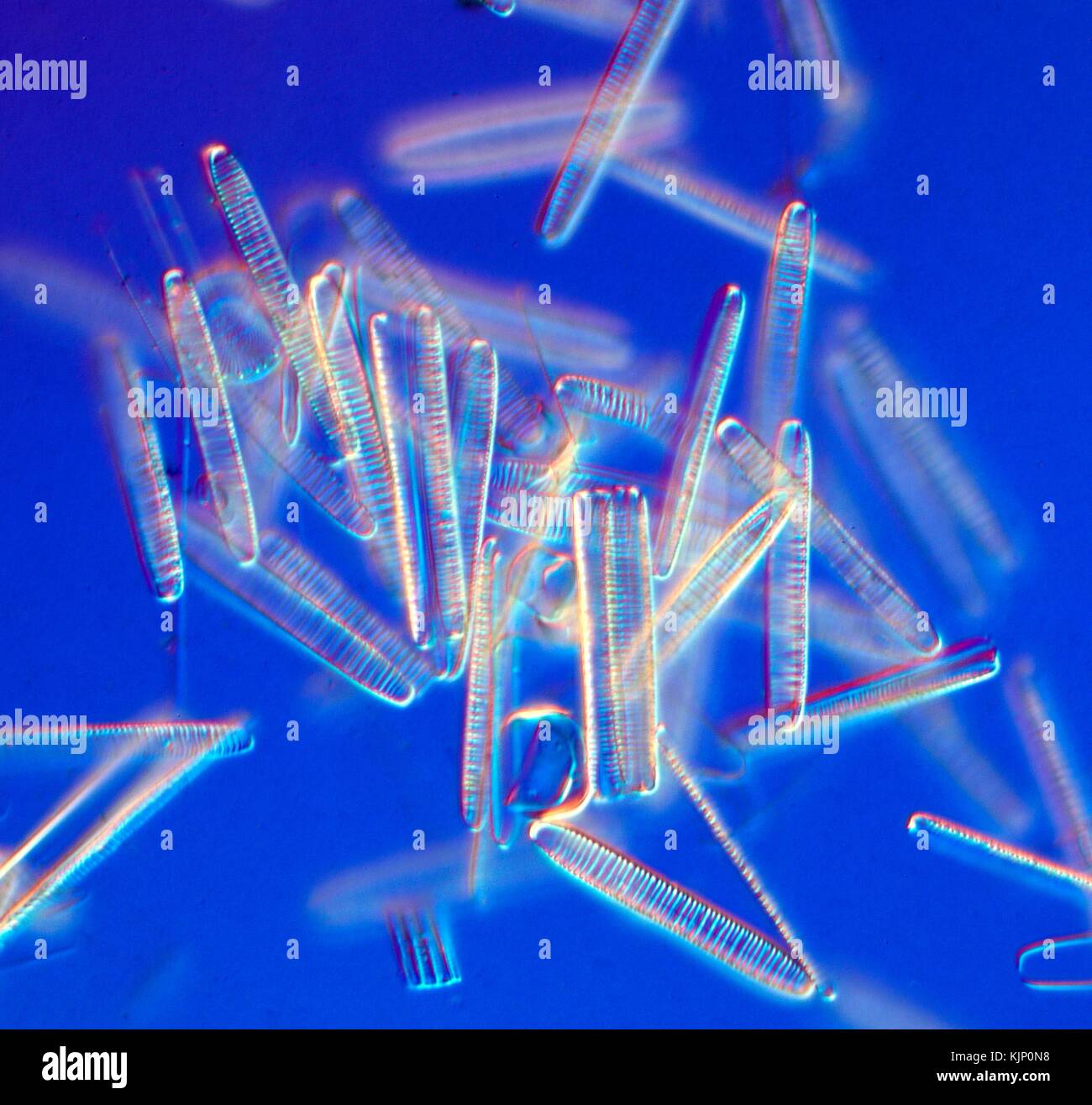 Dark field light micrograph (LM) of freshwater pennate diatoms. Diatoms represent about 25% of the plant biomass in the world. These microscopic unicellular plants are important biomass and oxygen producers. Through their photosynthesis, greenhouse gasses are converted to organic material and they therefore play a decisive role in the global carbon dioxide circulation. When individual cells die they remain as siliceous (silica dioxide) cell walls and sink to the bottom of the lake or ocean. Horizontal object size of this image section: 0.3 mm. Stock Photohttps://www.alamy.com/image-license-details/?v=1https://www.alamy.com/stock-image-dark-field-light-micrograph-lm-of-freshwater-pennate-diatoms-diatoms-166440660.html
RFKJP0N8–Dark field light micrograph (LM) of freshwater pennate diatoms. Diatoms represent about 25% of the plant biomass in the world. These microscopic unicellular plants are important biomass and oxygen producers. Through their photosynthesis, greenhouse gasses are converted to organic material and they therefore play a decisive role in the global carbon dioxide circulation. When individual cells die they remain as siliceous (silica dioxide) cell walls and sink to the bottom of the lake or ocean. Horizontal object size of this image section: 0.3 mm.
 Diatoms (Diatomea) from Ernst Haeckel's Kunstformen der Natur, 1904 Stock Photohttps://www.alamy.com/image-license-details/?v=1https://www.alamy.com/diatoms-diatomea-from-ernst-haeckels-kunstformen-der-natur-1904-image352829621.html
RF2BE0NNW–Diatoms (Diatomea) from Ernst Haeckel's Kunstformen der Natur, 1904
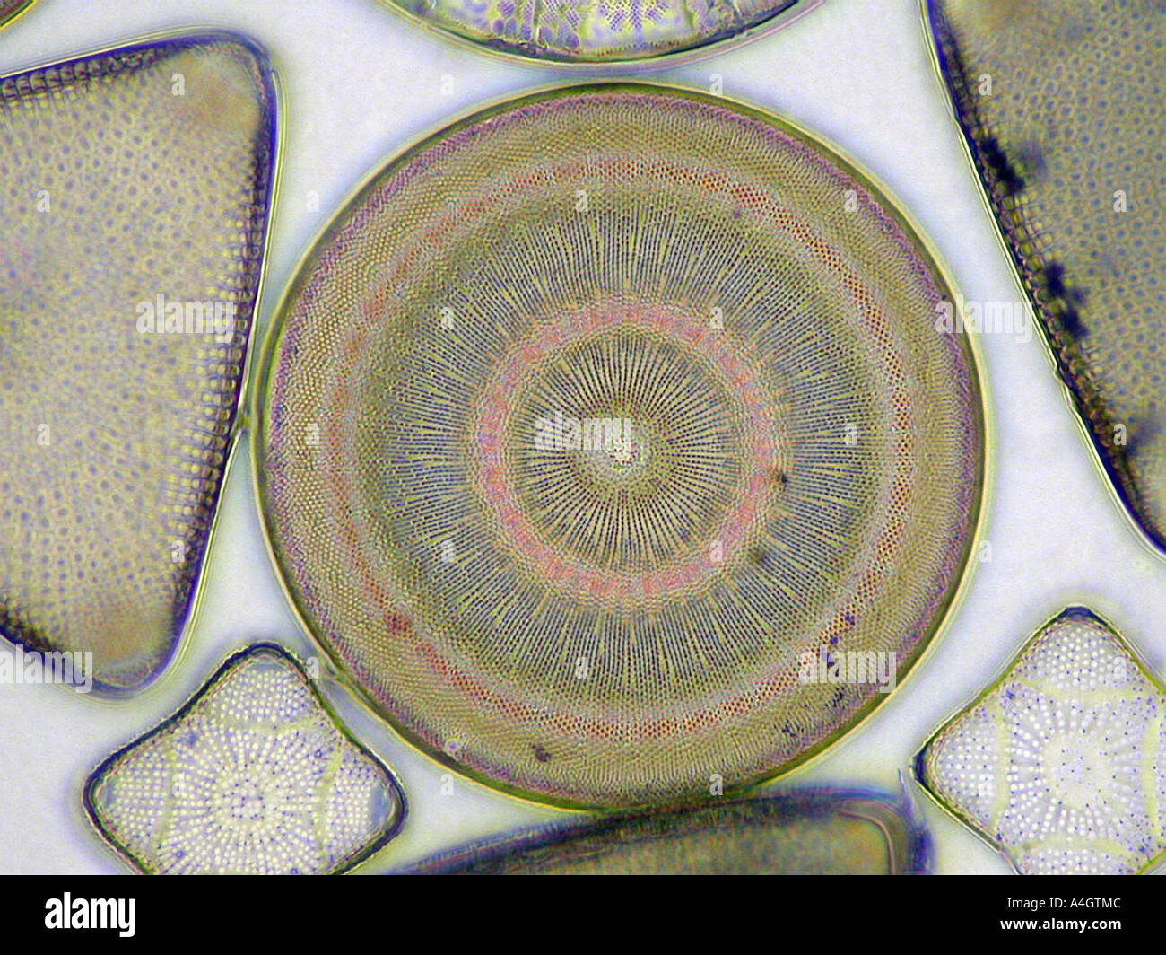 A photomicrograph of the diatom Actinocyclus Ehrebergii in an arranged pattern of other diatoms. Stock Photohttps://www.alamy.com/image-license-details/?v=1https://www.alamy.com/a-photomicrograph-of-the-diatom-actinocyclus-ehrebergii-in-an-arranged-image6312523.html
RMA4GTMC–A photomicrograph of the diatom Actinocyclus Ehrebergii in an arranged pattern of other diatoms.
 Six micro shots of cells, Numbers 1 to 6: potato starch, pine needle, uric acid crystals, diatoms, blood cells of a frog, mite of human scab., Albert Moitessier (mentioned on object), Armand Varroquier (mentioned on object), c. 1856 - in or before 1866, photographic support, albumen print, height 175 mm × width 110 mm Stock Photohttps://www.alamy.com/image-license-details/?v=1https://www.alamy.com/six-micro-shots-of-cells-numbers-1-to-6-potato-starch-pine-needle-uric-acid-crystals-diatoms-blood-cells-of-a-frog-mite-of-human-scab-albert-moitessier-mentioned-on-object-armand-varroquier-mentioned-on-object-c-1856-in-or-before-1866-photographic-support-albumen-print-height-175-mm-width-110-mm-image454392173.html
RM2HB79RW–Six micro shots of cells, Numbers 1 to 6: potato starch, pine needle, uric acid crystals, diatoms, blood cells of a frog, mite of human scab., Albert Moitessier (mentioned on object), Armand Varroquier (mentioned on object), c. 1856 - in or before 1866, photographic support, albumen print, height 175 mm × width 110 mm
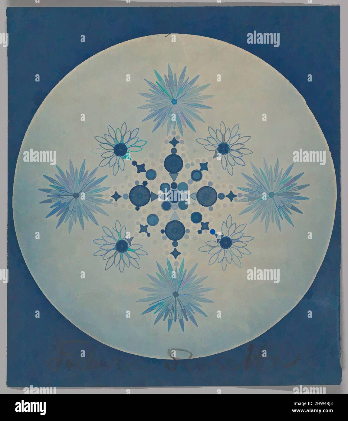 Art inspired by Frustules of Diatoms, ca. 1870, Cyanotype, 9.8 x 7.9 cm (3 7/8 x 3 1/8 in.), Photographs, Attributed to Julius Wiesner (Austrian, 1838–1916, Classic works modernized by Artotop with a splash of modernity. Shapes, color and value, eye-catching visual impact on art. Emotions through freedom of artworks in a contemporary way. A timeless message pursuing a wildly creative new direction. Artists turning to the digital medium and creating the Artotop NFT Stock Photohttps://www.alamy.com/image-license-details/?v=1https://www.alamy.com/art-inspired-by-frustules-of-diatoms-ca-1870-cyanotype-98-x-79-cm-3-78-x-3-18-in-photographs-attributed-to-julius-wiesner-austrian-18381916-classic-works-modernized-by-artotop-with-a-splash-of-modernity-shapes-color-and-value-eye-catching-visual-impact-on-art-emotions-through-freedom-of-artworks-in-a-contemporary-way-a-timeless-message-pursuing-a-wildly-creative-new-direction-artists-turning-to-the-digital-medium-and-creating-the-artotop-nft-image462942315.html
RF2HW4RJ3–Art inspired by Frustules of Diatoms, ca. 1870, Cyanotype, 9.8 x 7.9 cm (3 7/8 x 3 1/8 in.), Photographs, Attributed to Julius Wiesner (Austrian, 1838–1916, Classic works modernized by Artotop with a splash of modernity. Shapes, color and value, eye-catching visual impact on art. Emotions through freedom of artworks in a contemporary way. A timeless message pursuing a wildly creative new direction. Artists turning to the digital medium and creating the Artotop NFT
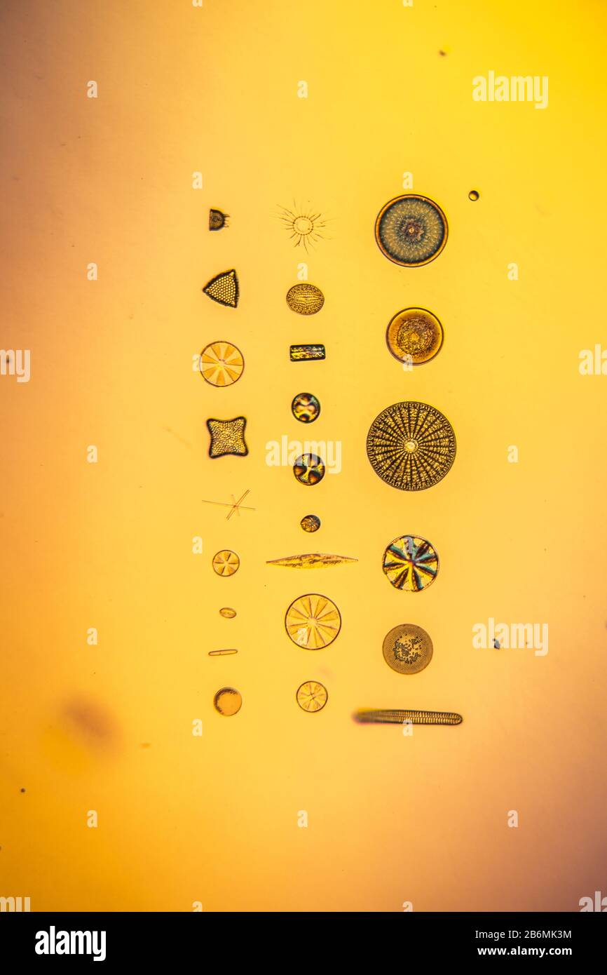 tiny microscopic diatoms in drops of water Stock Photohttps://www.alamy.com/image-license-details/?v=1https://www.alamy.com/tiny-microscopic-diatoms-in-drops-of-water-image348349336.html
RF2B6MK3M–tiny microscopic diatoms in drops of water
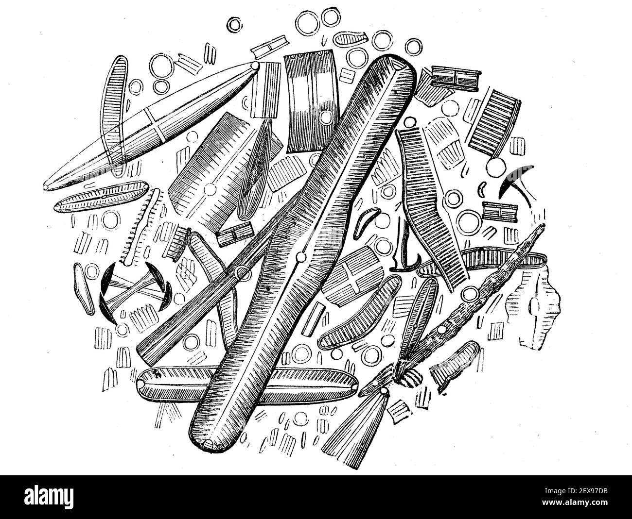 Diatoms or diatoms, Bacillariophyta, from the diatomaceous earth of Strafford in America, illustration from 1880 / Kieselalgen oder Diatomeen, Bacillariophyta, aus dem Kieselgur von Strafford in Amerika, Illustration aus 1880, Historisch, historical, digital improved reproduction of an original from the 19th century / digitale Reproduktion einer Originalvorlage aus dem 19. Jahrhundert, Stock Photohttps://www.alamy.com/image-license-details/?v=1https://www.alamy.com/diatoms-or-diatoms-bacillariophyta-from-the-diatomaceous-earth-of-strafford-in-america-illustration-from-1880-kieselalgen-oder-diatomeen-bacillariophyta-aus-dem-kieselgur-von-strafford-in-amerika-illustration-aus-1880-historisch-historical-digital-improved-reproduction-of-an-original-from-the-19th-century-digitale-reproduktion-einer-originalvorlage-aus-dem-19-jahrhundert-image412022951.html
RF2EX97DB–Diatoms or diatoms, Bacillariophyta, from the diatomaceous earth of Strafford in America, illustration from 1880 / Kieselalgen oder Diatomeen, Bacillariophyta, aus dem Kieselgur von Strafford in Amerika, Illustration aus 1880, Historisch, historical, digital improved reproduction of an original from the 19th century / digitale Reproduktion einer Originalvorlage aus dem 19. Jahrhundert,
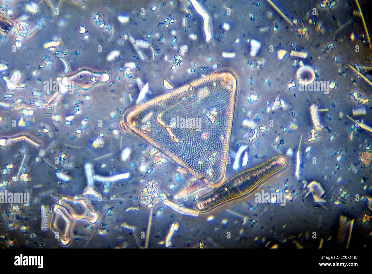 Assorted marine Diatoms magnified 100X in a microscopic view. Stock Photohttps://www.alamy.com/image-license-details/?v=1https://www.alamy.com/assorted-marine-diatoms-magnified-100x-in-a-microscopic-view-image602950219.html
RF2X0XN4B–Assorted marine Diatoms magnified 100X in a microscopic view.
![[Frustules of Diatoms]. Artist: Attributed to Julius Wiesner (Austrian, 1838-1916). Dimensions: 9.8 x 7.9 cm (3 7/8 x 3 1/8 in.). Date: ca. 1870. Museum: Metropolitan Museum of Art, New York, USA. Stock Photo [Frustules of Diatoms]. Artist: Attributed to Julius Wiesner (Austrian, 1838-1916). Dimensions: 9.8 x 7.9 cm (3 7/8 x 3 1/8 in.). Date: ca. 1870. Museum: Metropolitan Museum of Art, New York, USA. Stock Photo](https://c8.alamy.com/comp/PB9RH2/frustules-of-diatoms-artist-attributed-to-julius-wiesner-austrian-1838-1916-dimensions-98-x-79-cm-3-78-x-3-18-in-date-ca-1870-museum-metropolitan-museum-of-art-new-york-usa-PB9RH2.jpg) [Frustules of Diatoms]. Artist: Attributed to Julius Wiesner (Austrian, 1838-1916). Dimensions: 9.8 x 7.9 cm (3 7/8 x 3 1/8 in.). Date: ca. 1870. Museum: Metropolitan Museum of Art, New York, USA. Stock Photohttps://www.alamy.com/image-license-details/?v=1https://www.alamy.com/frustules-of-diatoms-artist-attributed-to-julius-wiesner-austrian-1838-1916-dimensions-98-x-79-cm-3-78-x-3-18-in-date-ca-1870-museum-metropolitan-museum-of-art-new-york-usa-image213501710.html
[Frustules of Diatoms]. Artist: Attributed to Julius Wiesner (Austrian, 1838-1916). Dimensions: 9.8 x 7.9 cm (3 7/8 x 3 1/8 in.). Date: ca. 1870. Museum: Metropolitan Museum of Art, New York, USA. Stock Photohttps://www.alamy.com/image-license-details/?v=1https://www.alamy.com/frustules-of-diatoms-artist-attributed-to-julius-wiesner-austrian-1838-1916-dimensions-98-x-79-cm-3-78-x-3-18-in-date-ca-1870-museum-metropolitan-museum-of-art-new-york-usa-image213501710.htmlRMPB9RH2–[Frustules of Diatoms]. Artist: Attributed to Julius Wiesner (Austrian, 1838-1916). Dimensions: 9.8 x 7.9 cm (3 7/8 x 3 1/8 in.). Date: ca. 1870. Museum: Metropolitan Museum of Art, New York, USA.
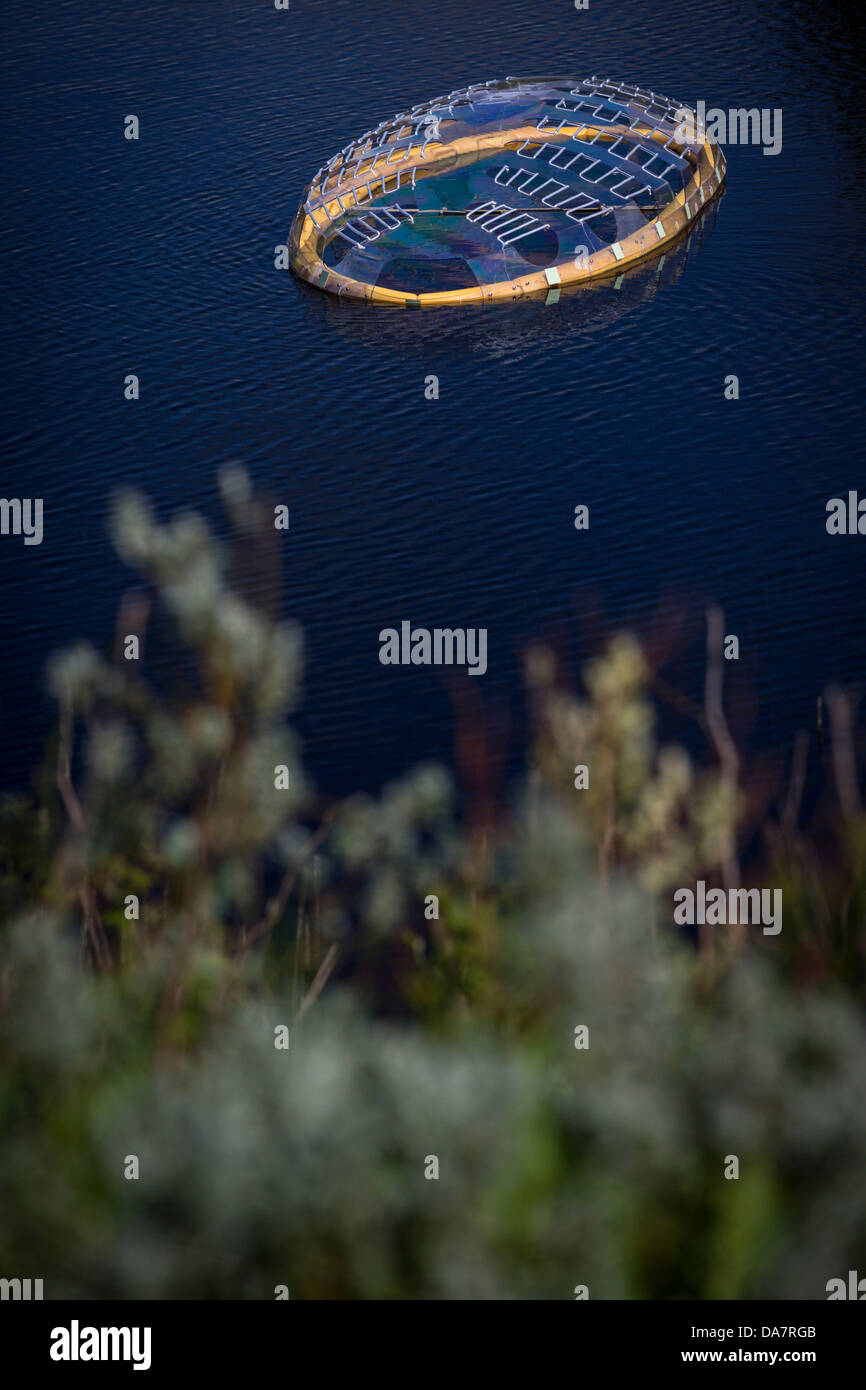 The Land Art work called Mégascospic Diatoms, made by Prisca Cosnier. A project within the Horizons 'Nature Arts' 2013 framework Stock Photohttps://www.alamy.com/image-license-details/?v=1https://www.alamy.com/stock-photo-the-land-art-work-called-mgascospic-diatoms-made-by-prisca-cosnier-57949819.html
RMDA7RGB–The Land Art work called Mégascospic Diatoms, made by Prisca Cosnier. A project within the Horizons 'Nature Arts' 2013 framework
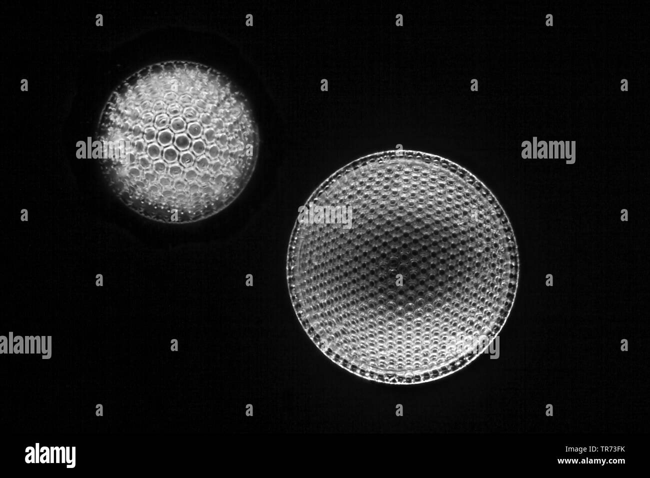 diatom (Diatomeae), fossile diatoms in darkfield Stock Photohttps://www.alamy.com/image-license-details/?v=1https://www.alamy.com/diatom-diatomeae-fossile-diatoms-in-darkfield-image255238695.html
RMTR73FK–diatom (Diatomeae), fossile diatoms in darkfield
 Thermal water, colored blue by diatoms, from the Svartsengi geothermal power plant in the Illahraun lava field near Grindavik on the Reykjanes peninsula. Stock Photohttps://www.alamy.com/image-license-details/?v=1https://www.alamy.com/thermal-water-colored-blue-by-diatoms-from-the-svartsengi-geothermal-power-plant-in-the-illahraun-lava-field-near-grindavik-on-the-reykjanes-peninsula-image357611120.html
RM2BNPGHM–Thermal water, colored blue by diatoms, from the Svartsengi geothermal power plant in the Illahraun lava field near Grindavik on the Reykjanes peninsula.
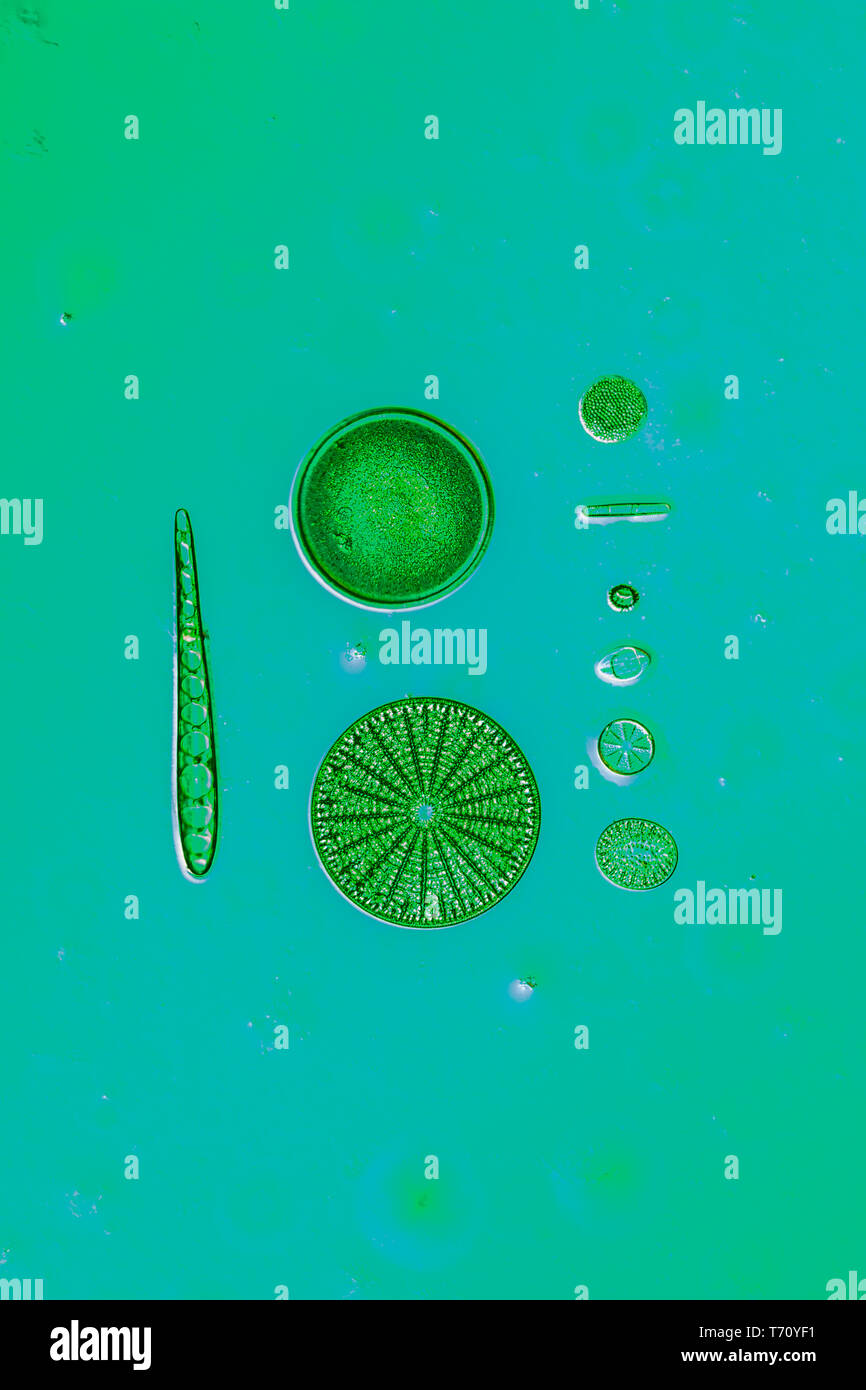 Diatoms in the water under the microskop Stock Photohttps://www.alamy.com/image-license-details/?v=1https://www.alamy.com/diatoms-in-the-water-under-the-microskop-image245269333.html
RMT70YF1–Diatoms in the water under the microskop
 Ernst Haeckel, Diatoms, Microalgae Stock Photohttps://www.alamy.com/image-license-details/?v=1https://www.alamy.com/ernst-haeckel-diatoms-microalgae-image416772777.html
RM2F61HX1–Ernst Haeckel, Diatoms, Microalgae
 Diatoms in a gerden pond, darkground illumination Stock Photohttps://www.alamy.com/image-license-details/?v=1https://www.alamy.com/diatoms-in-a-gerden-pond-darkground-illumination-image351084346.html
RM2BB57JJ–Diatoms in a gerden pond, darkground illumination
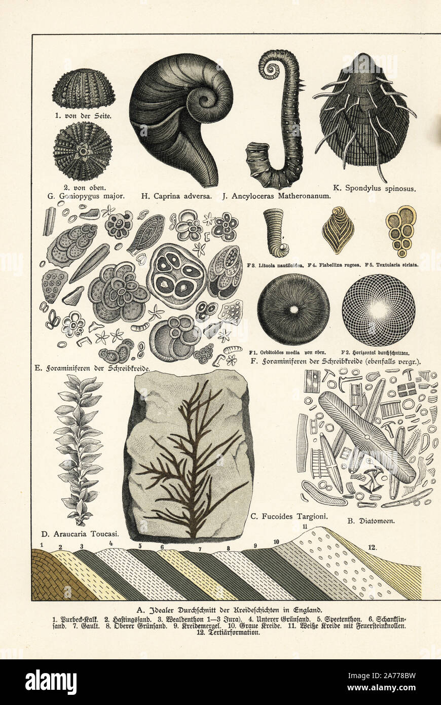 Diatoms, seaweed Fucoides targioni, conifer tree Araucaria toucasi, foraminifera in chalk, sea urchin Goniopygus major, mollusk Caprina adversa, ammonite cephalopod Ancyloceras matheronanum, and mollusk Spondylus spinosus. Chromolithograph from Dr. Fr. Rolle's 'Geology and Paleontology' section in Gotthilf Heinrich von Schubert's 'Naturgeschichte,' Schreiber, Munich, 1886. Stock Photohttps://www.alamy.com/image-license-details/?v=1https://www.alamy.com/diatoms-seaweed-fucoides-targioni-conifer-tree-araucaria-toucasi-foraminifera-in-chalk-sea-urchin-goniopygus-major-mollusk-caprina-adversa-ammonite-cephalopod-ancyloceras-matheronanum-and-mollusk-spondylus-spinosus-chromolithograph-from-dr-fr-rolles-geology-and-paleontology-section-in-gotthilf-heinrich-von-schuberts-naturgeschichte-schreiber-munich-1886-image331459853.html
RM2A778BW–Diatoms, seaweed Fucoides targioni, conifer tree Araucaria toucasi, foraminifera in chalk, sea urchin Goniopygus major, mollusk Caprina adversa, ammonite cephalopod Ancyloceras matheronanum, and mollusk Spondylus spinosus. Chromolithograph from Dr. Fr. Rolle's 'Geology and Paleontology' section in Gotthilf Heinrich von Schubert's 'Naturgeschichte,' Schreiber, Munich, 1886.
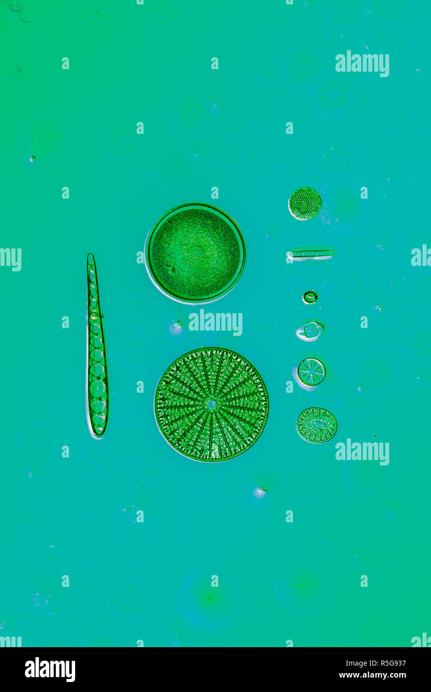 diatoms in the water Stock Photohttps://www.alamy.com/image-license-details/?v=1https://www.alamy.com/diatoms-in-the-water-image227166443.html
RFR5G937–diatoms in the water
 Diatoms - from marine plankton sample Stock Photohttps://www.alamy.com/image-license-details/?v=1https://www.alamy.com/diatoms-from-marine-plankton-sample-image614611666.html
RM2XKWYCJ–Diatoms - from marine plankton sample
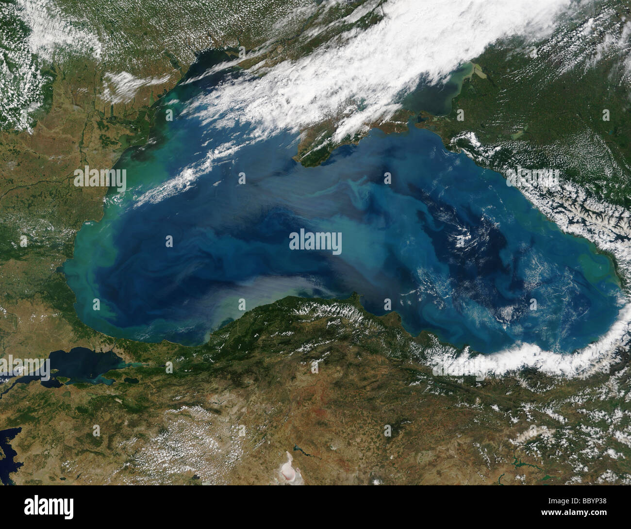 Satellite view of Phytoplankton organisms in 'bloom', Black Sea Russia Stock Photohttps://www.alamy.com/image-license-details/?v=1https://www.alamy.com/stock-photo-satellite-view-of-phytoplankton-organisms-in-bloom-black-sea-russia-24581628.html
RFBBYP38–Satellite view of Phytoplankton organisms in 'bloom', Black Sea Russia
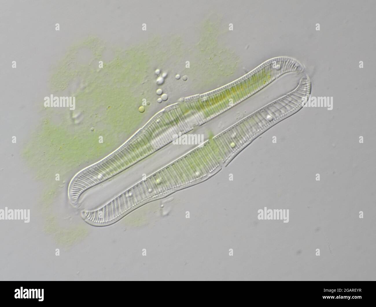 Freshwater diatom, Rhopalodia sp., about 80 microns in length, under the microscope Stock Photohttps://www.alamy.com/image-license-details/?v=1https://www.alamy.com/freshwater-diatom-rhopalodia-sp-about-80-microns-in-length-under-the-microscope-image436922411.html
RM2GAREYR–Freshwater diatom, Rhopalodia sp., about 80 microns in length, under the microscope
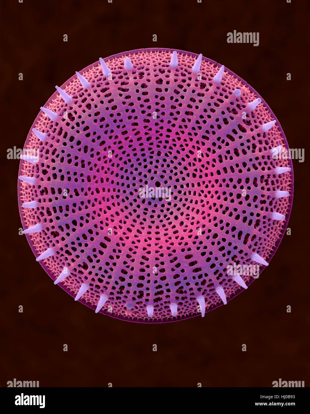 Coloured scanning electron micrograph (SEM) of centric fossil diatom frustule.This fresh water diatom frustule came from deposits found at Kalamath Falls,Oregon.This freshwater centric diatom is found in fresh water streams,ponds lakes.Diatoms are type of algae (Chromophyta,Bacillariophyceae).They Stock Photohttps://www.alamy.com/image-license-details/?v=1https://www.alamy.com/stock-photo-coloured-scanning-electron-micrograph-sem-of-centric-fossil-diatom-131545263.html
RFHJ0B93–Coloured scanning electron micrograph (SEM) of centric fossil diatom frustule.This fresh water diatom frustule came from deposits found at Kalamath Falls,Oregon.This freshwater centric diatom is found in fresh water streams,ponds lakes.Diatoms are type of algae (Chromophyta,Bacillariophyceae).They
![. Transactions of the Royal Society of New Zealand. finely and add smallquantities of some antiseptic, and dust it over the wound. Now as to the methods to be adopted to clean and mountthe diatoms in this deposit. The first thing to be done is todisintegrate the earth. This can be done (a very slow process)by soaking the earth a very long time in water and letting itcrumble, and then carefully washing out the clay. A muchquicker plan has been suggested by M. Parmentier, a pro-fessor of chemistry in Belgium, and was communicated by ]VLGuimard to the Quekett Club.* It consists in the supersatura Stock Photo . Transactions of the Royal Society of New Zealand. finely and add smallquantities of some antiseptic, and dust it over the wound. Now as to the methods to be adopted to clean and mountthe diatoms in this deposit. The first thing to be done is todisintegrate the earth. This can be done (a very slow process)by soaking the earth a very long time in water and letting itcrumble, and then carefully washing out the clay. A muchquicker plan has been suggested by M. Parmentier, a pro-fessor of chemistry in Belgium, and was communicated by ]VLGuimard to the Quekett Club.* It consists in the supersatura Stock Photo](https://c8.alamy.com/comp/2AG3BTE/transactions-of-the-royal-society-of-new-zealand-finely-and-add-smallquantities-of-some-antiseptic-and-dust-it-over-the-wound-now-as-to-the-methods-to-be-adopted-to-clean-and-mountthe-diatoms-in-this-deposit-the-first-thing-to-be-done-is-todisintegrate-the-earth-this-can-be-done-a-very-slow-processby-soaking-the-earth-a-very-long-time-in-water-and-letting-itcrumble-and-then-carefully-washing-out-the-clay-a-muchquicker-plan-has-been-suggested-by-m-parmentier-a-pro-fessor-of-chemistry-in-belgium-and-was-communicated-by-vlguimard-to-the-quekett-club-it-consists-in-the-supersatura-2AG3BTE.jpg) . Transactions of the Royal Society of New Zealand. finely and add smallquantities of some antiseptic, and dust it over the wound. Now as to the methods to be adopted to clean and mountthe diatoms in this deposit. The first thing to be done is todisintegrate the earth. This can be done (a very slow process)by soaking the earth a very long time in water and letting itcrumble, and then carefully washing out the clay. A muchquicker plan has been suggested by M. Parmentier, a pro-fessor of chemistry in Belgium, and was communicated by ]VLGuimard to the Quekett Club.* It consists in the supersatura Stock Photohttps://www.alamy.com/image-license-details/?v=1https://www.alamy.com/transactions-of-the-royal-society-of-new-zealand-finely-and-add-smallquantities-of-some-antiseptic-and-dust-it-over-the-wound-now-as-to-the-methods-to-be-adopted-to-clean-and-mountthe-diatoms-in-this-deposit-the-first-thing-to-be-done-is-todisintegrate-the-earth-this-can-be-done-a-very-slow-processby-soaking-the-earth-a-very-long-time-in-water-and-letting-itcrumble-and-then-carefully-washing-out-the-clay-a-muchquicker-plan-has-been-suggested-by-m-parmentier-a-pro-fessor-of-chemistry-in-belgium-and-was-communicated-by-vlguimard-to-the-quekett-club-it-consists-in-the-supersatura-image336906654.html
. Transactions of the Royal Society of New Zealand. finely and add smallquantities of some antiseptic, and dust it over the wound. Now as to the methods to be adopted to clean and mountthe diatoms in this deposit. The first thing to be done is todisintegrate the earth. This can be done (a very slow process)by soaking the earth a very long time in water and letting itcrumble, and then carefully washing out the clay. A muchquicker plan has been suggested by M. Parmentier, a pro-fessor of chemistry in Belgium, and was communicated by ]VLGuimard to the Quekett Club.* It consists in the supersatura Stock Photohttps://www.alamy.com/image-license-details/?v=1https://www.alamy.com/transactions-of-the-royal-society-of-new-zealand-finely-and-add-smallquantities-of-some-antiseptic-and-dust-it-over-the-wound-now-as-to-the-methods-to-be-adopted-to-clean-and-mountthe-diatoms-in-this-deposit-the-first-thing-to-be-done-is-todisintegrate-the-earth-this-can-be-done-a-very-slow-processby-soaking-the-earth-a-very-long-time-in-water-and-letting-itcrumble-and-then-carefully-washing-out-the-clay-a-muchquicker-plan-has-been-suggested-by-m-parmentier-a-pro-fessor-of-chemistry-in-belgium-and-was-communicated-by-vlguimard-to-the-quekett-club-it-consists-in-the-supersatura-image336906654.htmlRM2AG3BTE–. Transactions of the Royal Society of New Zealand. finely and add smallquantities of some antiseptic, and dust it over the wound. Now as to the methods to be adopted to clean and mountthe diatoms in this deposit. The first thing to be done is todisintegrate the earth. This can be done (a very slow process)by soaking the earth a very long time in water and letting itcrumble, and then carefully washing out the clay. A muchquicker plan has been suggested by M. Parmentier, a pro-fessor of chemistry in Belgium, and was communicated by ]VLGuimard to the Quekett Club.* It consists in the supersatura
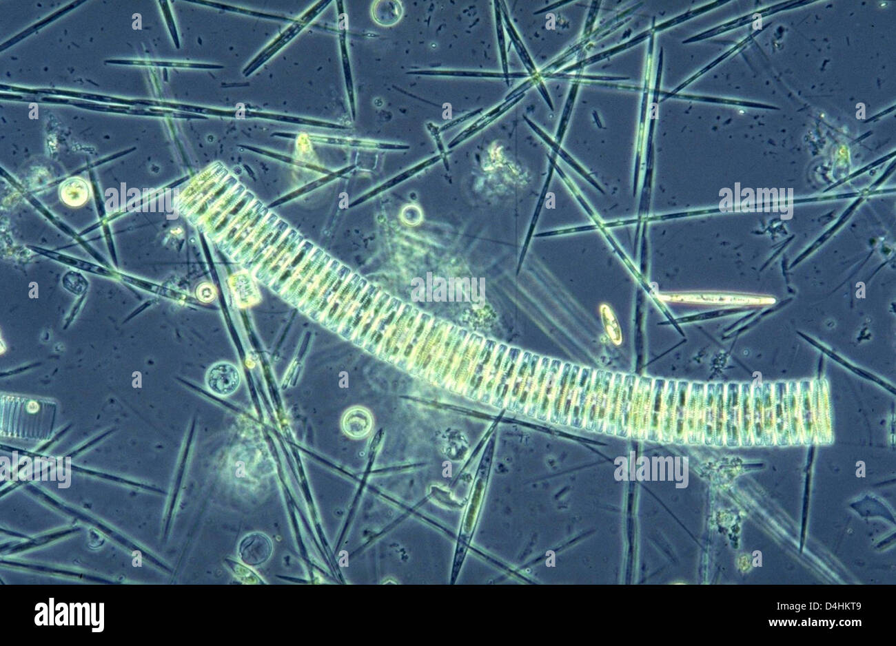 The undated microscope picture shows a plankton group three weeks after its fertilisation with iron on the research vessel ?Polarstern? of the Alfred Wegener Institute for Polar and Marine Research (AWI) in Bremerhaven, Germany. Typical Antarctic diatoms dominate the induced bloom. The AWI will decide whether or not to continue the controversial experiment on the ?Polarstern? on 26 Stock Photohttps://www.alamy.com/image-license-details/?v=1https://www.alamy.com/stock-photo-the-undated-microscope-picture-shows-a-plankton-group-three-weeks-54478489.html
RMD4HKT9–The undated microscope picture shows a plankton group three weeks after its fertilisation with iron on the research vessel ?Polarstern? of the Alfred Wegener Institute for Polar and Marine Research (AWI) in Bremerhaven, Germany. Typical Antarctic diatoms dominate the induced bloom. The AWI will decide whether or not to continue the controversial experiment on the ?Polarstern? on 26
 Inspired by Six micro shots of cells, Numbers 1 to 6: potato starch, pine needle, uric acid crystals, diatoms, blood cells of a frog, mite of human scab., Albert Moitessier, Armand Varroquier, c. 1856 - in or before 1866, albumen print, height 175 mm × width 110 mm, Reimagined by Artotop. Classic art reinvented with a modern twist. Design of warm cheerful glowing of brightness and light ray radiance. Photography inspired by surrealism and futurism, embracing dynamic energy of modern technology, movement, speed and revolutionize culture Stock Photohttps://www.alamy.com/image-license-details/?v=1https://www.alamy.com/inspired-by-six-micro-shots-of-cells-numbers-1-to-6-potato-starch-pine-needle-uric-acid-crystals-diatoms-blood-cells-of-a-frog-mite-of-human-scab-albert-moitessier-armand-varroquier-c-1856-in-or-before-1866-albumen-print-height-175-mm-width-110-mm-reimagined-by-artotop-classic-art-reinvented-with-a-modern-twist-design-of-warm-cheerful-glowing-of-brightness-and-light-ray-radiance-photography-inspired-by-surrealism-and-futurism-embracing-dynamic-energy-of-modern-technology-movement-speed-and-revolutionize-culture-image459449169.html
RF2HKDM2W–Inspired by Six micro shots of cells, Numbers 1 to 6: potato starch, pine needle, uric acid crystals, diatoms, blood cells of a frog, mite of human scab., Albert Moitessier, Armand Varroquier, c. 1856 - in or before 1866, albumen print, height 175 mm × width 110 mm, Reimagined by Artotop. Classic art reinvented with a modern twist. Design of warm cheerful glowing of brightness and light ray radiance. Photography inspired by surrealism and futurism, embracing dynamic energy of modern technology, movement, speed and revolutionize culture
 tiny microscopic diatoms in drops of water Stock Photohttps://www.alamy.com/image-license-details/?v=1https://www.alamy.com/tiny-microscopic-diatoms-in-drops-of-water-image348349174.html
RF2B6MJWX–tiny microscopic diatoms in drops of water
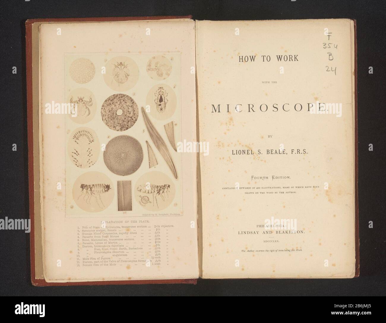 Thirteen enlarged photographs taken under a microscope (1) is the stem of a hydrangea, (2) is a mange mite, (3.) is a blood cell, (4) is a parasite , (5.) is a cross-sectional view of a bone, (6) is an aphid, (7), (8) and (9) are diatoms (11) is a flea (12) is a diatom (13) is a vlooi. Manufacturer : photographer: Richard Leach Maddox Dating: about 1865 - or for 1870 Material: paper Technique: albumen print dimensions: photo: h 135 mm × W 98 mmToelichtingFoto on frontispice. Subject: microscopeinsects: lousebones in general (human body) Stock Photohttps://www.alamy.com/image-license-details/?v=1https://www.alamy.com/thirteen-enlarged-photographs-taken-under-a-microscope-1-is-the-stem-of-a-hydrangea-2-is-a-mange-mite-3-is-a-blood-cell-4-is-a-parasite-5-is-a-cross-sectional-view-of-a-bone-6-is-an-aphid-7-8-and-9-are-diatoms-11-is-a-flea-12-is-a-diatom-13-is-a-vlooi-manufacturer-photographer-richard-leach-maddox-dating-about-1865-or-for-1870-material-paper-technique-albumen-print-dimensions-photo-h-135-mm-w-98-mmtoelichtingfoto-on-frontispice-subject-microscopeinsects-lousebones-in-general-human-body-image348306621.html
RM2B6JMJ5–Thirteen enlarged photographs taken under a microscope (1) is the stem of a hydrangea, (2) is a mange mite, (3.) is a blood cell, (4) is a parasite , (5.) is a cross-sectional view of a bone, (6) is an aphid, (7), (8) and (9) are diatoms (11) is a flea (12) is a diatom (13) is a vlooi. Manufacturer : photographer: Richard Leach Maddox Dating: about 1865 - or for 1870 Material: paper Technique: albumen print dimensions: photo: h 135 mm × W 98 mmToelichtingFoto on frontispice. Subject: microscopeinsects: lousebones in general (human body)
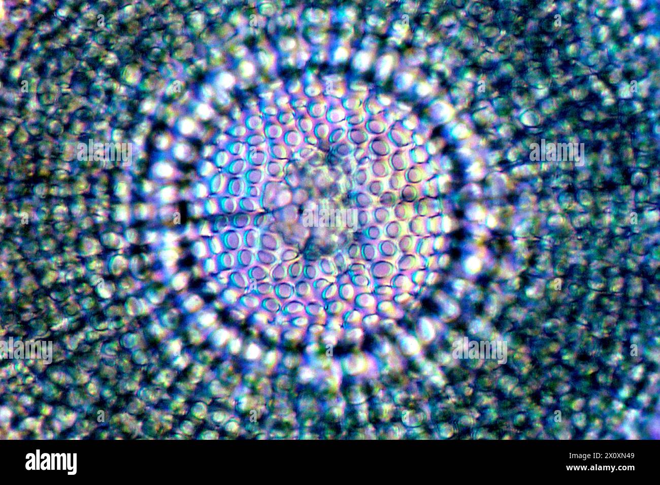 Foraminifera, magnified 400X in a microscopic view. Stock Photohttps://www.alamy.com/image-license-details/?v=1https://www.alamy.com/foraminifera-magnified-400x-in-a-microscopic-view-image602950217.html
RF2X0XN49–Foraminifera, magnified 400X in a microscopic view.
 Diatoms, seaweed Fucoides targioni, conifer tree Araucaria toucasi, foraminifera in chalk, sea urchin Goniopygus major, mollusk Caprina adversa, ammonite cephalopod Ancyloceras matheronanum, and mollusk Spondylus spinosus. Chromolithograph from Dr. Fr. Rolle's 'Geology and Paleontology' section in Gotthilf Heinrich von Schubert's 'Naturgeschichte,' Schreiber, Munich, 1886. Stock Photohttps://www.alamy.com/image-license-details/?v=1https://www.alamy.com/diatoms-seaweed-fucoides-targioni-conifer-tree-araucaria-toucasi-foraminifera-in-chalk-sea-urchin-goniopygus-major-mollusk-caprina-adversa-ammonite-cephalopod-ancyloceras-matheronanum-and-mollusk-spondylus-spinosus-chromolithograph-from-dr-fr-rolles-geology-and-paleontology-section-in-gotthilf-heinrich-von-schuberts-naturgeschichte-schreiber-munich-1886-image210548532.html
RMP6F8PC–Diatoms, seaweed Fucoides targioni, conifer tree Araucaria toucasi, foraminifera in chalk, sea urchin Goniopygus major, mollusk Caprina adversa, ammonite cephalopod Ancyloceras matheronanum, and mollusk Spondylus spinosus. Chromolithograph from Dr. Fr. Rolle's 'Geology and Paleontology' section in Gotthilf Heinrich von Schubert's 'Naturgeschichte,' Schreiber, Munich, 1886.
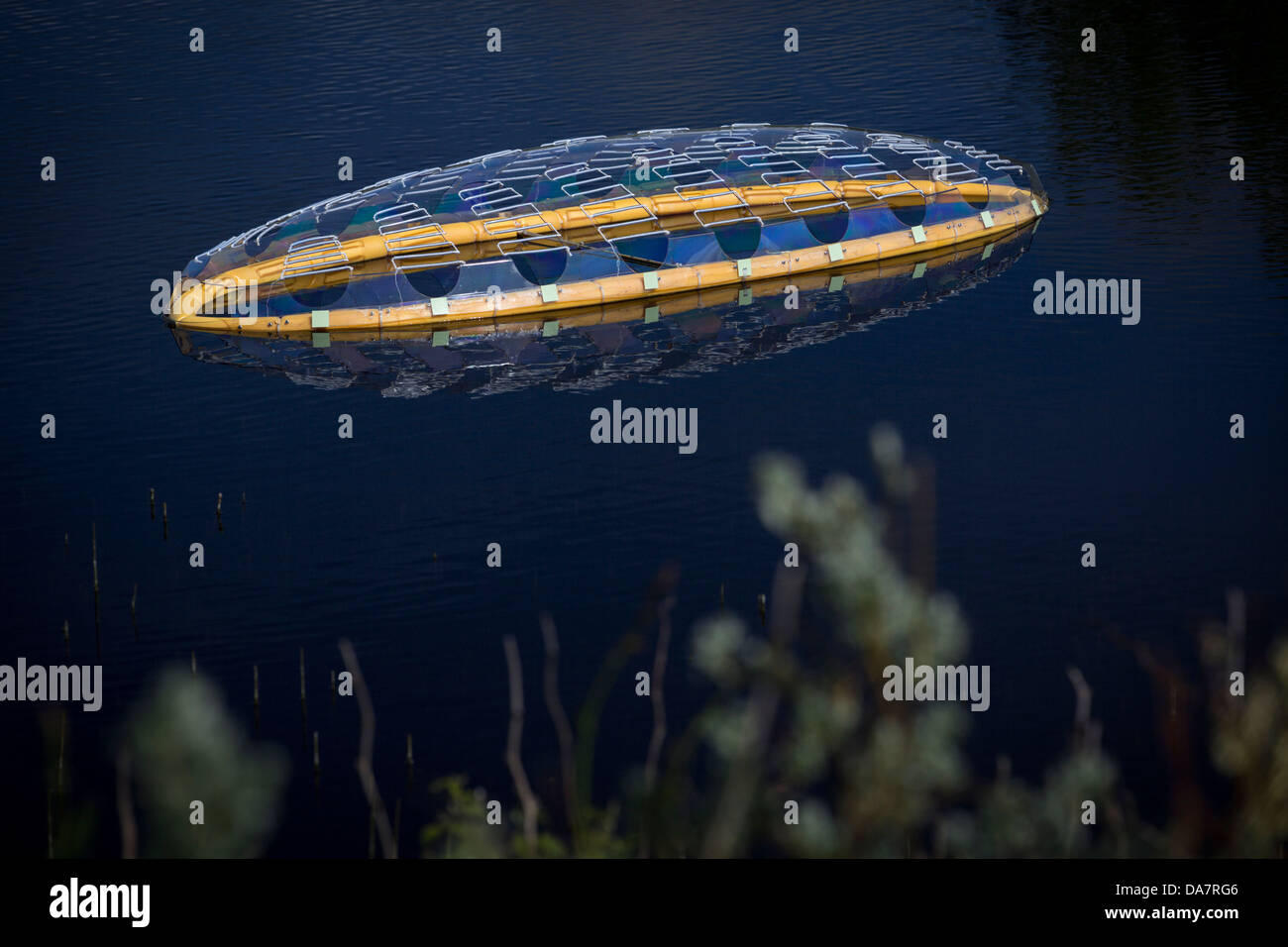 The Land Art work called Mégascospic Diatoms, made by Prisca Cosnier. A project within the Horizons 'Nature Arts' 2013 framework Stock Photohttps://www.alamy.com/image-license-details/?v=1https://www.alamy.com/stock-photo-the-land-art-work-called-mgascospic-diatoms-made-by-prisca-cosnier-57949814.html
RMDA7RG6–The Land Art work called Mégascospic Diatoms, made by Prisca Cosnier. A project within the Horizons 'Nature Arts' 2013 framework
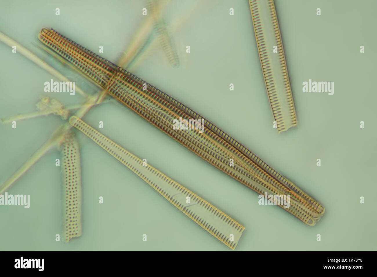 diatom (Diatomeae), diatoms in phase contrast and interference contrast, x 200 Stock Photohttps://www.alamy.com/image-license-details/?v=1https://www.alamy.com/diatom-diatomeae-diatoms-in-phase-contrast-and-interference-contrast-x-200-image255239020.html
RMTR73Y8–diatom (Diatomeae), diatoms in phase contrast and interference contrast, x 200
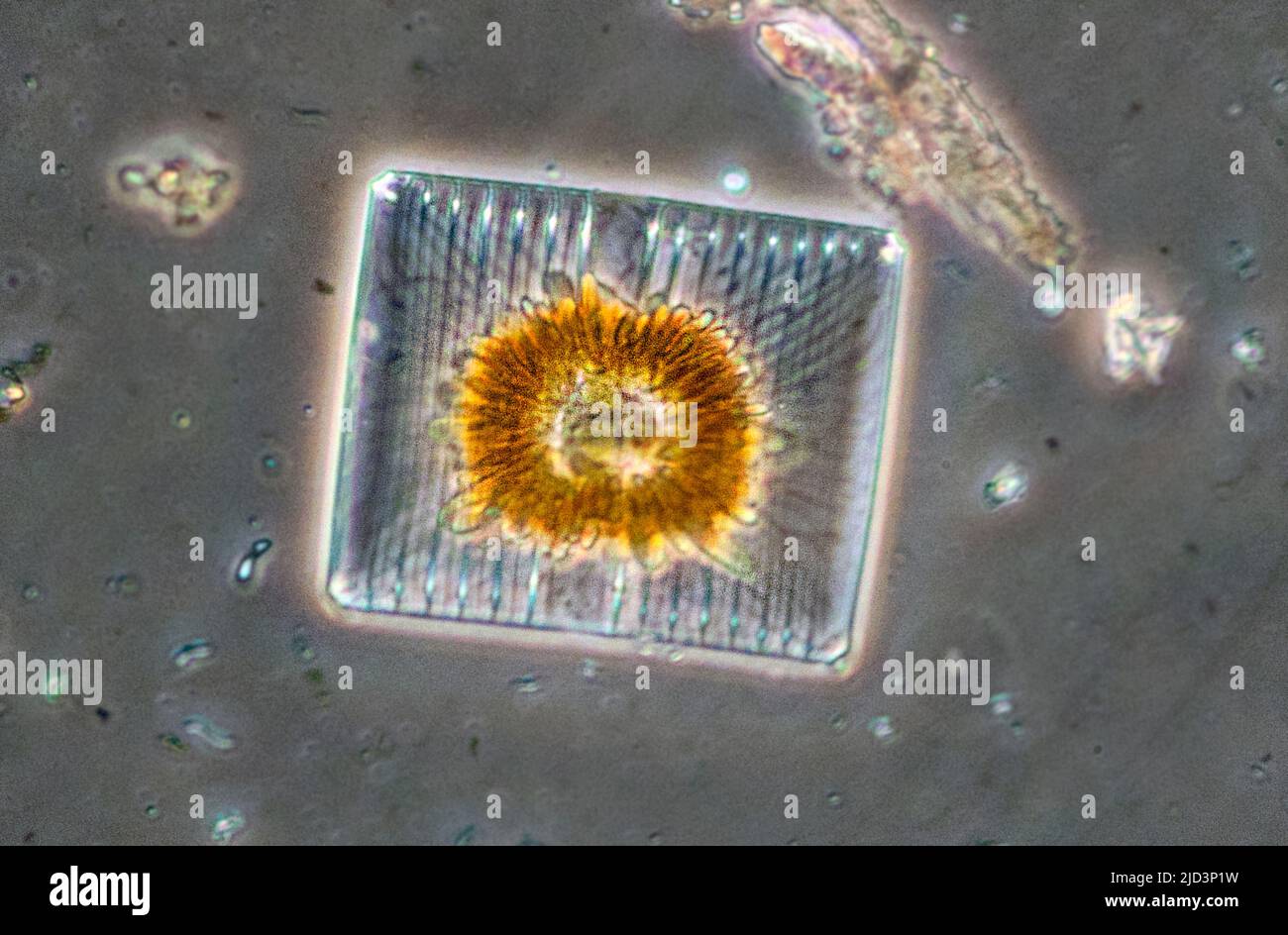 Diatoms from the genus Striatella. The square cell is about 0,070 x 0,056 mm in size. Stock Photohttps://www.alamy.com/image-license-details/?v=1https://www.alamy.com/diatoms-from-the-genus-striatella-the-square-cell-is-about-0070-x-0056-mm-in-size-image472753621.html
RM2JD3P1W–Diatoms from the genus Striatella. The square cell is about 0,070 x 0,056 mm in size.
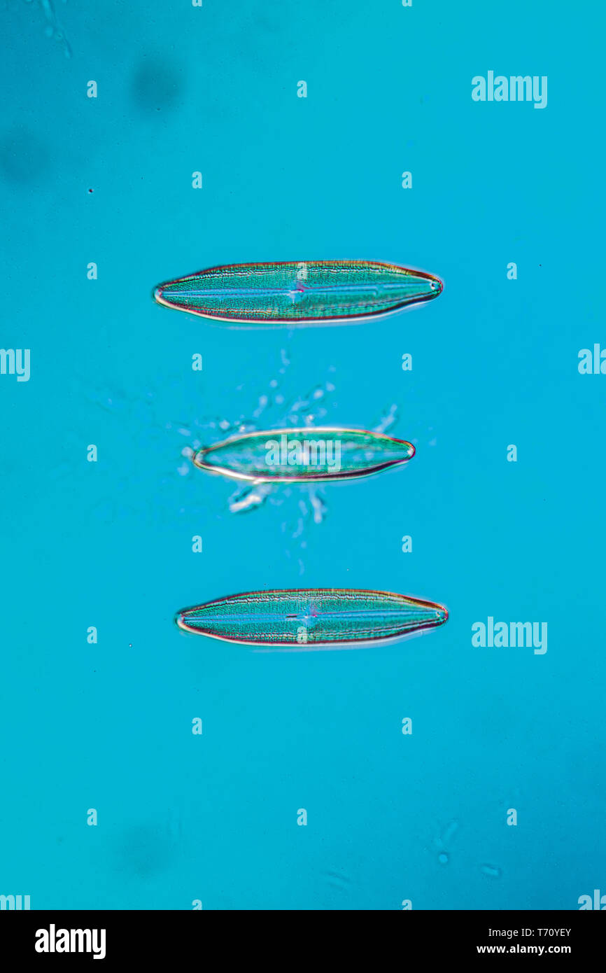 Diatoms in the water under the microskop Stock Photohttps://www.alamy.com/image-license-details/?v=1https://www.alamy.com/diatoms-in-the-water-under-the-microskop-image245269331.html
RMT70YEY–Diatoms in the water under the microskop
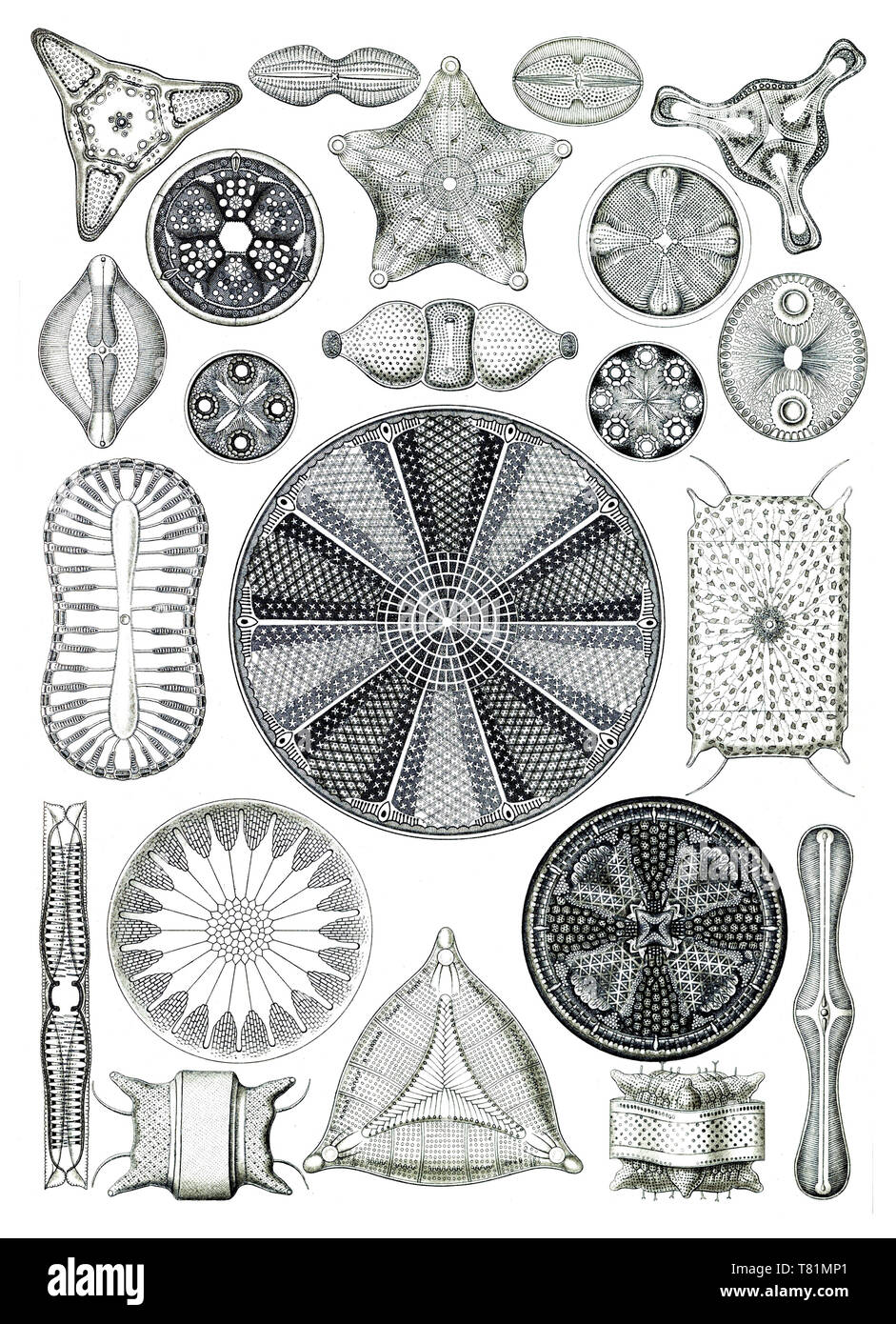 Ernst Haeckel, Diatoms, Microalgae Stock Photohttps://www.alamy.com/image-license-details/?v=1https://www.alamy.com/ernst-haeckel-diatoms-microalgae-image245900649.html
RMT81MP1–Ernst Haeckel, Diatoms, Microalgae
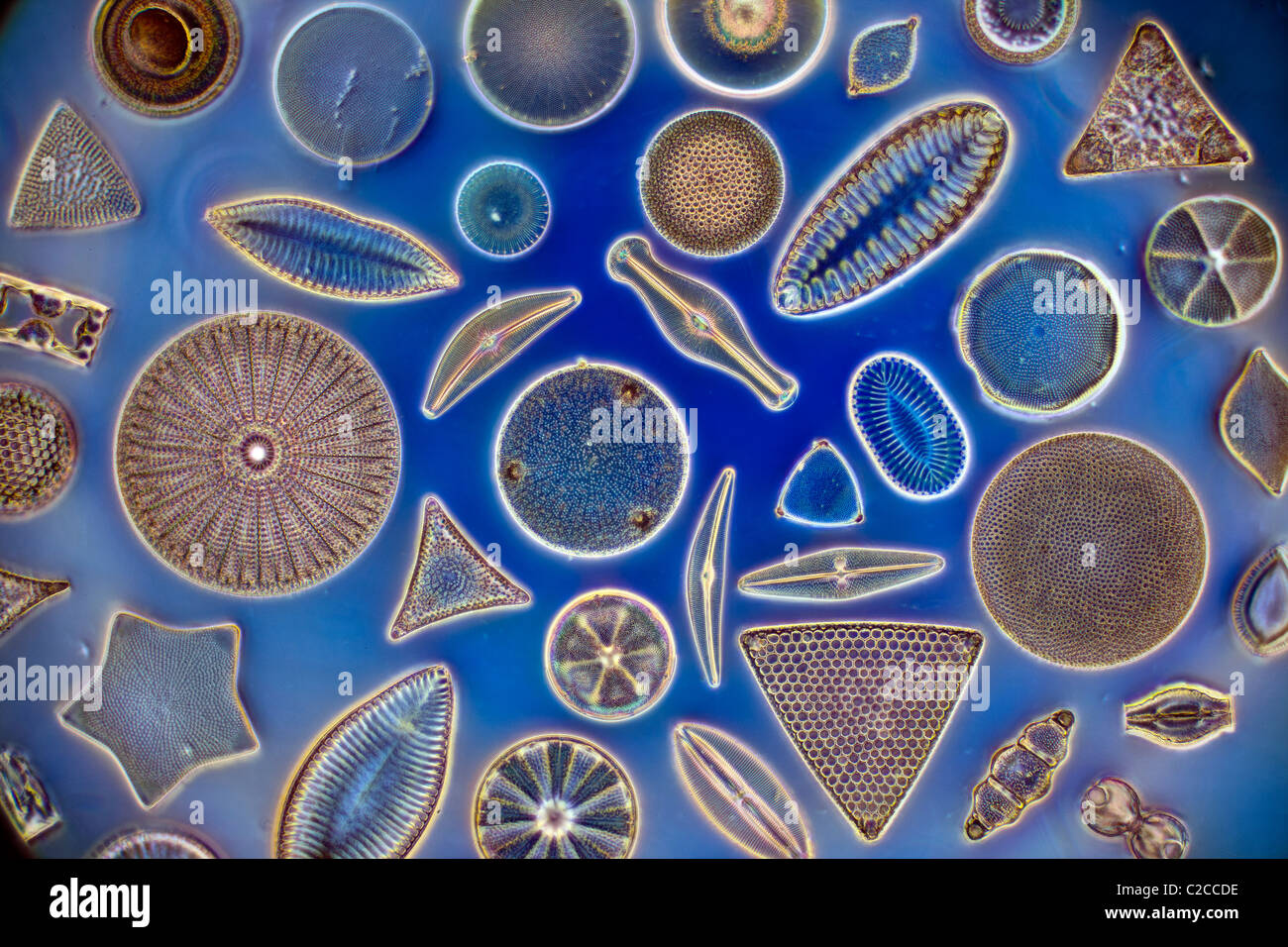 Diatoms, strewn variety of phytoplankton Stock Photohttps://www.alamy.com/image-license-details/?v=1https://www.alamy.com/stock-photo-diatoms-strewn-variety-of-phytoplankton-35923258.html
RMC2CCDE–Diatoms, strewn variety of phytoplankton
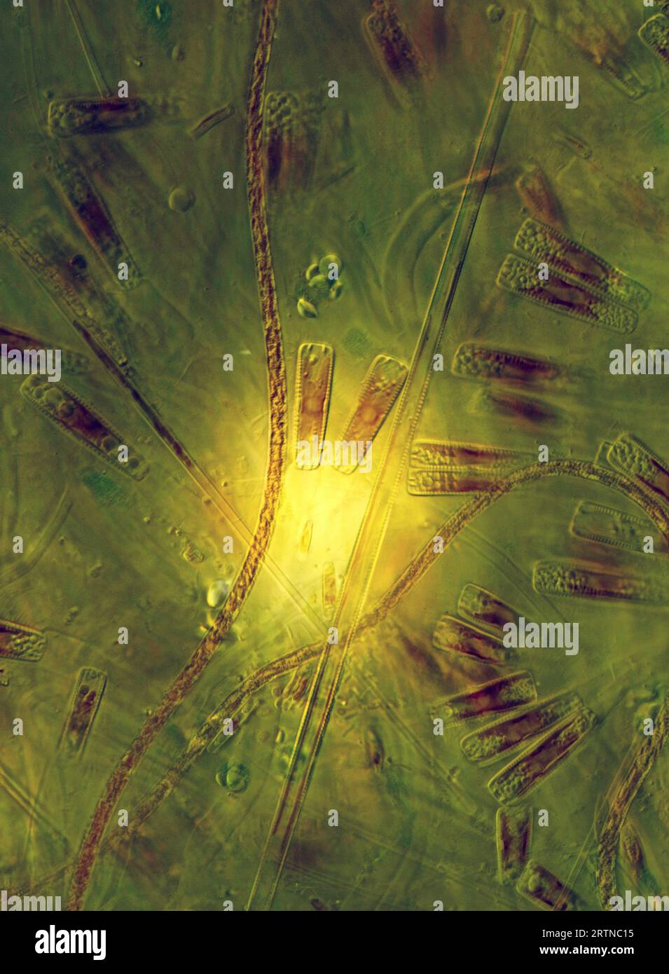 The image presents diatoms (mostly Gomphonema sp.) photographed through the microscope in polarized light at a magnification of 200X Stock Photohttps://www.alamy.com/image-license-details/?v=1https://www.alamy.com/the-image-presents-diatoms-mostly-gomphonema-sp-photographed-through-the-microscope-in-polarized-light-at-a-magnification-of-200x-image565953953.html
RM2RTNC15–The image presents diatoms (mostly Gomphonema sp.) photographed through the microscope in polarized light at a magnification of 200X
 diatoms in the water Stock Photohttps://www.alamy.com/image-license-details/?v=1https://www.alamy.com/diatoms-in-the-water-image227166457.html
RFR5G93N–diatoms in the water
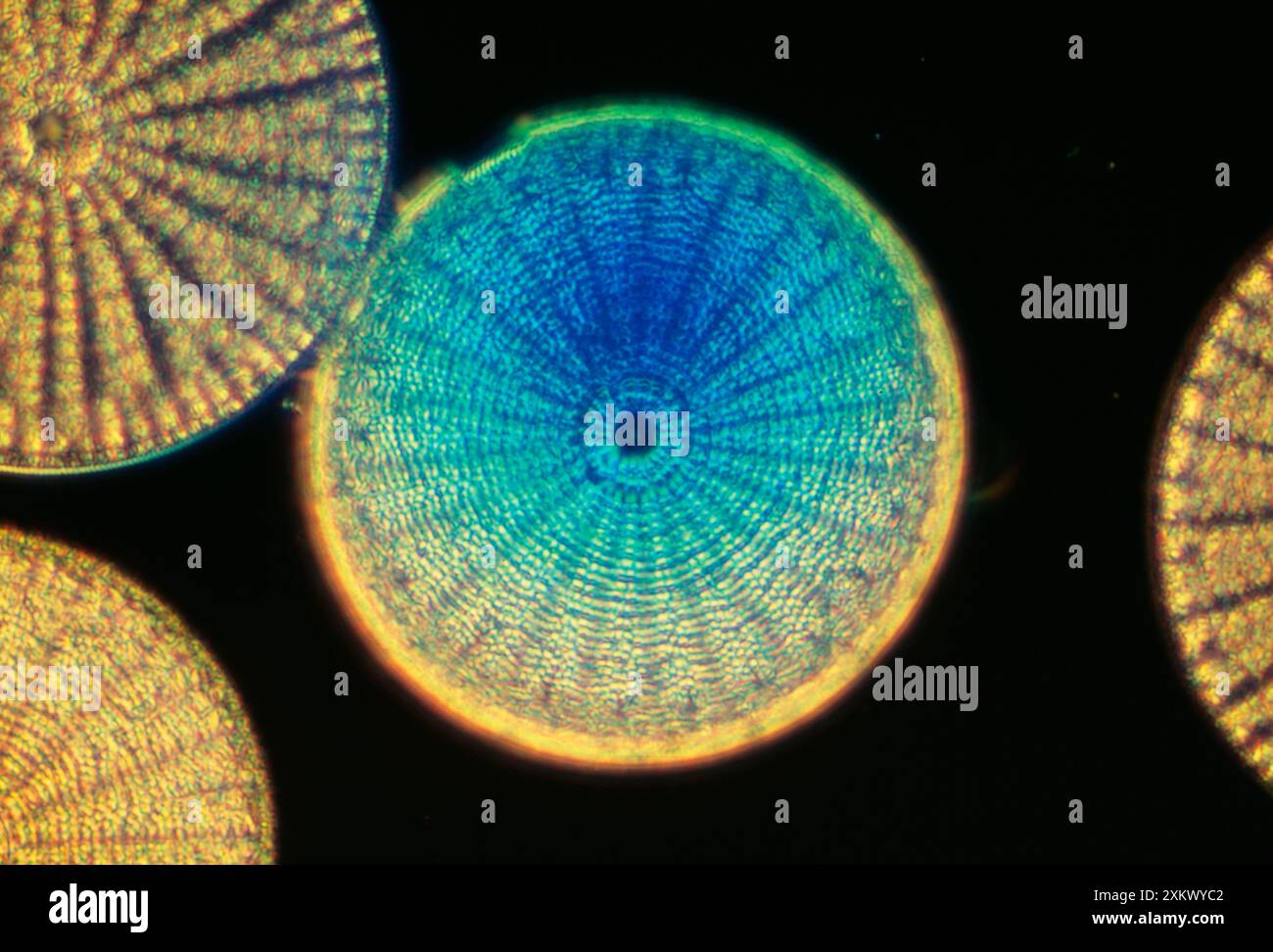 DIATOMS - From marine plankton sample. A type of Stock Photohttps://www.alamy.com/image-license-details/?v=1https://www.alamy.com/diatoms-from-marine-plankton-sample-a-type-of-image614611650.html
RM2XKWYC2–DIATOMS - From marine plankton sample. A type of
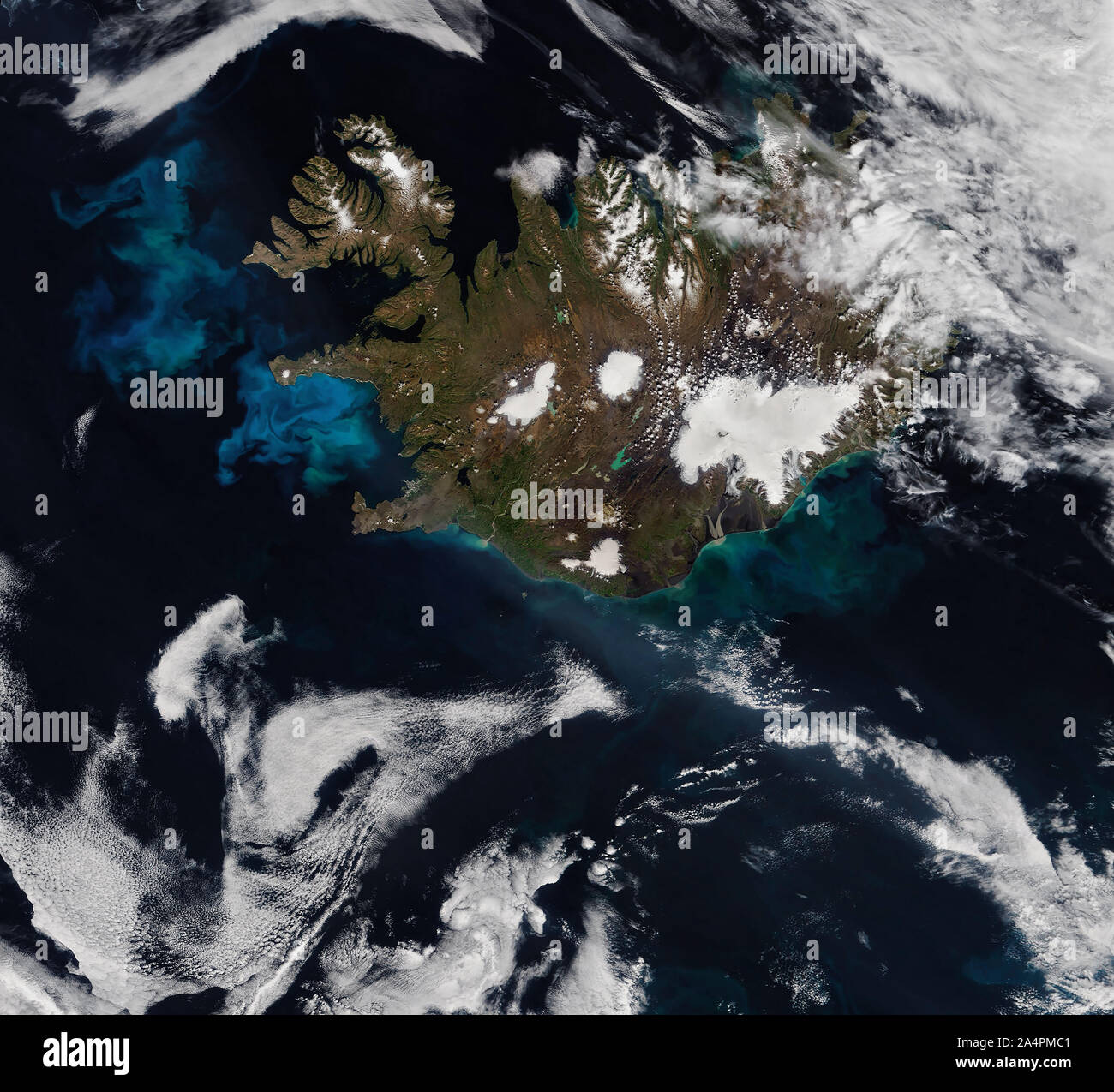 Satellite view of Iceland, Phytoplankton blooms in Atlantic ocean, July 6, 2019, by NASA/Joshua Stevens/DPA Stock Photohttps://www.alamy.com/image-license-details/?v=1https://www.alamy.com/satellite-view-of-iceland-phytoplankton-blooms-in-atlantic-ocean-july-6-2019-by-nasajoshua-stevensdpa-image329954577.html
RF2A4PMC1–Satellite view of Iceland, Phytoplankton blooms in Atlantic ocean, July 6, 2019, by NASA/Joshua Stevens/DPA
 Microscope intake of diatoms, or a single -cell whose, anonymous, c. 1880 - in or before 1899 photomechanical print paper collotype algae, seaweed. microscope Stock Photohttps://www.alamy.com/image-license-details/?v=1https://www.alamy.com/microscope-intake-of-diatoms-or-a-single-cell-whose-anonymous-c-1880-in-or-before-1899-photomechanical-print-paper-collotype-algae-seaweed-microscope-image591294085.html
RM2W9YNHW–Microscope intake of diatoms, or a single -cell whose, anonymous, c. 1880 - in or before 1899 photomechanical print paper collotype algae, seaweed. microscope
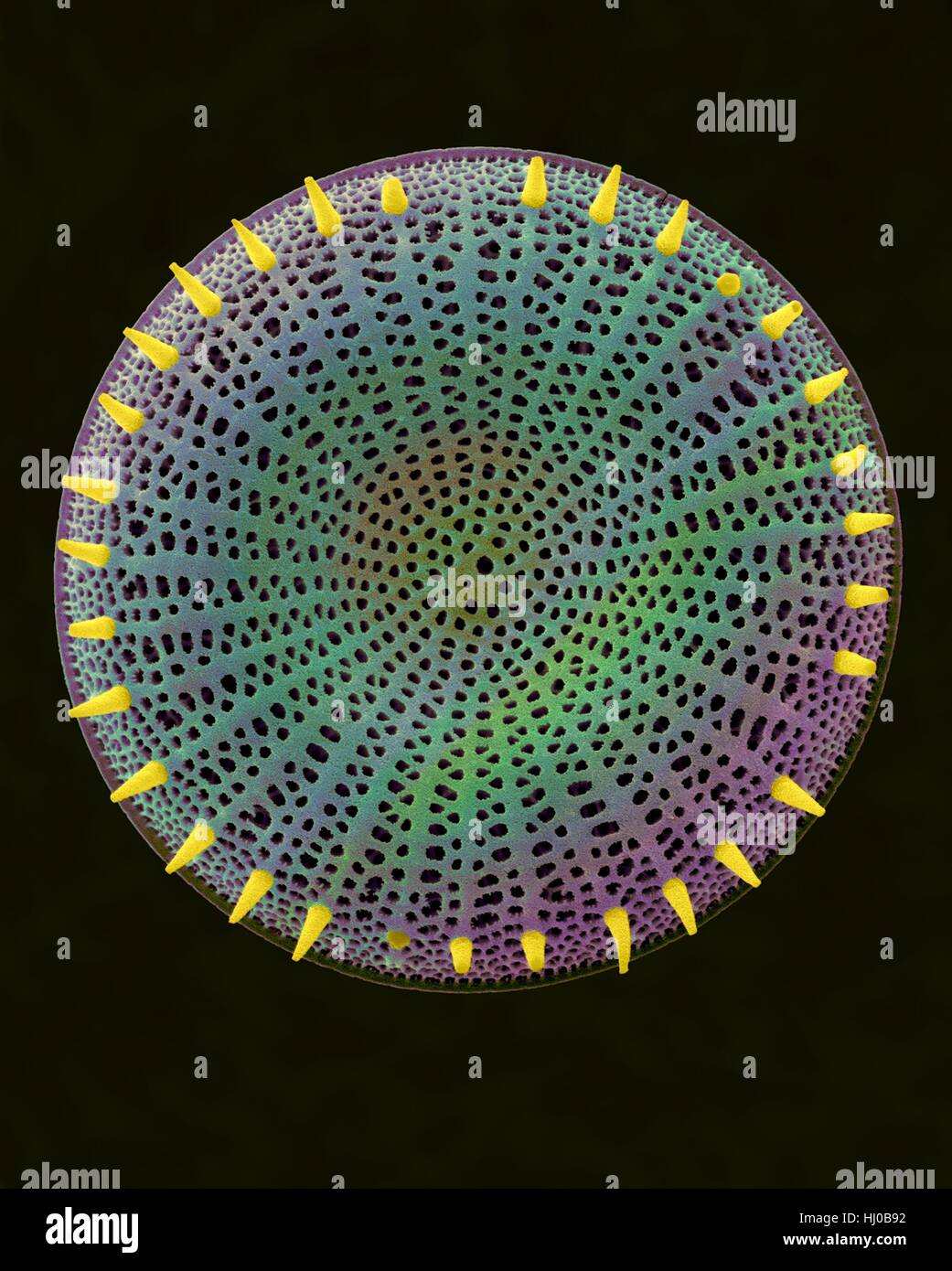 Coloured scanning electron micrograph (SEM) of centric fossil diatom frustule.This fresh water diatom frustule came from deposits found at Kalamath Falls,Oregon.This freshwater centric diatom is found in fresh water streams,ponds lakes.Diatoms are type of algae (Chromophyta,Bacillariophyceae).They Stock Photohttps://www.alamy.com/image-license-details/?v=1https://www.alamy.com/stock-photo-coloured-scanning-electron-micrograph-sem-of-centric-fossil-diatom-131545262.html
RFHJ0B92–Coloured scanning electron micrograph (SEM) of centric fossil diatom frustule.This fresh water diatom frustule came from deposits found at Kalamath Falls,Oregon.This freshwater centric diatom is found in fresh water streams,ponds lakes.Diatoms are type of algae (Chromophyta,Bacillariophyceae).They
![Hardwicke's science-gossip : an illustrated medium of interchange and gossip for students and lovers of nature . resting member ofthe same family, Cristalina lophophus—/. S. Plant Circulation.—A cyclosis, or circulationof protoplasm, may be very easily seen in the finerootlets of the common water-weed known by thename of Frog-bit. A power of from 250 to 300diameters shows the circulation well.—/. /. R. June 1, 18G6.] SCIENCE-GOSSIP 135 Toxonidea Gregoriana.—During last autumnI secured several gatherings of diatoms in whichT. Gregoriaua were more or less present. I obtainedthem in the outer har Stock Photo Hardwicke's science-gossip : an illustrated medium of interchange and gossip for students and lovers of nature . resting member ofthe same family, Cristalina lophophus—/. S. Plant Circulation.—A cyclosis, or circulationof protoplasm, may be very easily seen in the finerootlets of the common water-weed known by thename of Frog-bit. A power of from 250 to 300diameters shows the circulation well.—/. /. R. June 1, 18G6.] SCIENCE-GOSSIP 135 Toxonidea Gregoriana.—During last autumnI secured several gatherings of diatoms in whichT. Gregoriaua were more or less present. I obtainedthem in the outer har Stock Photo](https://c8.alamy.com/comp/2AKT2PD/hardwickes-science-gossip-an-illustrated-medium-of-interchange-and-gossip-for-students-and-lovers-of-nature-resting-member-ofthe-same-family-cristalina-lophophus-s-plant-circulationa-cyclosis-or-circulationof-protoplasm-may-be-very-easily-seen-in-the-finerootlets-of-the-common-water-weed-known-by-thename-of-frog-bit-a-power-of-from-250-to-300diameters-shows-the-circulation-well-r-june-1-18g6-science-gossip-135-toxonidea-gregorianaduring-last-autumni-secured-several-gatherings-of-diatoms-in-whicht-gregoriaua-were-more-or-less-present-i-obtainedthem-in-the-outer-har-2AKT2PD.jpg) Hardwicke's science-gossip : an illustrated medium of interchange and gossip for students and lovers of nature . resting member ofthe same family, Cristalina lophophus—/. S. Plant Circulation.—A cyclosis, or circulationof protoplasm, may be very easily seen in the finerootlets of the common water-weed known by thename of Frog-bit. A power of from 250 to 300diameters shows the circulation well.—/. /. R. June 1, 18G6.] SCIENCE-GOSSIP 135 Toxonidea Gregoriana.—During last autumnI secured several gatherings of diatoms in whichT. Gregoriaua were more or less present. I obtainedthem in the outer har Stock Photohttps://www.alamy.com/image-license-details/?v=1https://www.alamy.com/hardwickes-science-gossip-an-illustrated-medium-of-interchange-and-gossip-for-students-and-lovers-of-nature-resting-member-ofthe-same-family-cristalina-lophophus-s-plant-circulationa-cyclosis-or-circulationof-protoplasm-may-be-very-easily-seen-in-the-finerootlets-of-the-common-water-weed-known-by-thename-of-frog-bit-a-power-of-from-250-to-300diameters-shows-the-circulation-well-r-june-1-18g6-science-gossip-135-toxonidea-gregorianaduring-last-autumni-secured-several-gatherings-of-diatoms-in-whicht-gregoriaua-were-more-or-less-present-i-obtainedthem-in-the-outer-har-image339204501.html
Hardwicke's science-gossip : an illustrated medium of interchange and gossip for students and lovers of nature . resting member ofthe same family, Cristalina lophophus—/. S. Plant Circulation.—A cyclosis, or circulationof protoplasm, may be very easily seen in the finerootlets of the common water-weed known by thename of Frog-bit. A power of from 250 to 300diameters shows the circulation well.—/. /. R. June 1, 18G6.] SCIENCE-GOSSIP 135 Toxonidea Gregoriana.—During last autumnI secured several gatherings of diatoms in whichT. Gregoriaua were more or less present. I obtainedthem in the outer har Stock Photohttps://www.alamy.com/image-license-details/?v=1https://www.alamy.com/hardwickes-science-gossip-an-illustrated-medium-of-interchange-and-gossip-for-students-and-lovers-of-nature-resting-member-ofthe-same-family-cristalina-lophophus-s-plant-circulationa-cyclosis-or-circulationof-protoplasm-may-be-very-easily-seen-in-the-finerootlets-of-the-common-water-weed-known-by-thename-of-frog-bit-a-power-of-from-250-to-300diameters-shows-the-circulation-well-r-june-1-18g6-science-gossip-135-toxonidea-gregorianaduring-last-autumni-secured-several-gatherings-of-diatoms-in-whicht-gregoriaua-were-more-or-less-present-i-obtainedthem-in-the-outer-har-image339204501.htmlRM2AKT2PD–Hardwicke's science-gossip : an illustrated medium of interchange and gossip for students and lovers of nature . resting member ofthe same family, Cristalina lophophus—/. S. Plant Circulation.—A cyclosis, or circulationof protoplasm, may be very easily seen in the finerootlets of the common water-weed known by thename of Frog-bit. A power of from 250 to 300diameters shows the circulation well.—/. /. R. June 1, 18G6.] SCIENCE-GOSSIP 135 Toxonidea Gregoriana.—During last autumnI secured several gatherings of diatoms in whichT. Gregoriaua were more or less present. I obtainedthem in the outer har
 Relaxing in the thermal outdoor swimming pool Blaa Lonio on Reykjanes, Iceland, Stock Photohttps://www.alamy.com/image-license-details/?v=1https://www.alamy.com/relaxing-in-the-thermal-outdoor-swimming-pool-blaa-lonio-on-reykjanes-iceland-image183447035.html
RMMJCMFR–Relaxing in the thermal outdoor swimming pool Blaa Lonio on Reykjanes, Iceland,
 Conceptual image of Radiolarians with a skeletal frame. Radiolarians are tiny protozoans that live in the ocean Stock Photohttps://www.alamy.com/image-license-details/?v=1https://www.alamy.com/conceptual-image-of-radiolarians-with-a-skeletal-frame-radiolarians-are-tiny-protozoans-that-live-in-the-ocean-image619354215.html
RM2XYJ0HB–Conceptual image of Radiolarians with a skeletal frame. Radiolarians are tiny protozoans that live in the ocean
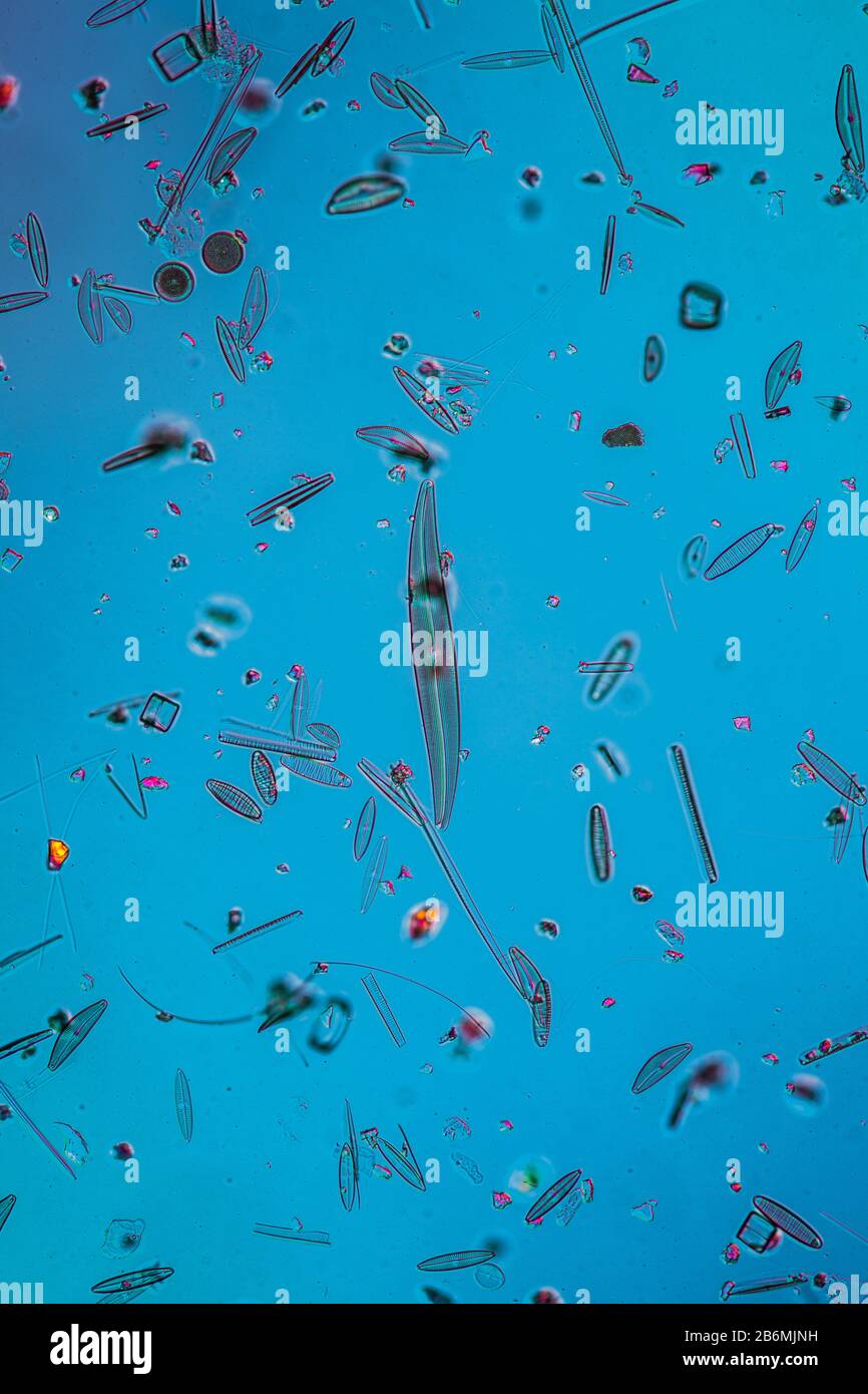 tiny microscopic diatoms in drops of water Stock Photohttps://www.alamy.com/image-license-details/?v=1https://www.alamy.com/tiny-microscopic-diatoms-in-drops-of-water-image348349053.html
RF2B6MJNH–tiny microscopic diatoms in drops of water
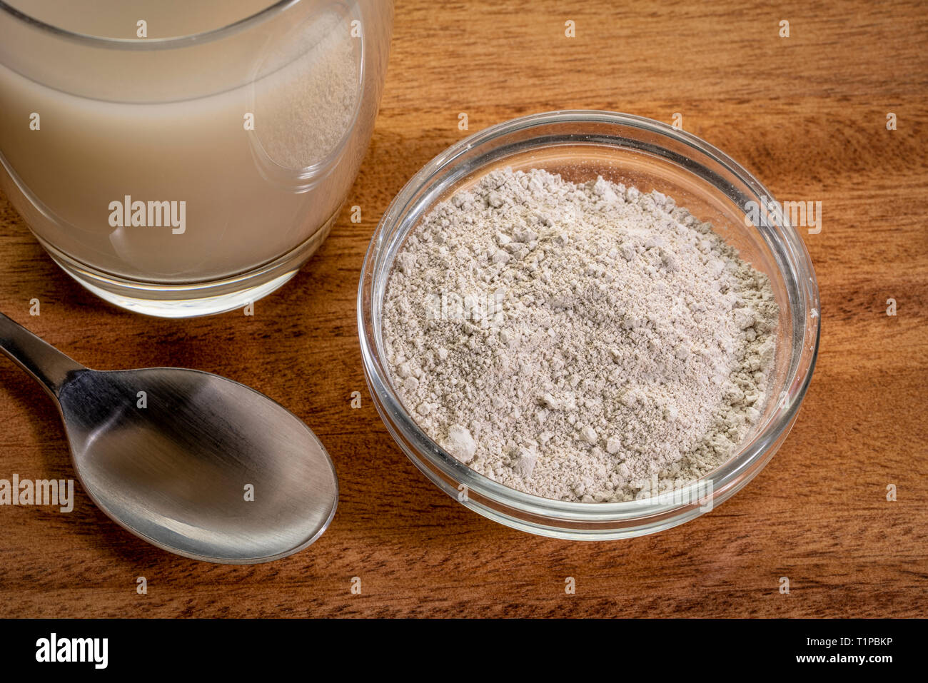 food grade diatomaceous earth supplement - powder and in a glass of water Stock Photohttps://www.alamy.com/image-license-details/?v=1https://www.alamy.com/food-grade-diatomaceous-earth-supplement-powder-and-in-a-glass-of-water-image242051930.html
RFT1PBKP–food grade diatomaceous earth supplement - powder and in a glass of water
 Humpback whale, Megaptera novaeangilae, showing its tail fluke while diving in Gerlache strait Antarctica Stock Photohttps://www.alamy.com/image-license-details/?v=1https://www.alamy.com/humpback-whale-megaptera-novaeangilae-showing-its-tail-fluke-while-diving-in-gerlache-strait-antarctica-image350728462.html
RM2BAH1ME–Humpback whale, Megaptera novaeangilae, showing its tail fluke while diving in Gerlache strait Antarctica
 The algae Didymo covering rocks in river, British Columbia Stock Photohttps://www.alamy.com/image-license-details/?v=1https://www.alamy.com/stock-photo-the-algae-didymo-covering-rocks-in-river-british-columbia-28241576.html
RMBHXEBM–The algae Didymo covering rocks in river, British Columbia
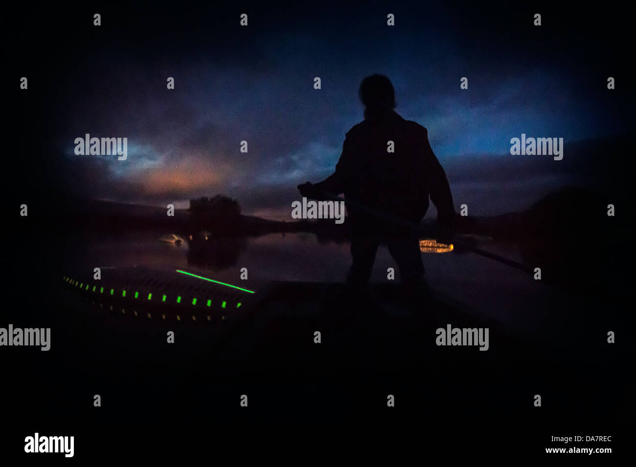 The Land Art work called Mégascospic Diatoms, made by Prisca Cosnier. A project within the Horizons 'Nature Arts' 2013 framework Stock Photohttps://www.alamy.com/image-license-details/?v=1https://www.alamy.com/stock-photo-the-land-art-work-called-mgascospic-diatoms-made-by-prisca-cosnier-57949764.html
RMDA7REC–The Land Art work called Mégascospic Diatoms, made by Prisca Cosnier. A project within the Horizons 'Nature Arts' 2013 framework
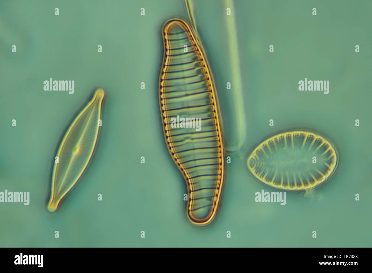 diatom (Diatomeae), diatoms in phase contrast and interference contrast, x 160 Stock Photohttps://www.alamy.com/image-license-details/?v=1https://www.alamy.com/diatom-diatomeae-diatoms-in-phase-contrast-and-interference-contrast-x-160-image255239010.html
RMTR73XX–diatom (Diatomeae), diatoms in phase contrast and interference contrast, x 160
 Flamingos in the Camargue Stock Photohttps://www.alamy.com/image-license-details/?v=1https://www.alamy.com/flamingos-in-the-camargue-image479986551.html
RF2JTW7MR–Flamingos in the Camargue
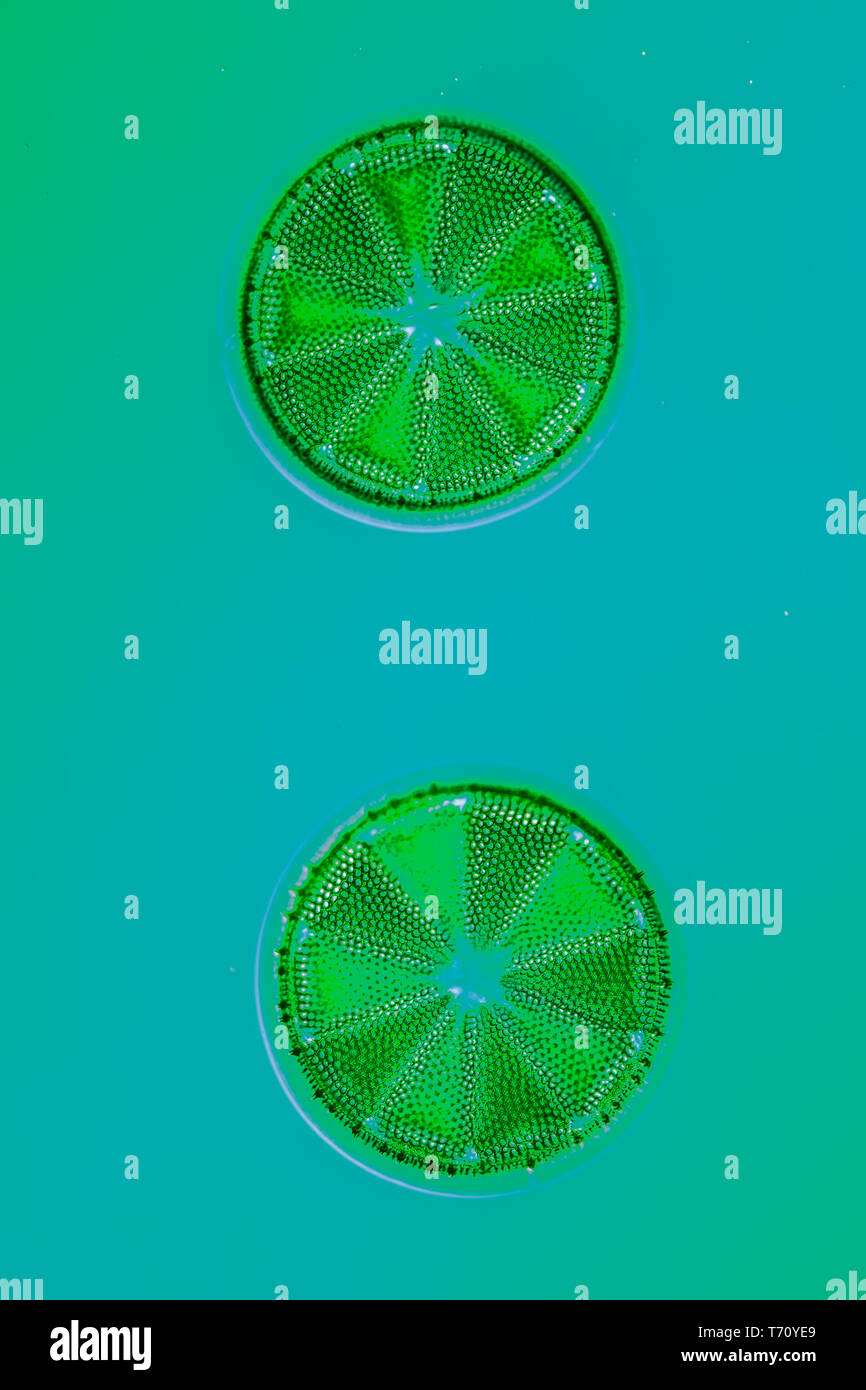 Diatoms in the water under the microskop Stock Photohttps://www.alamy.com/image-license-details/?v=1https://www.alamy.com/diatoms-in-the-water-under-the-microskop-image245269313.html
RMT70YE9–Diatoms in the water under the microskop
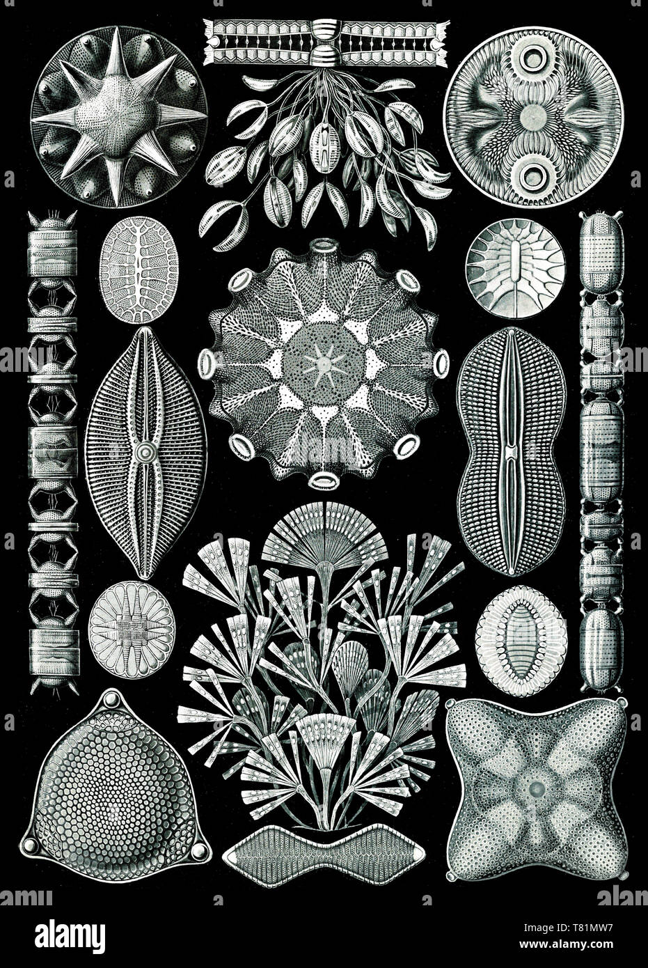 Ernst Haeckel, Diatoms Stock Photohttps://www.alamy.com/image-license-details/?v=1https://www.alamy.com/ernst-haeckel-diatoms-image245900739.html
RMT81MW7–Ernst Haeckel, Diatoms
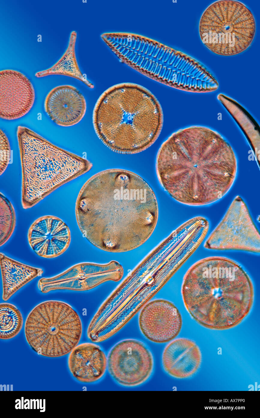 A selection of diatom species showing variety in shape and detail, darkfield blue background photomicrograph Stock Photohttps://www.alamy.com/image-license-details/?v=1https://www.alamy.com/a-selection-of-diatom-species-showing-variety-in-shape-and-detail-image9575391.html
RMAX7PP0–A selection of diatom species showing variety in shape and detail, darkfield blue background photomicrograph
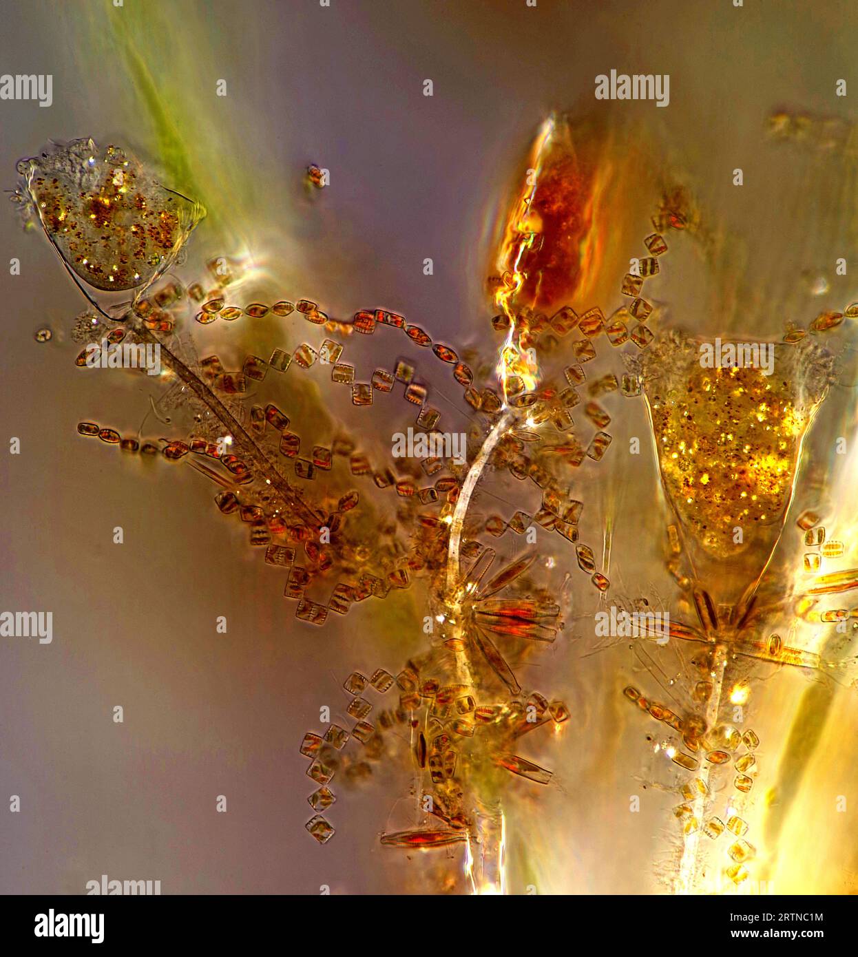 The image presentstwo suctorians ( a kind of ciliate) and tiny diatoms, photographed through the microscope in polarized light at a magnification of 2 Stock Photohttps://www.alamy.com/image-license-details/?v=1https://www.alamy.com/the-image-presentstwo-suctorians-a-kind-of-ciliate-and-tiny-diatoms-photographed-through-the-microscope-in-polarized-light-at-a-magnification-of-2-image565953968.html
RM2RTNC1M–The image presentstwo suctorians ( a kind of ciliate) and tiny diatoms, photographed through the microscope in polarized light at a magnification of 2
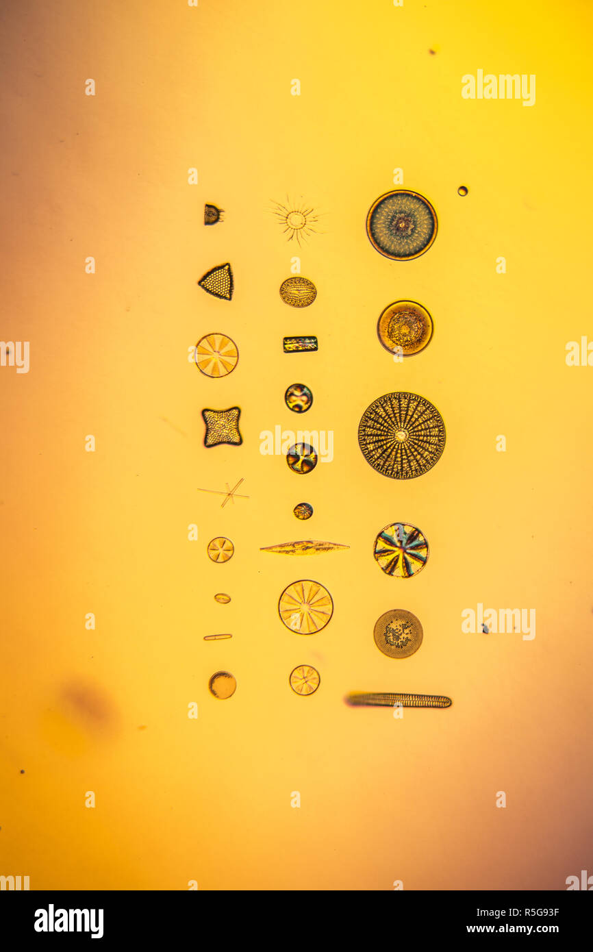 diatoms in the water Stock Photohttps://www.alamy.com/image-license-details/?v=1https://www.alamy.com/diatoms-in-the-water-image227166451.html
RFR5G93F–diatoms in the water
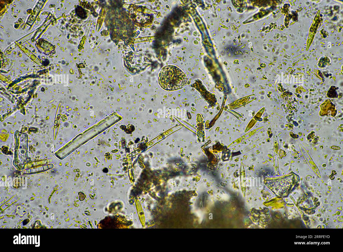 diatom and water microorganisms under the microscope in australia Stock Photohttps://www.alamy.com/image-license-details/?v=1https://www.alamy.com/diatom-and-water-microorganisms-under-the-microscope-in-australia-image565209889.html
RF2RRFEYD–diatom and water microorganisms under the microscope in australia
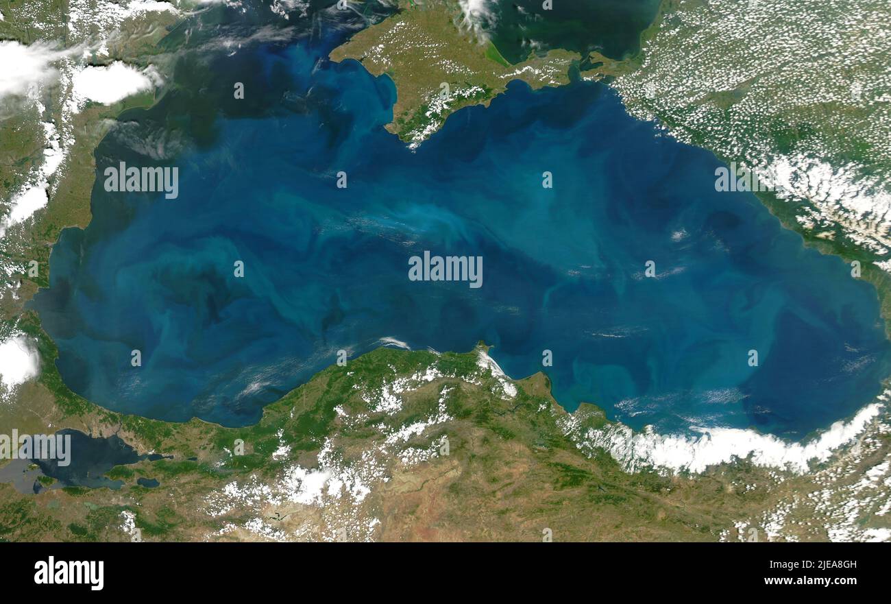 Phytoplankton blooms in the Black Sea, as seen from space, June 20, 2006, by the MODIS on the Aqua satellite, by NASA/DPA Stock Photohttps://www.alamy.com/image-license-details/?v=1https://www.alamy.com/phytoplankton-blooms-in-the-black-sea-as-seen-from-space-june-20-2006-by-the-modis-on-the-aqua-satellite-by-nasadpa-image473511377.html
RF2JEA8GH–Phytoplankton blooms in the Black Sea, as seen from space, June 20, 2006, by the MODIS on the Aqua satellite, by NASA/DPA
 Thirteen photo excuses, made under a microscope, Richard Leach Maddox, c. 1865 - in or before 1870 photograph (1.) Is the tribe of a hydrangea, (2.) is a scab mite, (3.) is a blood body, (4.) is a parasite, (5.) is a cross -section of a bone, (6.) is a louse, (7.), (8.) and (9.) are diatoms, (11.) is a flea, (12.) is a dialome, (13.) is a fleas. photographic support albumen print microscope. insects: louse. bones in general (human body) Stock Photohttps://www.alamy.com/image-license-details/?v=1https://www.alamy.com/thirteen-photo-excuses-made-under-a-microscope-richard-leach-maddox-c-1865-in-or-before-1870-photograph-1-is-the-tribe-of-a-hydrangea-2-is-a-scab-mite-3-is-a-blood-body-4-is-a-parasite-5-is-a-cross-section-of-a-bone-6-is-a-louse-7-8-and-9-are-diatoms-11-is-a-flea-12-is-a-dialome-13-is-a-fleas-photographic-support-albumen-print-microscope-insects-louse-bones-in-general-human-body-image591161828.html
RM2W9NMXC–Thirteen photo excuses, made under a microscope, Richard Leach Maddox, c. 1865 - in or before 1870 photograph (1.) Is the tribe of a hydrangea, (2.) is a scab mite, (3.) is a blood body, (4.) is a parasite, (5.) is a cross -section of a bone, (6.) is a louse, (7.), (8.) and (9.) are diatoms, (11.) is a flea, (12.) is a dialome, (13.) is a fleas. photographic support albumen print microscope. insects: louse. bones in general (human body)
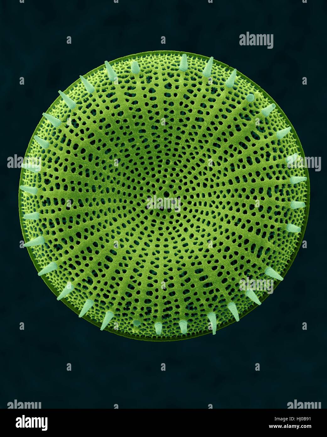 Coloured scanning electron micrograph (SEM) of centric fossil diatom frustule (Stephanodiscus sp.).This fresh water diatom frustule came from deposits found at Kalamath Falls,Oregon.This freshwater centric diatom is found in fresh water streams,ponds lakes.Diatoms are type of algae Stock Photohttps://www.alamy.com/image-license-details/?v=1https://www.alamy.com/stock-photo-coloured-scanning-electron-micrograph-sem-of-centric-fossil-diatom-131545261.html
RFHJ0B91–Coloured scanning electron micrograph (SEM) of centric fossil diatom frustule (Stephanodiscus sp.).This fresh water diatom frustule came from deposits found at Kalamath Falls,Oregon.This freshwater centric diatom is found in fresh water streams,ponds lakes.Diatoms are type of algae
 Nature . of unequal size, VictoriaLand and Edward VII. Land, separ-ated by a great barrier of ice, andof two seas extending far to thesouth, the Ross Sea and the WeddellSea. The papers by Dr. Harvey Pirieand Mr. Mossman contain manypoints of great interest, although inthe nature of things the material1 1 llei ted requires further elabor-ation, and comparison with that ofthe other expeditions, before its fullvalue becomes apparent. Dr. Harvey Piries observations givemuch additional information bear-ing on the variations in the relativeamounts of diatoms in the surfacewaters and in the deposits, Stock Photohttps://www.alamy.com/image-license-details/?v=1https://www.alamy.com/nature-of-unequal-size-victorialand-and-edward-vii-land-separ-ated-by-a-great-barrier-of-ice-andof-two-seas-extending-far-to-thesouth-the-ross-sea-and-the-weddellsea-the-papers-by-dr-harvey-pirieand-mr-mossman-contain-manypoints-of-great-interest-although-inthe-nature-of-things-the-material1-1-llei-ted-requires-further-elabor-ation-and-comparison-with-that-ofthe-other-expeditions-before-its-fullvalue-becomes-apparent-dr-harvey-piries-observations-givemuch-additional-information-bear-ing-on-the-variations-in-the-relativeamounts-of-diatoms-in-the-surfacewaters-and-in-the-deposits-image339468373.html
RM2AM83AD–Nature . of unequal size, VictoriaLand and Edward VII. Land, separ-ated by a great barrier of ice, andof two seas extending far to thesouth, the Ross Sea and the WeddellSea. The papers by Dr. Harvey Pirieand Mr. Mossman contain manypoints of great interest, although inthe nature of things the material1 1 llei ted requires further elabor-ation, and comparison with that ofthe other expeditions, before its fullvalue becomes apparent. Dr. Harvey Piries observations givemuch additional information bear-ing on the variations in the relativeamounts of diatoms in the surfacewaters and in the deposits,
 Relaxation in the thermal outdoor swimming pool Blaa Lonio on Reykjanes, Iceland, Stock Photohttps://www.alamy.com/image-license-details/?v=1https://www.alamy.com/relaxation-in-the-thermal-outdoor-swimming-pool-blaa-lonio-on-reykjanes-iceland-image183444922.html
RMMJCHTA–Relaxation in the thermal outdoor swimming pool Blaa Lonio on Reykjanes, Iceland,
 House by a geothermal spring near Reykjavik in Iceland Stock Photohttps://www.alamy.com/image-license-details/?v=1https://www.alamy.com/house-by-a-geothermal-spring-near-reykjavik-in-iceland-image619576492.html
RM2Y0043T–House by a geothermal spring near Reykjavik in Iceland
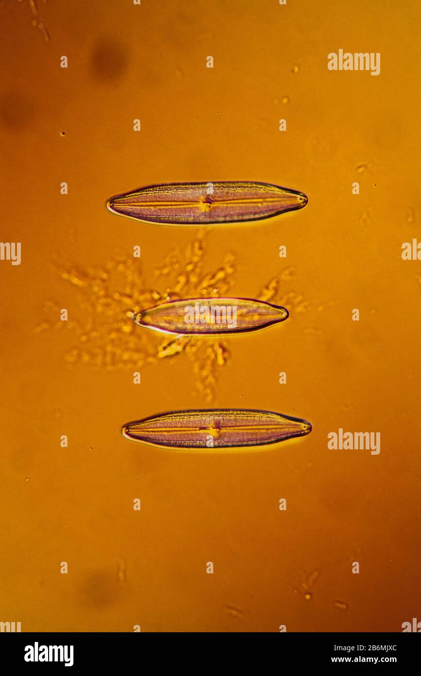 tiny microscopic diatoms in drops of water Stock Photohttps://www.alamy.com/image-license-details/?v=1https://www.alamy.com/tiny-microscopic-diatoms-in-drops-of-water-image348349188.html
RF2B6MJXC–tiny microscopic diatoms in drops of water
 food grade diatomaceous earth supplement - powder and in a glass of water Stock Photohttps://www.alamy.com/image-license-details/?v=1https://www.alamy.com/food-grade-diatomaceous-earth-supplement-powder-and-in-a-glass-of-water-image259634554.html
RFW2BAEJ–food grade diatomaceous earth supplement - powder and in a glass of water
 Vegetative division in a pinnate diatom from the genus Nitzschia or Cylindrotheca. The cell is about 0,050mm long. Stock Photohttps://www.alamy.com/image-license-details/?v=1https://www.alamy.com/vegetative-division-in-a-pinnate-diatom-from-the-genus-nitzschia-or-cylindrotheca-the-cell-is-about-0050mm-long-image472753616.html
RM2JD3P1M–Vegetative division in a pinnate diatom from the genus Nitzschia or Cylindrotheca. The cell is about 0,050mm long.
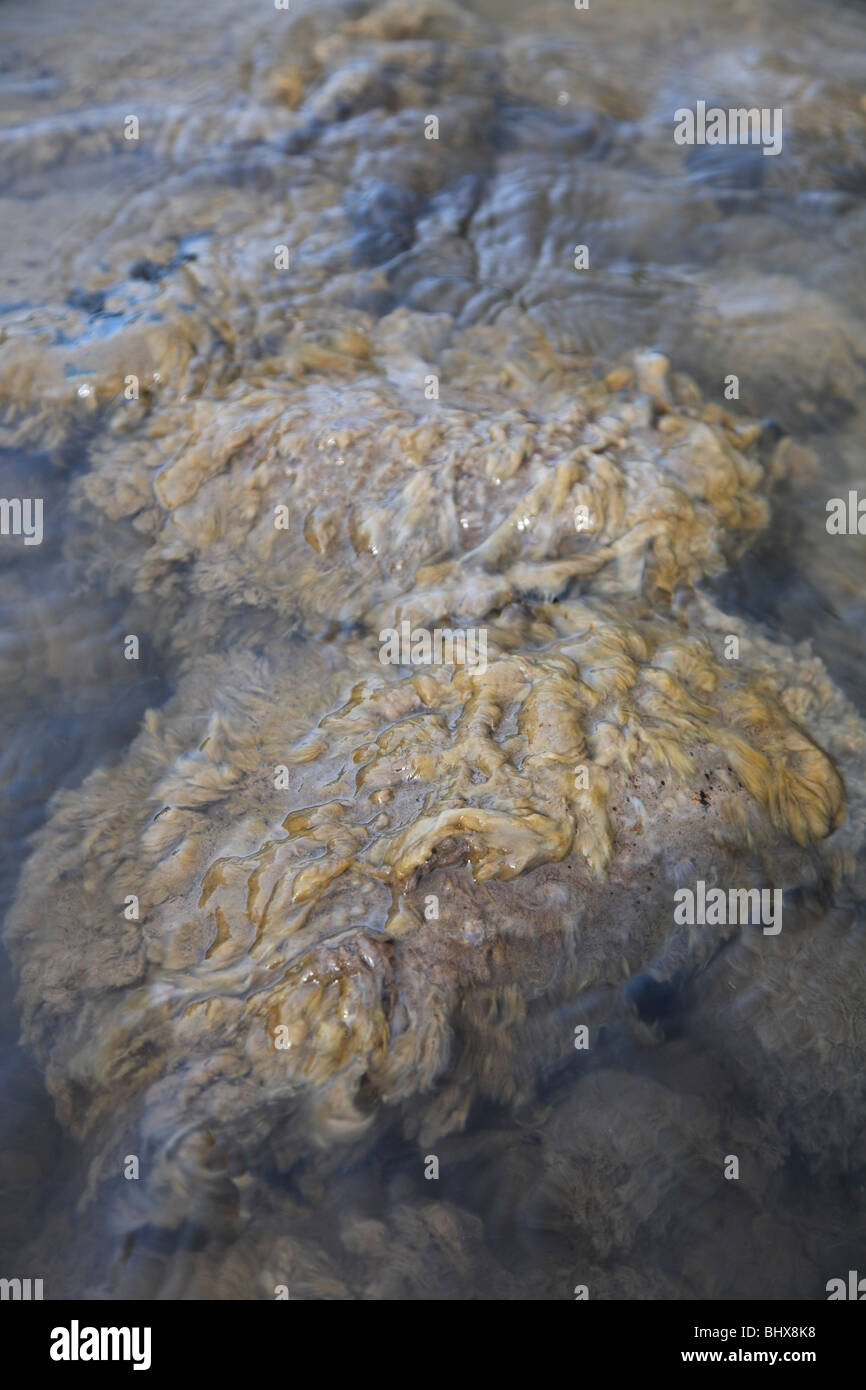 The algae Didymo covering rocks in river, British Columbia Stock Photohttps://www.alamy.com/image-license-details/?v=1https://www.alamy.com/stock-photo-the-algae-didymo-covering-rocks-in-river-british-columbia-28237084.html
RMBHX8K8–The algae Didymo covering rocks in river, British Columbia
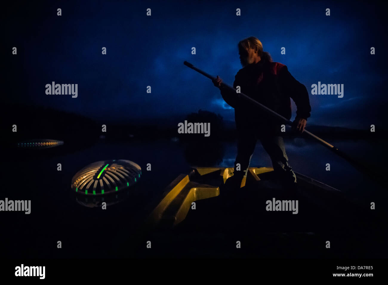 The Land Art work called Mégascospic Diatoms, made by Prisca Cosnier. A project within the Horizons 'Nature Arts' 2013 framework Stock Photohttps://www.alamy.com/image-license-details/?v=1https://www.alamy.com/stock-photo-the-land-art-work-called-mgascospic-diatoms-made-by-prisca-cosnier-57949757.html
RMDA7RE5–The Land Art work called Mégascospic Diatoms, made by Prisca Cosnier. A project within the Horizons 'Nature Arts' 2013 framework
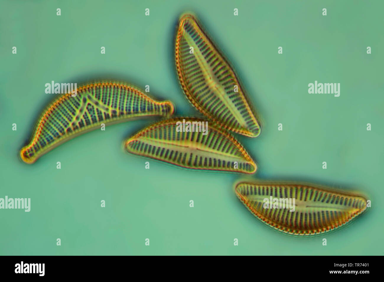 diatom (Diatomeae), diatoms in phase contrast and interference contrast, x 200 Stock Photohttps://www.alamy.com/image-license-details/?v=1https://www.alamy.com/diatom-diatomeae-diatoms-in-phase-contrast-and-interference-contrast-x-200-image255239041.html
RMTR7401–diatom (Diatomeae), diatoms in phase contrast and interference contrast, x 200
 Flamingos in the Camargue Stock Photohttps://www.alamy.com/image-license-details/?v=1https://www.alamy.com/flamingos-in-the-camargue-image479986632.html
RF2JTW7RM–Flamingos in the Camargue
 Diatoms in the water under the microskop Stock Photohttps://www.alamy.com/image-license-details/?v=1https://www.alamy.com/diatoms-in-the-water-under-the-microskop-image245269347.html
RMT70YFF–Diatoms in the water under the microskop
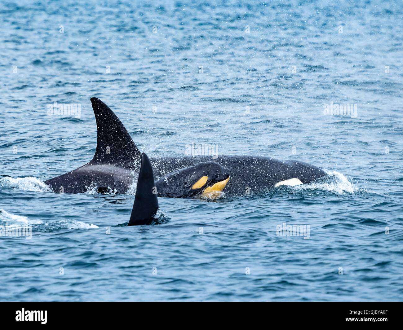 Young calf discolored by diatonsm Transiant Killer Whales (Orca orcinus) family pod in Monterey Bay, Monterey Bay National Marine Refuge, California Stock Photohttps://www.alamy.com/image-license-details/?v=1https://www.alamy.com/young-calf-discolored-by-diatonsm-transiant-killer-whales-orca-orcinus-family-pod-in-monterey-bay-monterey-bay-national-marine-refuge-california-image472041711.html
RF2JBYA0F–Young calf discolored by diatonsm Transiant Killer Whales (Orca orcinus) family pod in Monterey Bay, Monterey Bay National Marine Refuge, California
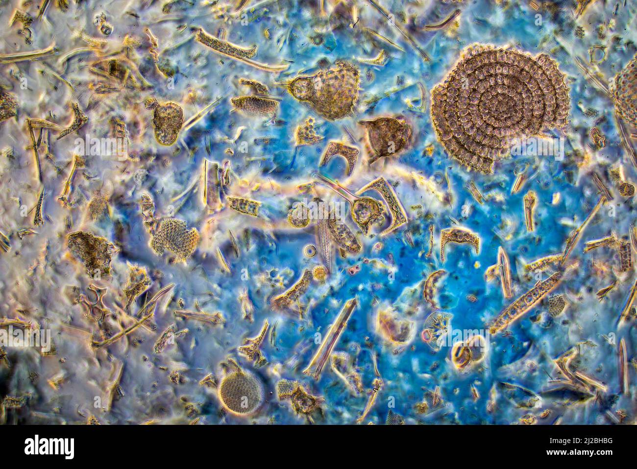 Fossil diatoms,radiolaria,sponge spicules, Barbados 1800's diversity Stock Photohttps://www.alamy.com/image-license-details/?v=1https://www.alamy.com/fossil-diatomsradiolariasponge-spicules-barbados-1800s-diversity-image466164372.html
RM2J2BHBG–Fossil diatoms,radiolaria,sponge spicules, Barbados 1800's diversity
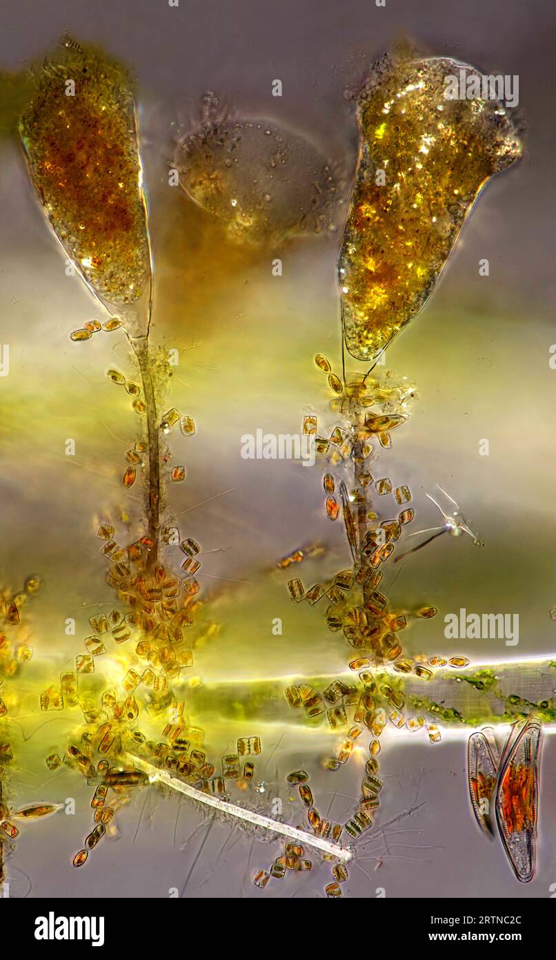 The image presentstwo suctorians ( a kind of ciliate) and tiny diatoms, photographed through the microscope in polarized light at a magnification of 2 Stock Photohttps://www.alamy.com/image-license-details/?v=1https://www.alamy.com/the-image-presentstwo-suctorians-a-kind-of-ciliate-and-tiny-diatoms-photographed-through-the-microscope-in-polarized-light-at-a-magnification-of-2-image565953988.html
RM2RTNC2C–The image presentstwo suctorians ( a kind of ciliate) and tiny diatoms, photographed through the microscope in polarized light at a magnification of 2
 diatoms in the water Stock Photohttps://www.alamy.com/image-license-details/?v=1https://www.alamy.com/diatoms-in-the-water-image227166419.html
RFR5G92B–diatoms in the water
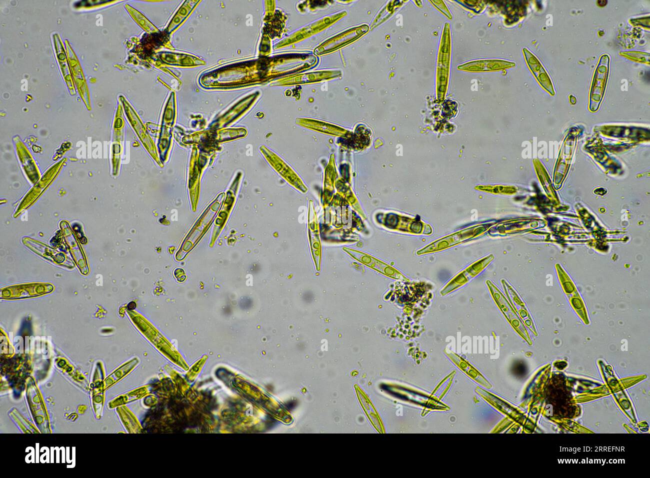 diatom and water microorganisms under the microscope in australia Stock Photohttps://www.alamy.com/image-license-details/?v=1https://www.alamy.com/diatom-and-water-microorganisms-under-the-microscope-in-australia-image565188563.html
RF2RREFNR–diatom and water microorganisms under the microscope in australia
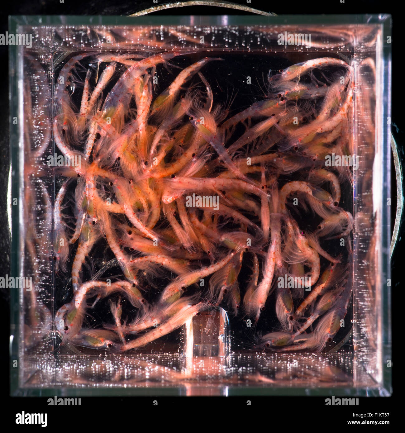 Antarctic krill Euphausia superba in Antarctica Stock Photohttps://www.alamy.com/image-license-details/?v=1https://www.alamy.com/stock-photo-antarctic-krill-euphausia-superba-in-antarctica-87102547.html
RMF1KT57–Antarctic krill Euphausia superba in Antarctica
 . Fresh-water biology. Freshwater biology. FREE-LIVING NEMATODES SOI 6i (60) Form of cavity various, closed in front, amphids opposite it. 62 62 (63) Lateral organs or amphids inconspicuous Trilobus Bastian. Fresh-water genus of which about half a dozen species are known. Known to feed upon diatoms in one case and upon rotifers in another. Hermapfiroditism occurs. Representative species. Trilobus longus (Leidy) i85r. source of nourishment of tills species, about one-fourth as wide as the body. The lips bear papillae but their number is not „. known. The intestine frequently contains diatoms in Stock Photohttps://www.alamy.com/image-license-details/?v=1https://www.alamy.com/fresh-water-biology-freshwater-biology-free-living-nematodes-soi-6i-60-form-of-cavity-various-closed-in-front-amphids-opposite-it-62-62-63-lateral-organs-or-amphids-inconspicuous-trilobus-bastian-fresh-water-genus-of-which-about-half-a-dozen-species-are-known-known-to-feed-upon-diatoms-in-one-case-and-upon-rotifers-in-another-hermapfiroditism-occurs-representative-species-trilobus-longus-leidy-i85r-source-of-nourishment-of-tills-species-about-one-fourth-as-wide-as-the-body-the-lips-bear-papillae-but-their-number-is-not-known-the-intestine-frequently-contains-diatoms-in-image216351638.html
RMPFYJM6–. Fresh-water biology. Freshwater biology. FREE-LIVING NEMATODES SOI 6i (60) Form of cavity various, closed in front, amphids opposite it. 62 62 (63) Lateral organs or amphids inconspicuous Trilobus Bastian. Fresh-water genus of which about half a dozen species are known. Known to feed upon diatoms in one case and upon rotifers in another. Hermapfiroditism occurs. Representative species. Trilobus longus (Leidy) i85r. source of nourishment of tills species, about one-fourth as wide as the body. The lips bear papillae but their number is not „. known. The intestine frequently contains diatoms in
 Plankton. Coloured scanning electron micrograph (SEM) of plankton containing mainly Ceratium dinoflagellates. A few diatoms are also present. Dinoflagellates are unicellular protozoans. About 90% are found in marine environments as plankton. They range in size from 2 millimetres to less than a hundredth of a milimetre across. These specimens were found off the coast of Scotland. Magnification: x200 when printed at 10 centimetres wide. Stock Photohttps://www.alamy.com/image-license-details/?v=1https://www.alamy.com/plankton-coloured-scanning-electron-micrograph-sem-of-plankton-containing-mainly-ceratium-dinoflagellates-a-few-diatoms-are-also-present-dinoflagellates-are-unicellular-protozoans-about-90-are-found-in-marine-environments-as-plankton-they-range-in-size-from-2-millimetres-to-less-than-a-hundredth-of-a-milimetre-across-these-specimens-were-found-off-the-coast-of-scotland-magnification-x200-when-printed-at-10-centimetres-wide-image364972776.html
RF2C5NXE0–Plankton. Coloured scanning electron micrograph (SEM) of plankton containing mainly Ceratium dinoflagellates. A few diatoms are also present. Dinoflagellates are unicellular protozoans. About 90% are found in marine environments as plankton. They range in size from 2 millimetres to less than a hundredth of a milimetre across. These specimens were found off the coast of Scotland. Magnification: x200 when printed at 10 centimetres wide.
 . A year of Costa Rican natural history. 4- I, 2. Thaumatoncura inopinata, male and iemale x4. 200. Pile a sp.Myrincarpa sp. To IlUC p. 20I JUAN VINAS—THE WATERFALLS 20l transportation that goes a long way to explaining the suddenappearance of large numbers of diatoms in new localities.I therefore look upon this rather abundant flora on theminute leg of this aquatic insect as of some scientific impor-tance. Of course in this case the power to fly does not existuntil a later period of development, but many of the waterbeetles and other insects are doubtless coated with livingdiatoms in the same Stock Photohttps://www.alamy.com/image-license-details/?v=1https://www.alamy.com/a-year-of-costa-rican-natural-history-4-i-2-thaumatoncura-inopinata-male-and-iemale-x4-200-pile-a-spmyrincarpa-sp-to-iluc-p-20i-juan-vinasthe-waterfalls-20l-transportation-that-goes-a-long-way-to-explaining-the-suddenappearance-of-large-numbers-of-diatoms-in-new-localitiesi-therefore-look-upon-this-rather-abundant-flora-on-theminute-leg-of-this-aquatic-insect-as-of-some-scientific-impor-tance-of-course-in-this-case-the-power-to-fly-does-not-existuntil-a-later-period-of-development-but-many-of-the-waterbeetles-and-other-insects-are-doubtless-coated-with-livingdiatoms-in-the-same-image336965192.html
RM2AG62F4–. A year of Costa Rican natural history. 4- I, 2. Thaumatoncura inopinata, male and iemale x4. 200. Pile a sp.Myrincarpa sp. To IlUC p. 20I JUAN VINAS—THE WATERFALLS 20l transportation that goes a long way to explaining the suddenappearance of large numbers of diatoms in new localities.I therefore look upon this rather abundant flora on theminute leg of this aquatic insect as of some scientific impor-tance. Of course in this case the power to fly does not existuntil a later period of development, but many of the waterbeetles and other insects are doubtless coated with livingdiatoms in the same
 Diatom - from marine plankton sample Stock Photohttps://www.alamy.com/image-license-details/?v=1https://www.alamy.com/diatom-from-marine-plankton-sample-image614611660.html
RM2XKWYCC–Diatom - from marine plankton sample
 A pacific chorus frog tadpole spends the first part of its life in an ephemeral vernal pool Feb. 13, 2017 at Travis Air Force Base, Calif. The adult frog will lay an egg mass in shallow temporary ponds which limit predators like fish and turtles. The tadpoles feed on periphyton, filamentous algae, diatoms and pollen in or on the surface of the water. (U.S. Air Force photo/ Heide Couch) Stock Photohttps://www.alamy.com/image-license-details/?v=1https://www.alamy.com/a-pacific-chorus-frog-tadpole-spends-the-first-part-of-its-life-in-an-ephemeral-vernal-pool-feb-13-2017-at-travis-air-force-base-calif-the-adult-frog-will-lay-an-egg-mass-in-shallow-temporary-ponds-which-limit-predators-like-fish-and-turtles-the-tadpoles-feed-on-periphyton-filamentous-algae-diatoms-and-pollen-in-or-on-the-surface-of-the-water-us-air-force-photo-heide-couch-image185947889.html
RMMPEJC1–A pacific chorus frog tadpole spends the first part of its life in an ephemeral vernal pool Feb. 13, 2017 at Travis Air Force Base, Calif. The adult frog will lay an egg mass in shallow temporary ponds which limit predators like fish and turtles. The tadpoles feed on periphyton, filamentous algae, diatoms and pollen in or on the surface of the water. (U.S. Air Force photo/ Heide Couch)
 tiny microscopic diatoms in drops of water Stock Photohttps://www.alamy.com/image-license-details/?v=1https://www.alamy.com/tiny-microscopic-diatoms-in-drops-of-water-image348349105.html
RF2B6MJRD–tiny microscopic diatoms in drops of water
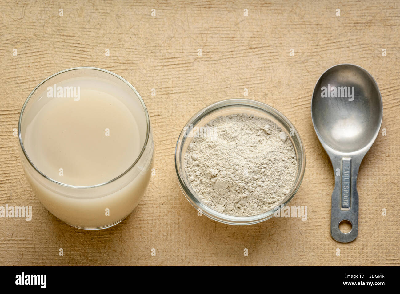 food grade diatomaceous earth supplement - powder and in a glass of water with measuring scoop Stock Photohttps://www.alamy.com/image-license-details/?v=1https://www.alamy.com/food-grade-diatomaceous-earth-supplement-powder-and-in-a-glass-of-water-with-measuring-scoop-image242472967.html
RFT2DGMR–food grade diatomaceous earth supplement - powder and in a glass of water with measuring scoop
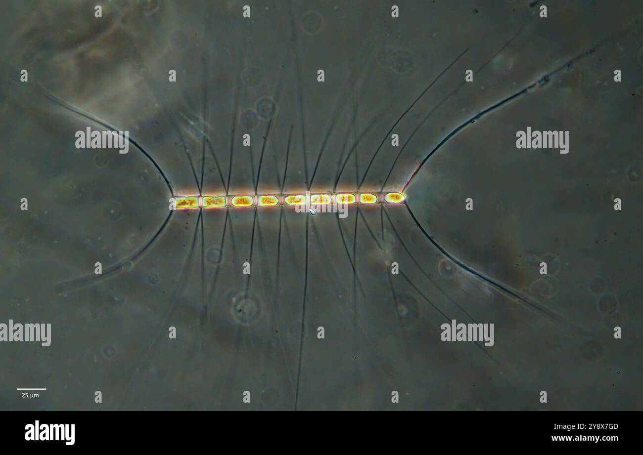 Diatom from the genus Chaetoceros. Collected in surface plankton sample from Hidra, south-western Norway in October. Stock Photohttps://www.alamy.com/image-license-details/?v=1https://www.alamy.com/diatom-from-the-genus-chaetoceros-collected-in-surface-plankton-sample-from-hidra-south-western-norway-in-october-image625067197.html
RM2Y8X7GD–Diatom from the genus Chaetoceros. Collected in surface plankton sample from Hidra, south-western Norway in October.
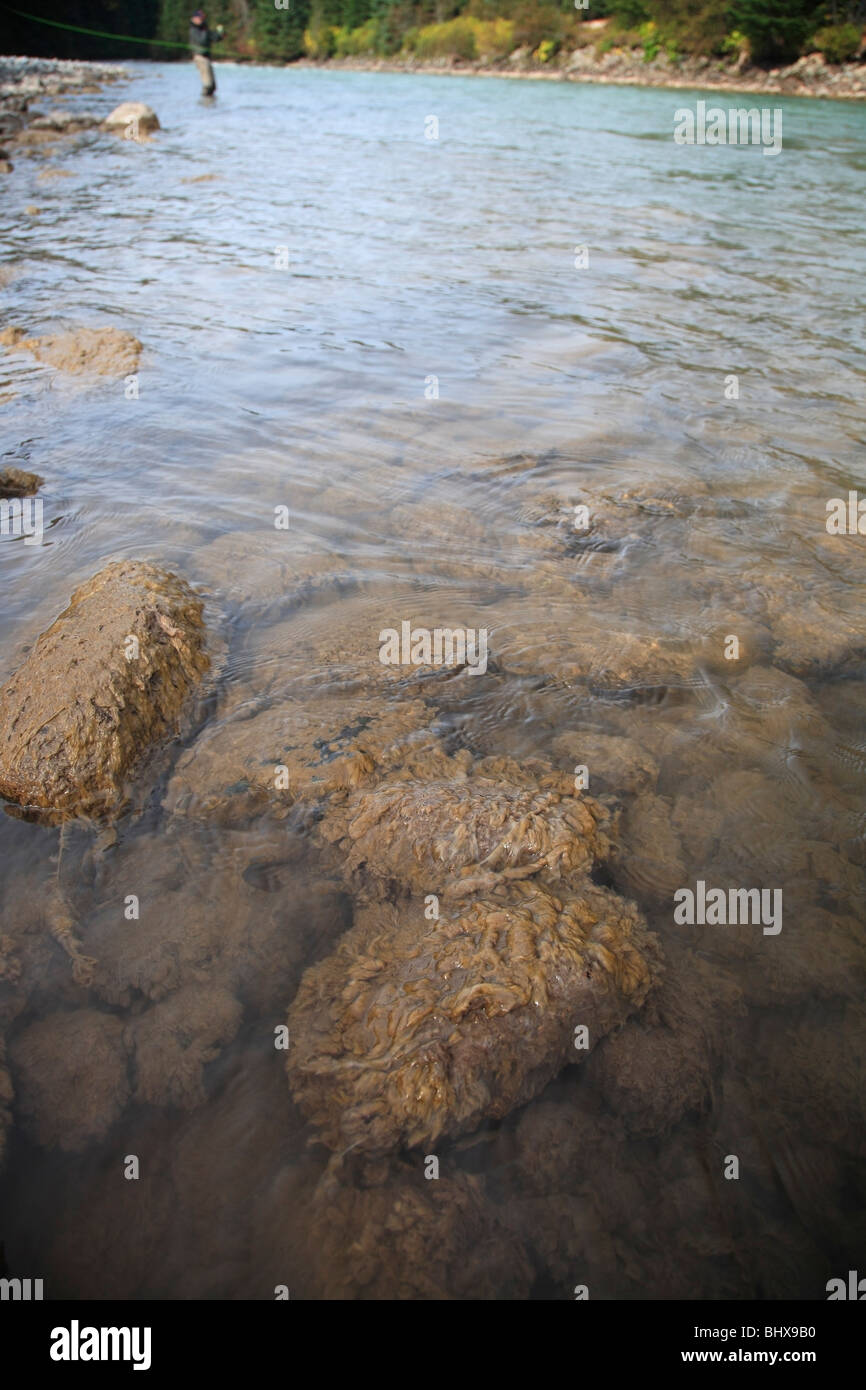 The invasive algae Didymo covering rocks in river, British Columbia Stock Photohttps://www.alamy.com/image-license-details/?v=1https://www.alamy.com/stock-photo-the-invasive-algae-didymo-covering-rocks-in-river-british-columbia-28237636.html
RMBHX9B0–The invasive algae Didymo covering rocks in river, British Columbia
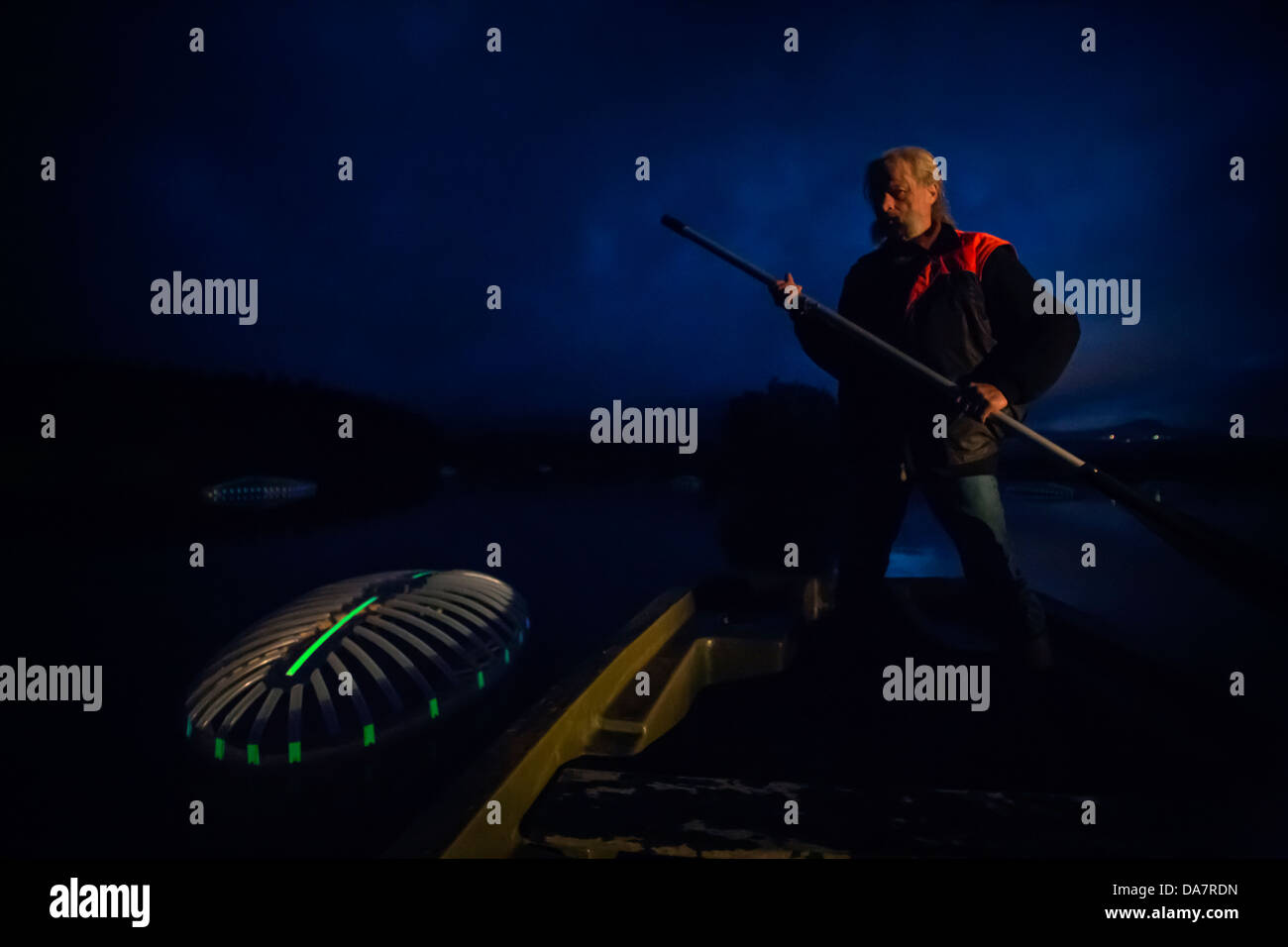 The Land Art work called Mégascospic Diatoms, made by Prisca Cosnier. A project within the Horizons 'Nature Arts' 2013 framework Stock Photohttps://www.alamy.com/image-license-details/?v=1https://www.alamy.com/stock-photo-the-land-art-work-called-mgascospic-diatoms-made-by-prisca-cosnier-57949745.html
RMDA7RDN–The Land Art work called Mégascospic Diatoms, made by Prisca Cosnier. A project within the Horizons 'Nature Arts' 2013 framework
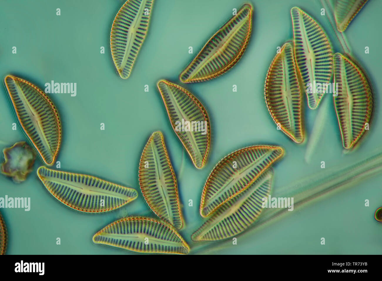 diatom (Diatomeae), diatoms in phase contrast and interference contrast, x 200 Stock Photohttps://www.alamy.com/image-license-details/?v=1https://www.alamy.com/diatom-diatomeae-diatoms-in-phase-contrast-and-interference-contrast-x-200-image255239023.html
RMTR73YB–diatom (Diatomeae), diatoms in phase contrast and interference contrast, x 200
 Flamingos in the Camargue 2 Stock Photohttps://www.alamy.com/image-license-details/?v=1https://www.alamy.com/flamingos-in-the-camargue-2-image479986554.html
RF2JTW7MX–Flamingos in the Camargue 2
 Diatoms in the water under the microskop Stock Photohttps://www.alamy.com/image-license-details/?v=1https://www.alamy.com/diatoms-in-the-water-under-the-microskop-image245269250.html
RMT70YC2–Diatoms in the water under the microskop
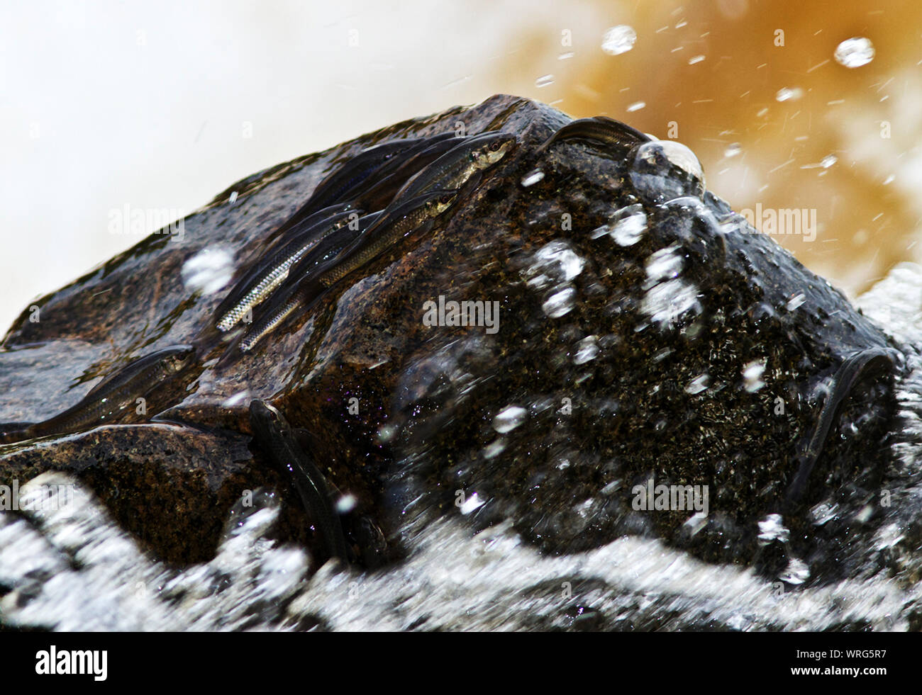 The Red-eyed Labeo is a small fish of rocky and clean fereshwater in East Africa and as far South as the Pondola River. Stock Photohttps://www.alamy.com/image-license-details/?v=1https://www.alamy.com/the-red-eyed-labeo-is-a-small-fish-of-rocky-and-clean-fereshwater-in-east-africa-and-as-far-south-as-the-pondola-river-image272648411.html
RMWRG5R7–The Red-eyed Labeo is a small fish of rocky and clean fereshwater in East Africa and as far South as the Pondola River.
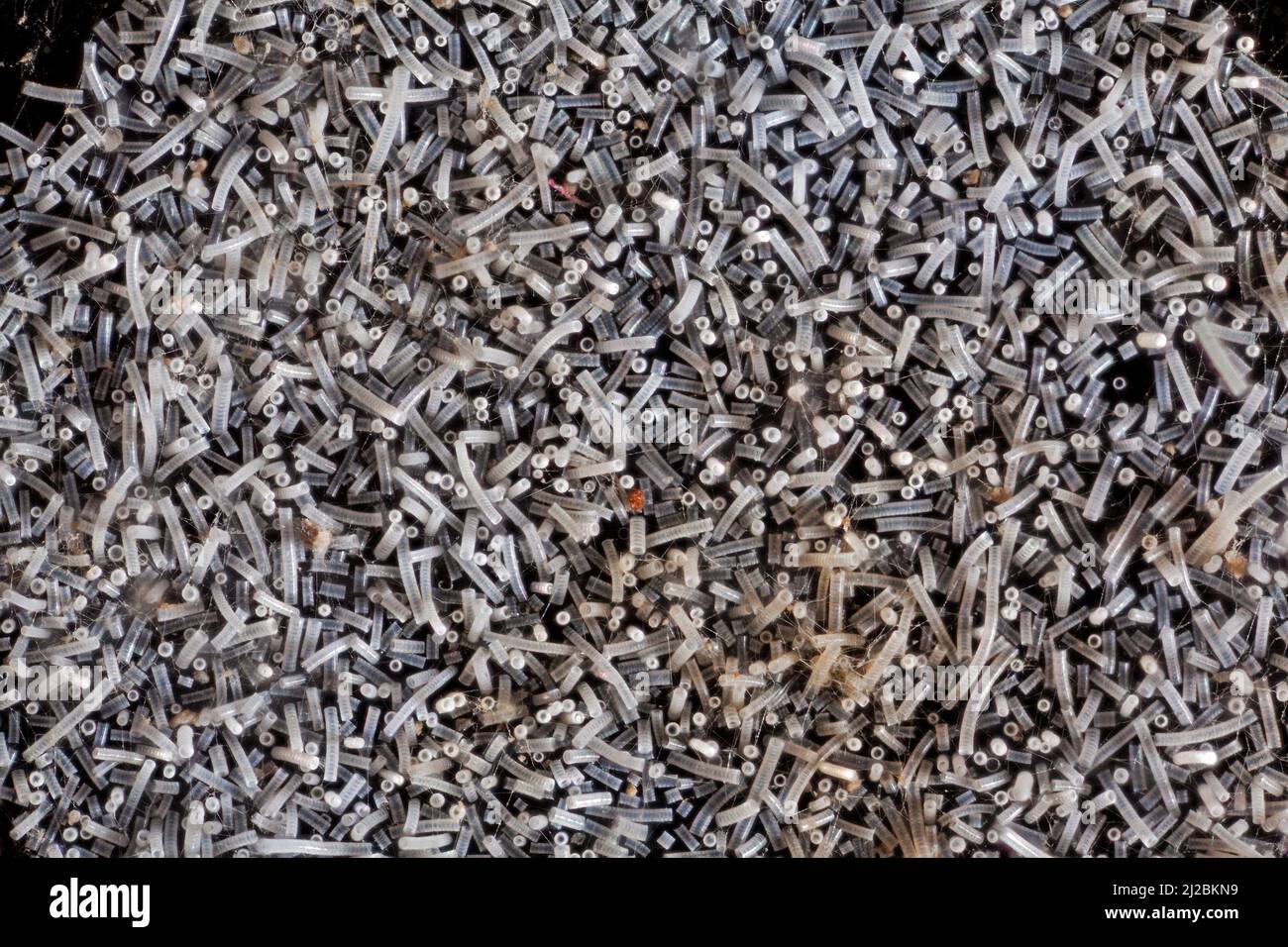 Fresh water diatoms, Orthoseira, Orthosira arenaria, high macro view Stock Photohttps://www.alamy.com/image-license-details/?v=1https://www.alamy.com/fresh-water-diatoms-orthoseira-orthosira-arenaria-high-macro-view-image466166213.html
RM2J2BKN9–Fresh water diatoms, Orthoseira, Orthosira arenaria, high macro view