Quick filters:
Dracunculus medinensis Stock Photos and Images
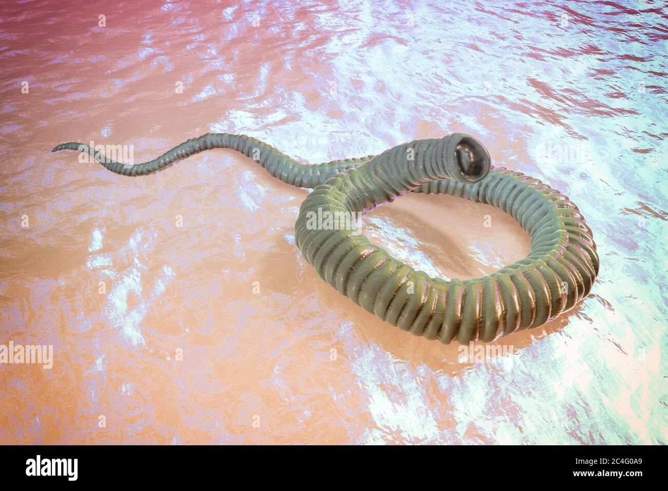 Guinea worm (Dracunculus medinensis) first-stage larva, computer illustration. Larvae are excreted from female worm parasitising under the skin of human extremities in patient's with dracunculiasis. Stock Photohttps://www.alamy.com/image-license-details/?v=1https://www.alamy.com/guinea-worm-dracunculus-medinensis-first-stage-larva-computer-illustration-larvae-are-excreted-from-female-worm-parasitising-under-the-skin-of-human-extremities-in-patients-with-dracunculiasis-image364227873.html
RF2C4G0A9–Guinea worm (Dracunculus medinensis) first-stage larva, computer illustration. Larvae are excreted from female worm parasitising under the skin of human extremities in patient's with dracunculiasis.
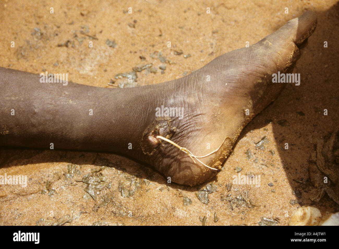 Thread tied around Emerging Guinea Worm, Niger. Stock Photohttps://www.alamy.com/image-license-details/?v=1https://www.alamy.com/thread-tied-around-emerging-guinea-worm-niger-image6323344.html
RMA4JTW1–Thread tied around Emerging Guinea Worm, Niger.
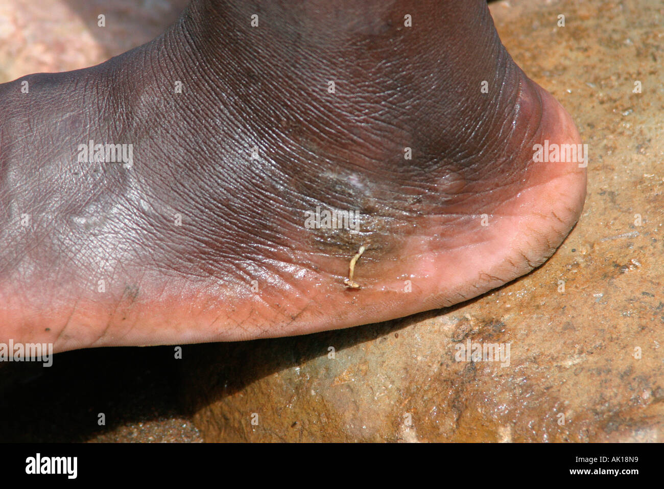 Foot with Guinea-Worm / Nyanyagachor Stock Photohttps://www.alamy.com/image-license-details/?v=1https://www.alamy.com/foot-with-guinea-worm-nyanyagachor-image8486744.html
RMAK18N9–Foot with Guinea-Worm / Nyanyagachor
 European Doctor Treating African Man Infected by and Being Treated for Guinea Worm, Dracunculiasis medinensis. The doctor extracts the worm by winding it around a stick which may be the origin of the medical symbol of the Rod of Asclepius. (Engraving 1879) Stock Photohttps://www.alamy.com/image-license-details/?v=1https://www.alamy.com/stock-photo-european-doctor-treating-african-man-infected-by-and-being-treated-174598140.html
RMM41HKT–European Doctor Treating African Man Infected by and Being Treated for Guinea Worm, Dracunculiasis medinensis. The doctor extracts the worm by winding it around a stick which may be the origin of the medical symbol of the Rod of Asclepius. (Engraving 1879)
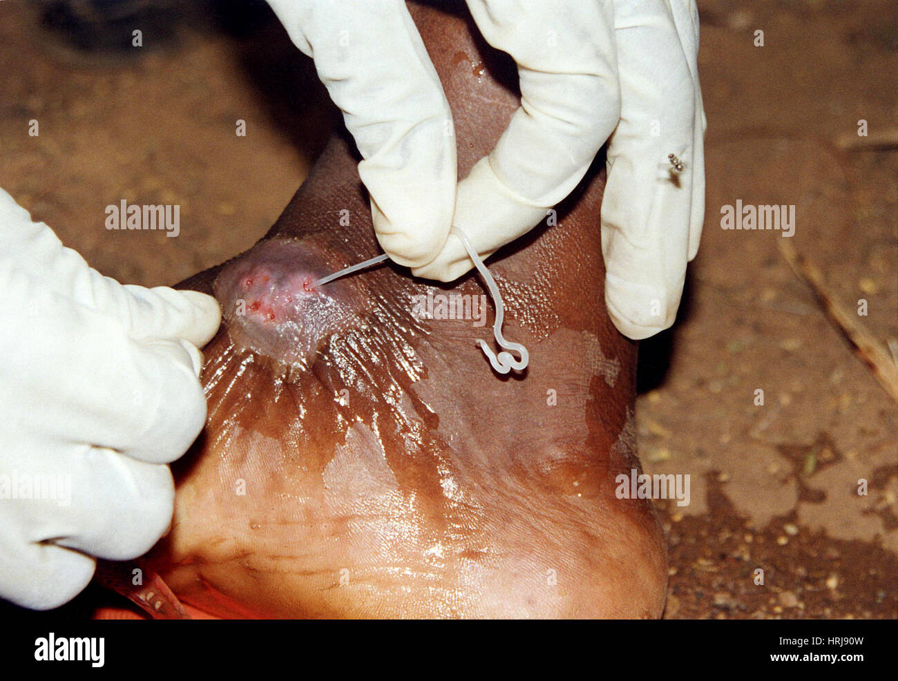 Dracunculus medinensis, Guinea Worm Extraction Stock Photohttps://www.alamy.com/image-license-details/?v=1https://www.alamy.com/stock-photo-dracunculus-medinensis-guinea-worm-extraction-135011881.html
RMHRJ90W–Dracunculus medinensis, Guinea Worm Extraction
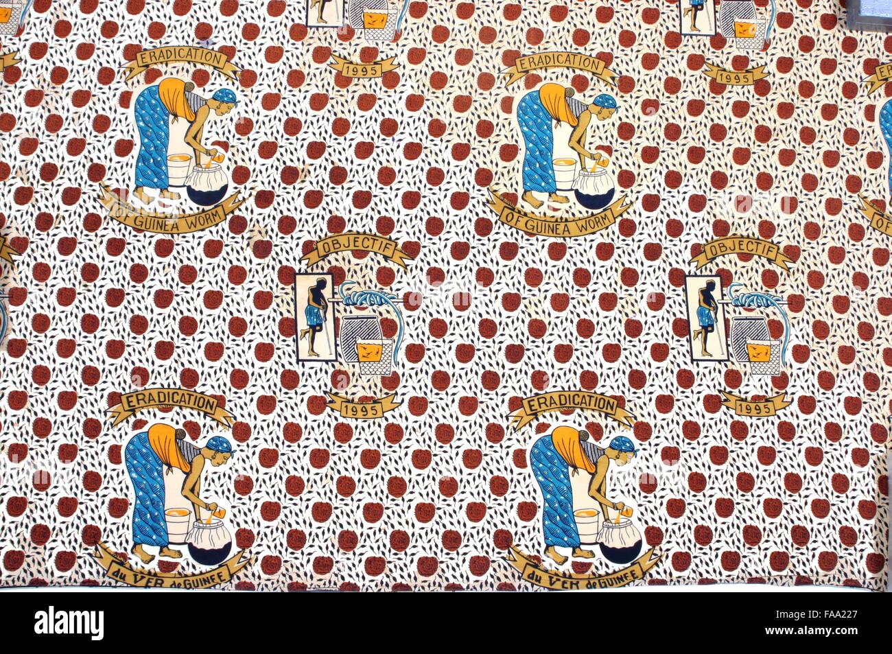 Cloth that can be used as a skirt from the country of Togo, Africa. Stock Photohttps://www.alamy.com/image-license-details/?v=1https://www.alamy.com/stock-photo-cloth-that-can-be-used-as-a-skirt-from-the-country-of-togo-africa-92419551.html
RMFAA227–Cloth that can be used as a skirt from the country of Togo, Africa.
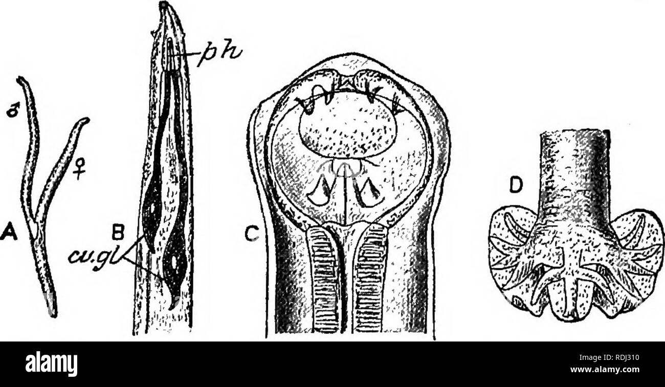 . A manual of elementary zoology . Zoology. Fig. 211.—The Corn-cockle Worm.—From Theobald. A, Cockle gall; C, larvae; in D, gall cut open ; E, larvae magnified. 5. Larva and adults parasitic in different animals, with a free stage. —The Guinea Worm, Dracunculus medinensis. The female, about 90 cm. long, encysts beneath the skin of man, usually in the leg, with the head in the host's foot, causing an abscess. She is viviparous.. Fig. 212.—The Miners' Worm {Ancylostomum duodenale).—From Parker and Haswell, after Leuckart. A, Male and female in caitu ; B, anterior end; C, mouth, with spines; D, h Stock Photohttps://www.alamy.com/image-license-details/?v=1https://www.alamy.com/a-manual-of-elementary-zoology-zoology-fig-211the-corn-cockle-wormfrom-theobald-a-cockle-gall-c-larvae-in-d-gall-cut-open-e-larvae-magnified-5-larva-and-adults-parasitic-in-different-animals-with-a-free-stage-the-guinea-worm-dracunculus-medinensis-the-female-about-90-cm-long-encysts-beneath-the-skin-of-man-usually-in-the-leg-with-the-head-in-the-hosts-foot-causing-an-abscess-she-is-viviparous-fig-212the-miners-worm-ancylostomum-duodenalefrom-parker-and-haswell-after-leuckart-a-male-and-female-in-caitu-b-anterior-end-c-mouth-with-spines-d-h-image232122828.html
RMRDJ310–. A manual of elementary zoology . Zoology. Fig. 211.—The Corn-cockle Worm.—From Theobald. A, Cockle gall; C, larvae; in D, gall cut open ; E, larvae magnified. 5. Larva and adults parasitic in different animals, with a free stage. —The Guinea Worm, Dracunculus medinensis. The female, about 90 cm. long, encysts beneath the skin of man, usually in the leg, with the head in the host's foot, causing an abscess. She is viviparous.. Fig. 212.—The Miners' Worm {Ancylostomum duodenale).—From Parker and Haswell, after Leuckart. A, Male and female in caitu ; B, anterior end; C, mouth, with spines; D, h
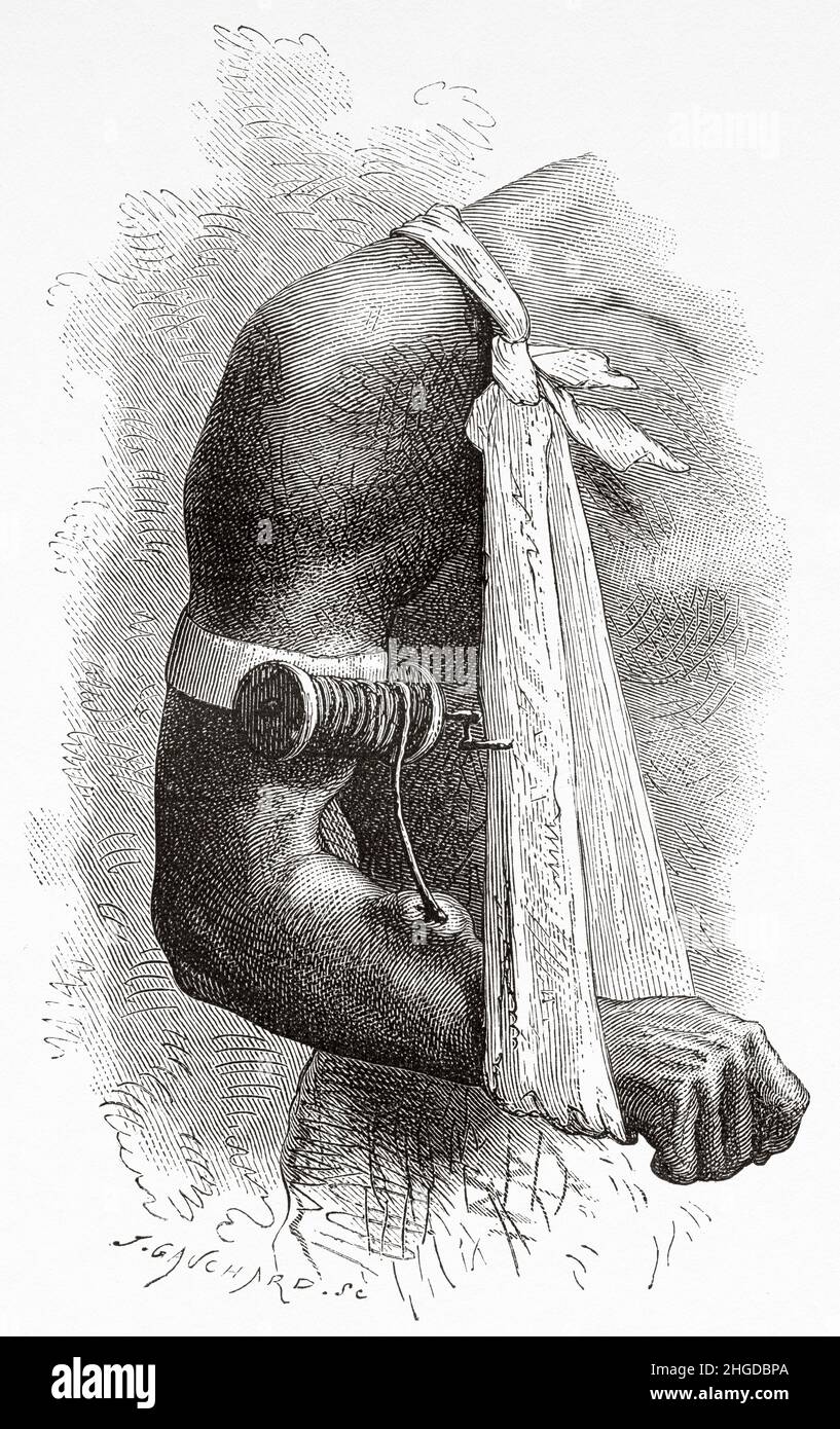 Guinea worm disease. Dracunculiasis. A disease caused by nematode roundworm Dracunculus medinensis, it occurs by drinking unfiltered water containing copepods infected with larvae. Old 19th century engraved illustration from Four months in Florida by Achille Poussielgue, Le Tour du Monde 1870 Stock Photohttps://www.alamy.com/image-license-details/?v=1https://www.alamy.com/guinea-worm-disease-dracunculiasis-a-disease-caused-by-nematode-roundworm-dracunculus-medinensis-it-occurs-by-drinking-unfiltered-water-containing-copepods-infected-with-larvae-old-19th-century-engraved-illustration-from-four-months-in-florida-by-achille-poussielgue-le-tour-du-monde-1870-image457598690.html
RM2HGDBPA–Guinea worm disease. Dracunculiasis. A disease caused by nematode roundworm Dracunculus medinensis, it occurs by drinking unfiltered water containing copepods infected with larvae. Old 19th century engraved illustration from Four months in Florida by Achille Poussielgue, Le Tour du Monde 1870
 FILARIA Stock Photohttps://www.alamy.com/image-license-details/?v=1https://www.alamy.com/stock-photo-filaria-49178268.html
RMCT07AM–FILARIA
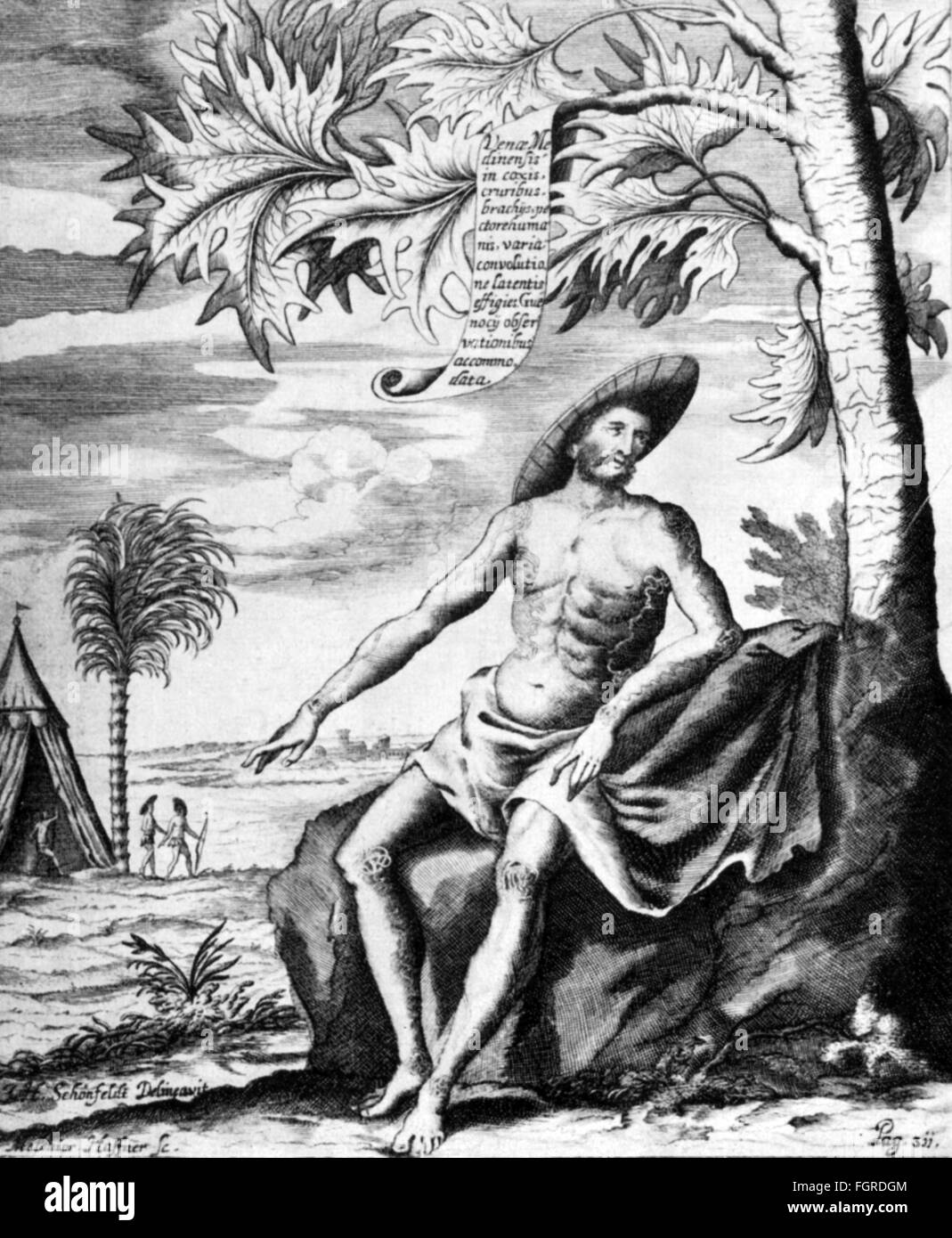 medicine, sick persons, man with Guinea worms, after drawing by J.H.Schönfeldt, copper engraving by Melchior Haffner, out of: Georg Hieronymus Welsch, 'Exercitatio de vena Medinensi', 1674, Artist's Copyright has not to be cleared Stock Photohttps://www.alamy.com/image-license-details/?v=1https://www.alamy.com/stock-photo-medicine-sick-persons-man-with-guinea-worms-after-drawing-by-jhschnfeldt-96401892.html
RMFGRDGM–medicine, sick persons, man with Guinea worms, after drawing by J.H.Schönfeldt, copper engraving by Melchior Haffner, out of: Georg Hieronymus Welsch, 'Exercitatio de vena Medinensi', 1674, Artist's Copyright has not to be cleared
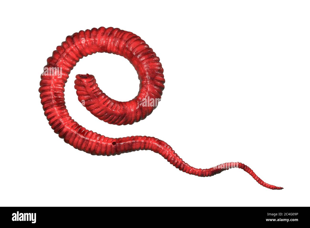 Guinea worm (Dracunculus medinensis) first-stage larva, computer illustration. Larvae are excreted from female worm parasitising under the skin of human extremities in patient's with dracunculiasis. Stock Photohttps://www.alamy.com/image-license-details/?v=1https://www.alamy.com/guinea-worm-dracunculus-medinensis-first-stage-larva-computer-illustration-larvae-are-excreted-from-female-worm-parasitising-under-the-skin-of-human-extremities-in-patients-with-dracunculiasis-image364227858.html
RF2C4G09P–Guinea worm (Dracunculus medinensis) first-stage larva, computer illustration. Larvae are excreted from female worm parasitising under the skin of human extremities in patient's with dracunculiasis.
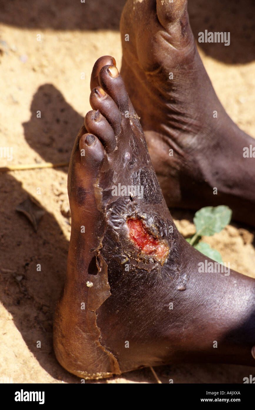 Secondary Infection Caused by Guinea Worm Wound, Niger Stock Photohttps://www.alamy.com/image-license-details/?v=1https://www.alamy.com/secondary-infection-caused-by-guinea-worm-wound-niger-image6323753.html
RMA4JXXA–Secondary Infection Caused by Guinea Worm Wound, Niger
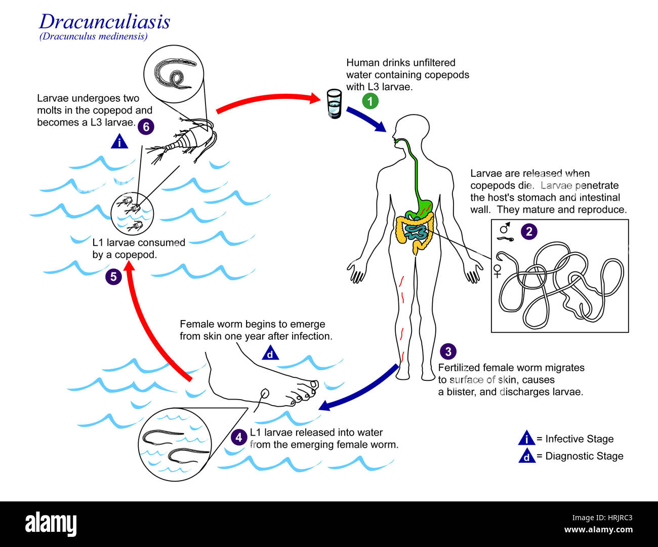 Dracunculus medinensis, Guinea Worm Life Cycle Stock Photohttps://www.alamy.com/image-license-details/?v=1https://www.alamy.com/stock-photo-dracunculus-medinensis-guinea-worm-life-cycle-135023171.html
RMHRJRC3–Dracunculus medinensis, Guinea Worm Life Cycle
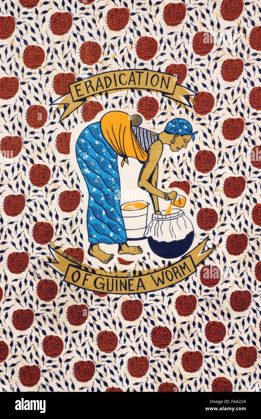 Detail of a cloth (from Togo, Africa) that depicts a woman filtering water to prevent Guinea Worm Disease GWD Stock Photohttps://www.alamy.com/image-license-details/?v=1https://www.alamy.com/stock-photo-detail-of-a-cloth-from-togo-africa-that-depicts-a-woman-filtering-92419548.html
RMFAA224–Detail of a cloth (from Togo, Africa) that depicts a woman filtering water to prevent Guinea Worm Disease GWD
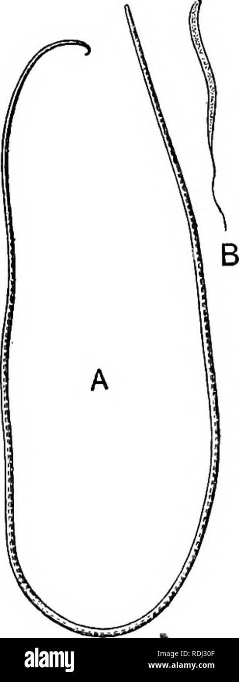 . A manual of elementary zoology . Zoology. 3°4 MANUAL OF ELEMENTARY ZOOLOGY and larvse escape into the host's tissues, sepsis, fever, and even death may result. . 6. Larva and adult parasitic, without free stage, in animals 0} unlike kinds.—The supposed cause of elephantiasis, Filaria bancrofti,. Fig. 213.—The Guinea Worm (Dracunculus medinensis). A, Adult female, reduced; J3, larva, much magnified.. Please note that these images are extracted from scanned page images that may have been digitally enhanced for readability - coloration and appearance of these illustrations may not perfectly res Stock Photohttps://www.alamy.com/image-license-details/?v=1https://www.alamy.com/a-manual-of-elementary-zoology-zoology-34-manual-of-elementary-zoology-and-larvse-escape-into-the-hosts-tissues-sepsis-fever-and-even-death-may-result-6-larva-and-adult-parasitic-without-free-stage-in-animals-0-unlike-kindsthe-supposed-cause-of-elephantiasis-filaria-bancrofti-fig-213the-guinea-worm-dracunculus-medinensis-a-adult-female-reduced-j3-larva-much-magnified-please-note-that-these-images-are-extracted-from-scanned-page-images-that-may-have-been-digitally-enhanced-for-readability-coloration-and-appearance-of-these-illustrations-may-not-perfectly-res-image232122815.html
RMRDJ30F–. A manual of elementary zoology . Zoology. 3°4 MANUAL OF ELEMENTARY ZOOLOGY and larvse escape into the host's tissues, sepsis, fever, and even death may result. . 6. Larva and adult parasitic, without free stage, in animals 0} unlike kinds.—The supposed cause of elephantiasis, Filaria bancrofti,. Fig. 213.—The Guinea Worm (Dracunculus medinensis). A, Adult female, reduced; J3, larva, much magnified.. Please note that these images are extracted from scanned page images that may have been digitally enhanced for readability - coloration and appearance of these illustrations may not perfectly res
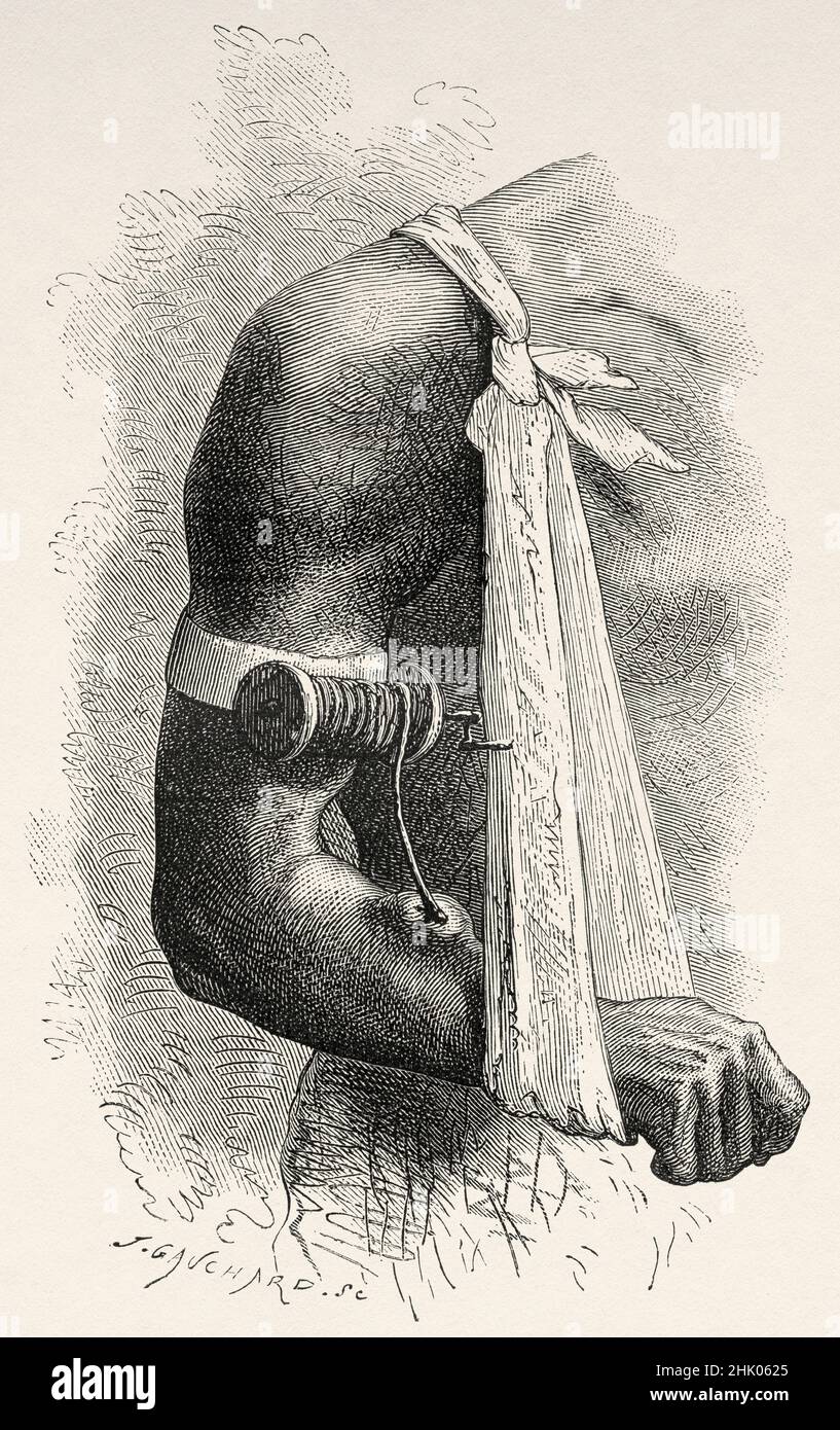 Guinea worm disease. Dracunculiasis. A disease caused by nematode roundworm Dracunculus medinensis, it occurs by drinking unfiltered water containing copepods infected with larvae. Old 19th century engraved illustration from Four months in Florida by Achille Poussielgue, Le Tour du Monde 1870 Stock Photohttps://www.alamy.com/image-license-details/?v=1https://www.alamy.com/guinea-worm-disease-dracunculiasis-a-disease-caused-by-nematode-roundworm-dracunculus-medinensis-it-occurs-by-drinking-unfiltered-water-containing-copepods-infected-with-larvae-old-19th-century-engraved-illustration-from-four-months-in-florida-by-achille-poussielgue-le-tour-du-monde-1870-image459152797.html
RM2HK0625–Guinea worm disease. Dracunculiasis. A disease caused by nematode roundworm Dracunculus medinensis, it occurs by drinking unfiltered water containing copepods infected with larvae. Old 19th century engraved illustration from Four months in Florida by Achille Poussielgue, Le Tour du Monde 1870
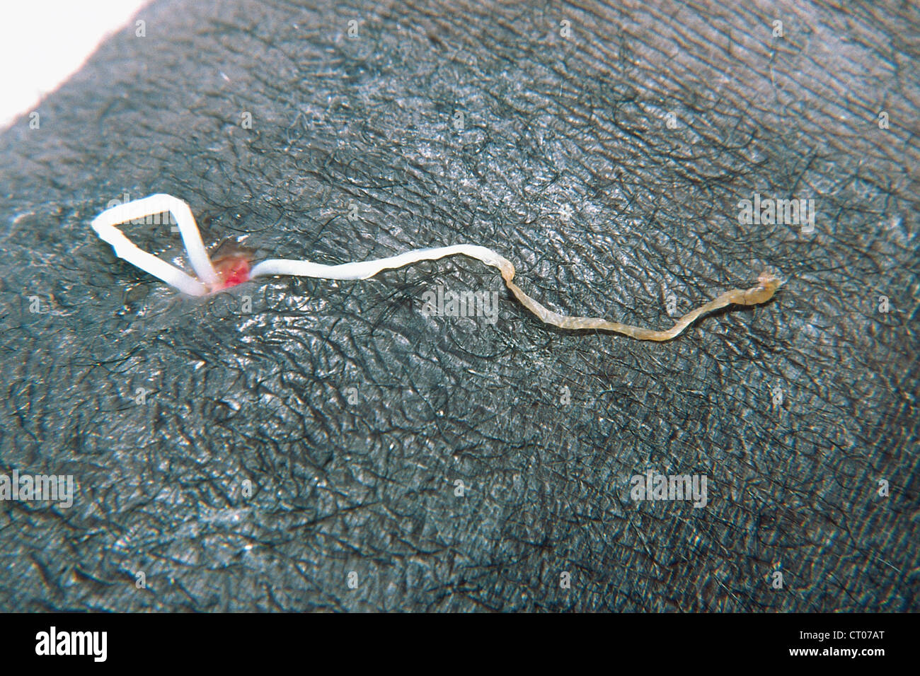 FILARIA Stock Photohttps://www.alamy.com/image-license-details/?v=1https://www.alamy.com/stock-photo-filaria-49178272.html
RMCT07AT–FILARIA
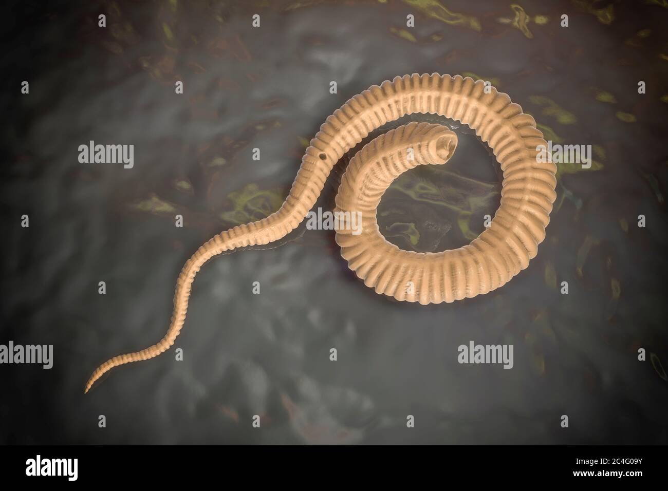 Guinea worm (Dracunculus medinensis) first-stage larva, computer illustration. Larvae are excreted from female worm parasitising under the skin of human extremities in patient's with dracunculiasis. Stock Photohttps://www.alamy.com/image-license-details/?v=1https://www.alamy.com/guinea-worm-dracunculus-medinensis-first-stage-larva-computer-illustration-larvae-are-excreted-from-female-worm-parasitising-under-the-skin-of-human-extremities-in-patients-with-dracunculiasis-image364227863.html
RF2C4G09Y–Guinea worm (Dracunculus medinensis) first-stage larva, computer illustration. Larvae are excreted from female worm parasitising under the skin of human extremities in patient's with dracunculiasis.
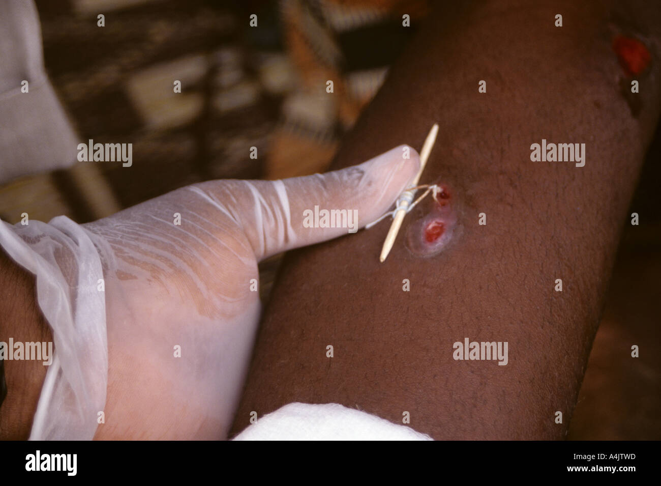 Looping Thread around Emerging Guinea Worm, Niger. Stock Photohttps://www.alamy.com/image-license-details/?v=1https://www.alamy.com/looping-thread-around-emerging-guinea-worm-niger-image6323356.html
RMA4JTWD–Looping Thread around Emerging Guinea Worm, Niger.
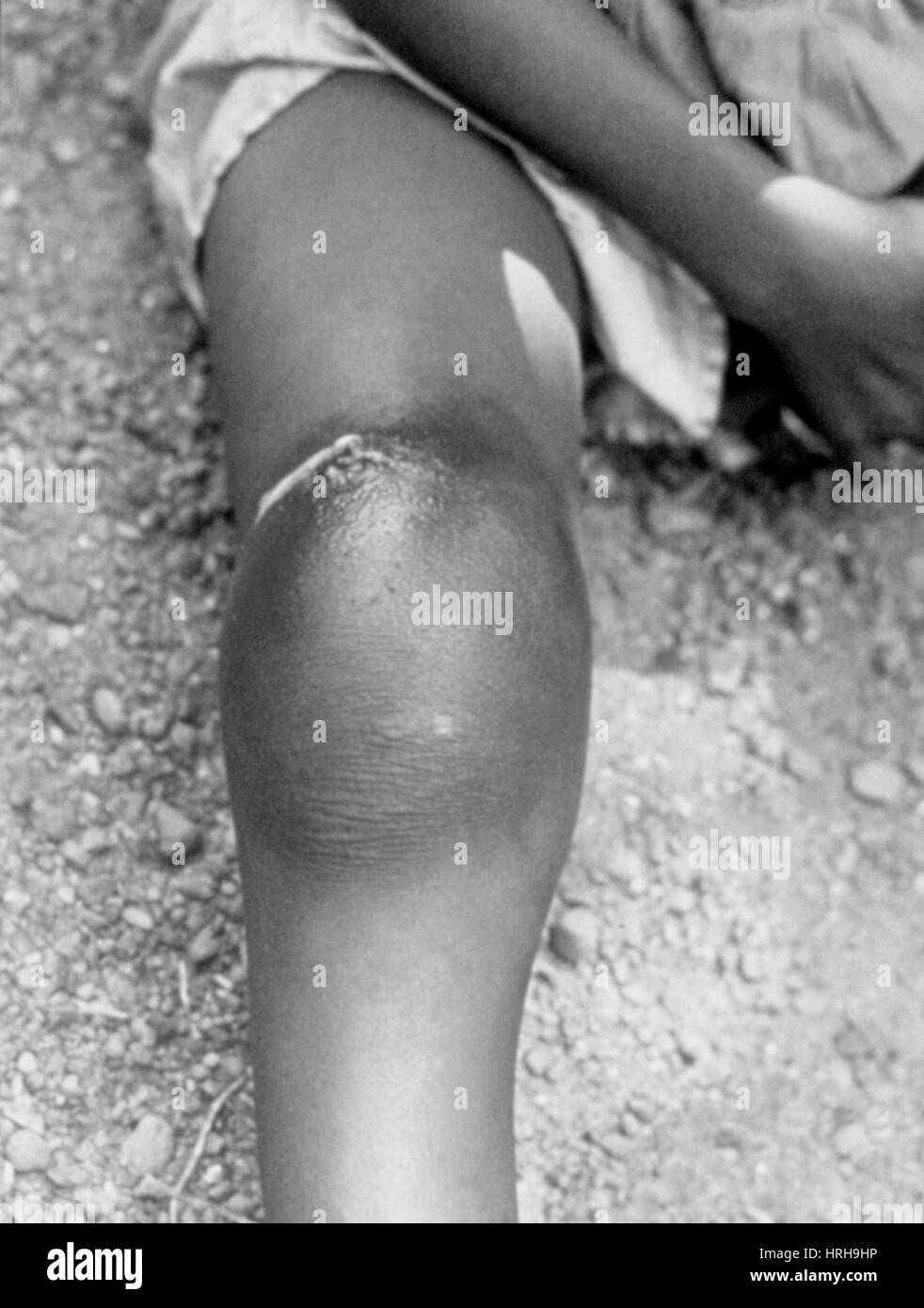 Guinea Worm Infection Stock Photohttps://www.alamy.com/image-license-details/?v=1https://www.alamy.com/stock-photo-guinea-worm-infection-134990402.html
RMHRH9HP–Guinea Worm Infection
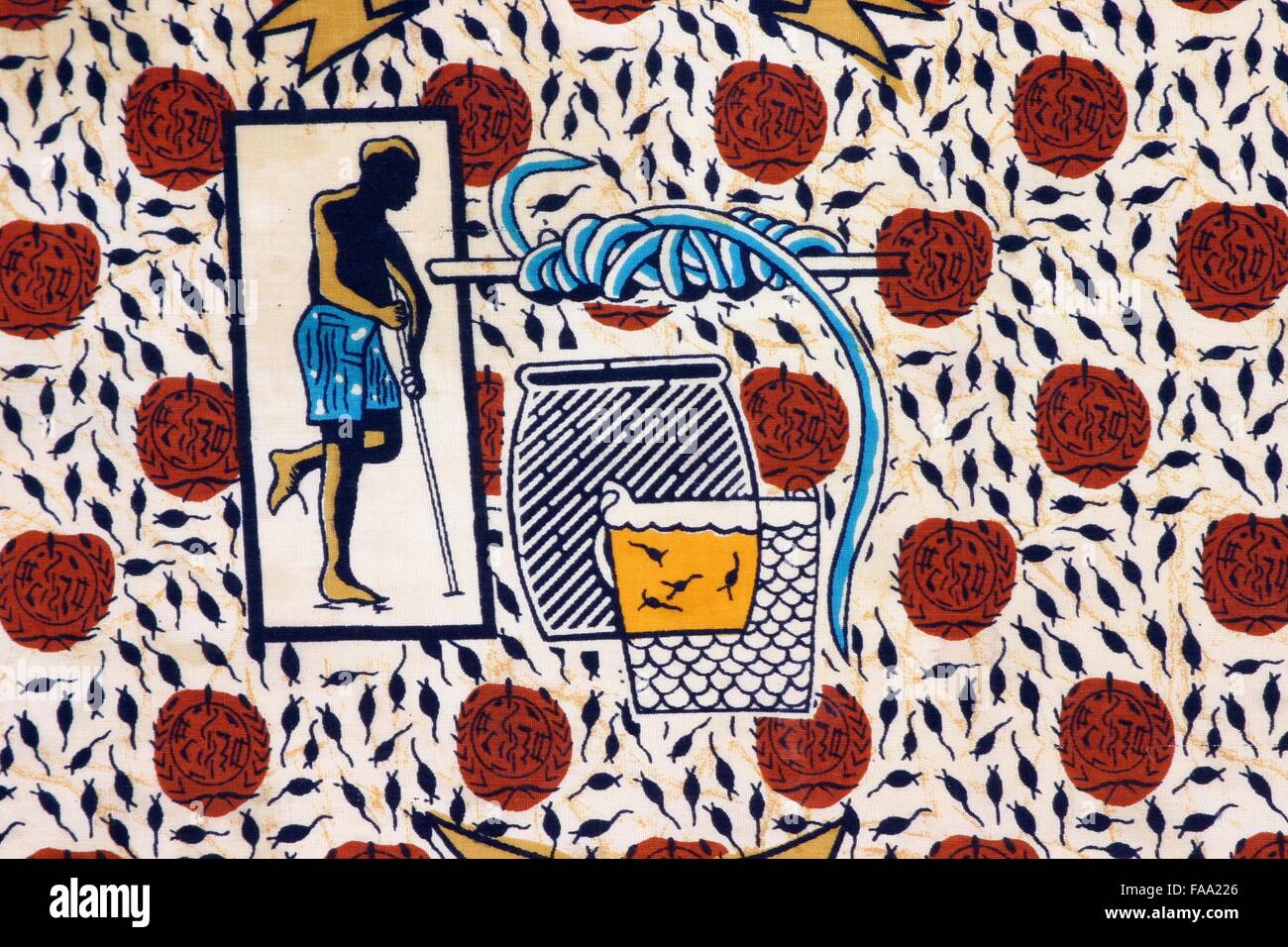 Detail of a cloth (from Togo, Africa) that depicts Guinea Worm Disease GWD due to the lack of clean water Stock Photohttps://www.alamy.com/image-license-details/?v=1https://www.alamy.com/stock-photo-detail-of-a-cloth-from-togo-africa-that-depicts-guinea-worm-disease-92419550.html
RMFAA226–Detail of a cloth (from Togo, Africa) that depicts Guinea Worm Disease GWD due to the lack of clean water
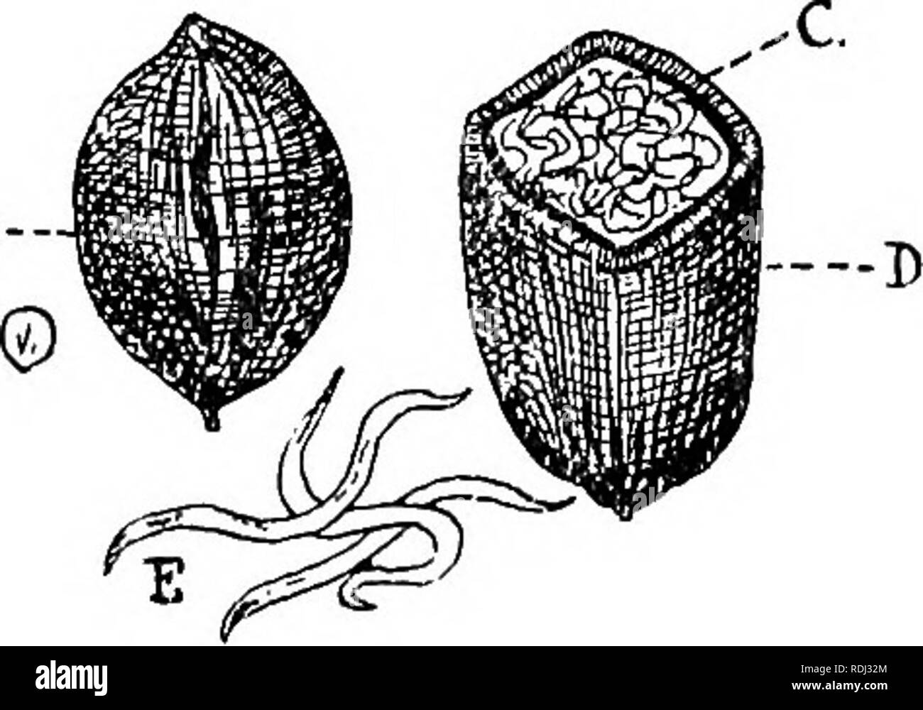 . A manual of elementary zoology . Zoology. THE NEMATODA. PARASITISM 3°3 and live in the body cavity till they become adult, when they escape into damp earth, become sexually mature, and pair. After rain the adults sometimes climb the stems of plants in such numbers as to give rise to the legend of " showers of worms.". Fig. 211.—The Corn-cockle Worm.—From Theobald. A, Cockle gall; C, larvae; in D, gall cut open ; E, larvae magnified. 5. Larva and adults parasitic in different animals, with a free stage. —The Guinea Worm, Dracunculus medinensis. The female, about 90 cm. long, encysts Stock Photohttps://www.alamy.com/image-license-details/?v=1https://www.alamy.com/a-manual-of-elementary-zoology-zoology-the-nematoda-parasitism-33-and-live-in-the-body-cavity-till-they-become-adult-when-they-escape-into-damp-earth-become-sexually-mature-and-pair-after-rain-the-adults-sometimes-climb-the-stems-of-plants-in-such-numbers-as-to-give-rise-to-the-legend-of-quot-showers-of-wormsquot-fig-211the-corn-cockle-wormfrom-theobald-a-cockle-gall-c-larvae-in-d-gall-cut-open-e-larvae-magnified-5-larva-and-adults-parasitic-in-different-animals-with-a-free-stage-the-guinea-worm-dracunculus-medinensis-the-female-about-90-cm-long-encysts-image232122876.html
RMRDJ32M–. A manual of elementary zoology . Zoology. THE NEMATODA. PARASITISM 3°3 and live in the body cavity till they become adult, when they escape into damp earth, become sexually mature, and pair. After rain the adults sometimes climb the stems of plants in such numbers as to give rise to the legend of " showers of worms.". Fig. 211.—The Corn-cockle Worm.—From Theobald. A, Cockle gall; C, larvae; in D, gall cut open ; E, larvae magnified. 5. Larva and adults parasitic in different animals, with a free stage. —The Guinea Worm, Dracunculus medinensis. The female, about 90 cm. long, encysts
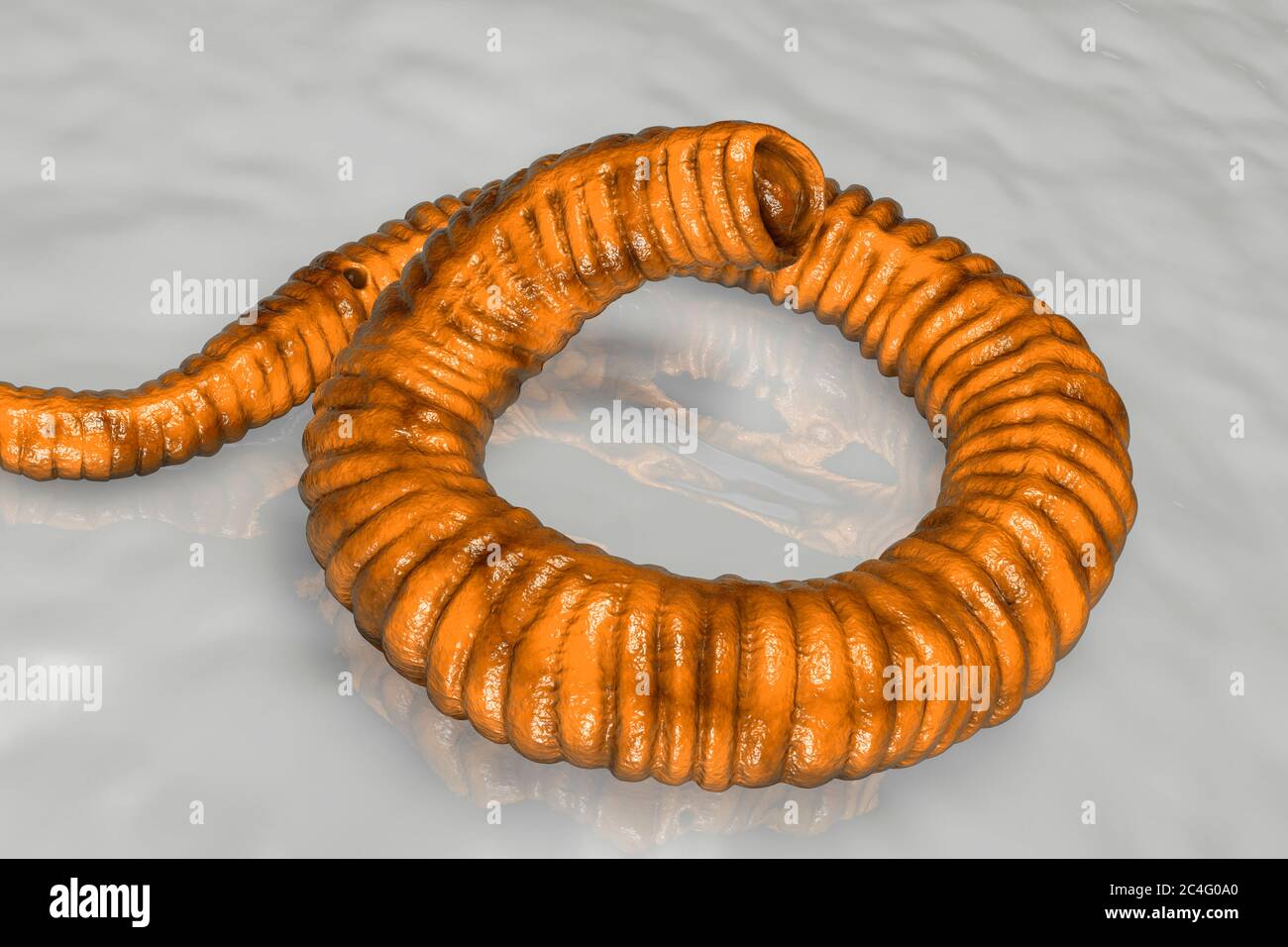 Guinea worm (Dracunculus medinensis) first-stage larva, computer illustration. Larvae are excreted from female worm parasitising under the skin of human extremities in patient's with dracunculiasis. Stock Photohttps://www.alamy.com/image-license-details/?v=1https://www.alamy.com/guinea-worm-dracunculus-medinensis-first-stage-larva-computer-illustration-larvae-are-excreted-from-female-worm-parasitising-under-the-skin-of-human-extremities-in-patients-with-dracunculiasis-image364227864.html
RF2C4G0A0–Guinea worm (Dracunculus medinensis) first-stage larva, computer illustration. Larvae are excreted from female worm parasitising under the skin of human extremities in patient's with dracunculiasis.
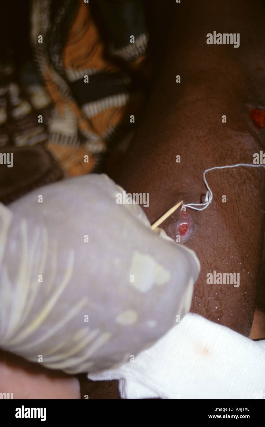 Looping Thread around Emerging Guinea Worm, Niger. Stock Photohttps://www.alamy.com/image-license-details/?v=1https://www.alamy.com/looping-thread-around-emerging-guinea-worm-niger-image6323373.html
RMA4JTXE–Looping Thread around Emerging Guinea Worm, Niger.
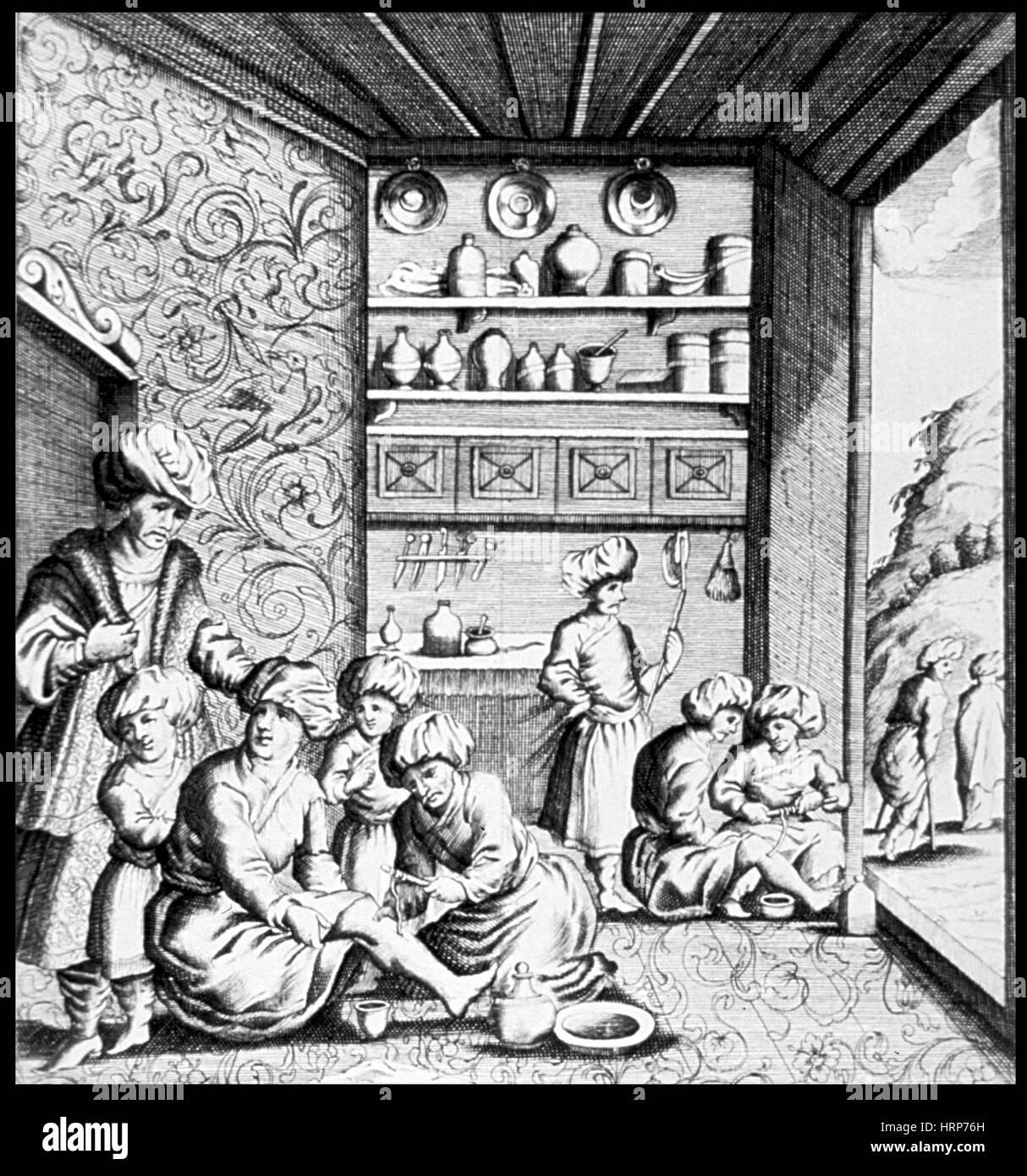 Guinea Worm Extraction, 1674 Stock Photohttps://www.alamy.com/image-license-details/?v=1https://www.alamy.com/stock-photo-guinea-worm-extraction-1674-135098281.html
RMHRP76H–Guinea Worm Extraction, 1674
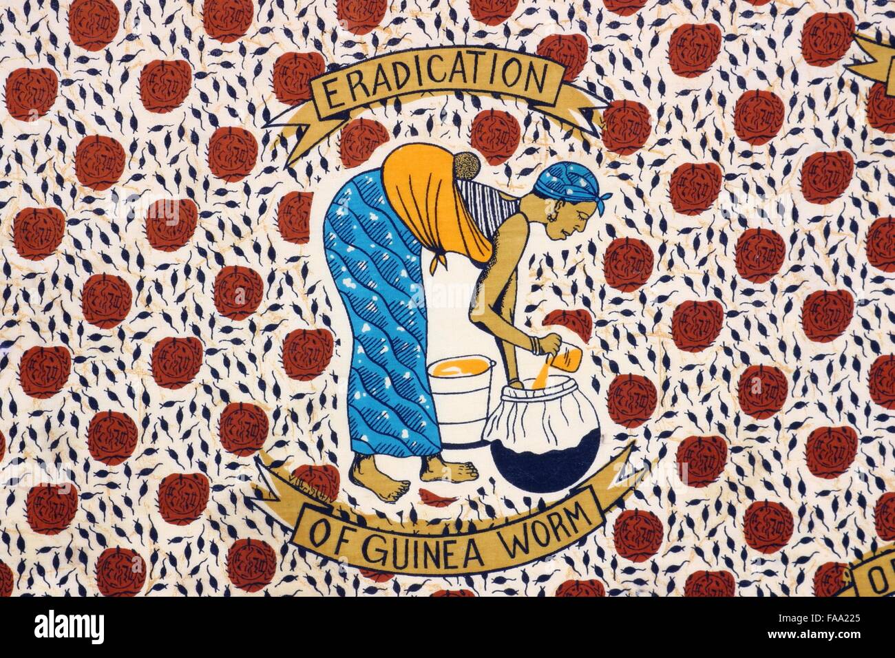 Detail of a cloth (from Togo, Africa) that depicts a woman filtering water to prevent Guinea Worm Disease GWD Stock Photohttps://www.alamy.com/image-license-details/?v=1https://www.alamy.com/stock-photo-detail-of-a-cloth-from-togo-africa-that-depicts-a-woman-filtering-92419549.html
RMFAA225–Detail of a cloth (from Togo, Africa) that depicts a woman filtering water to prevent Guinea Worm Disease GWD
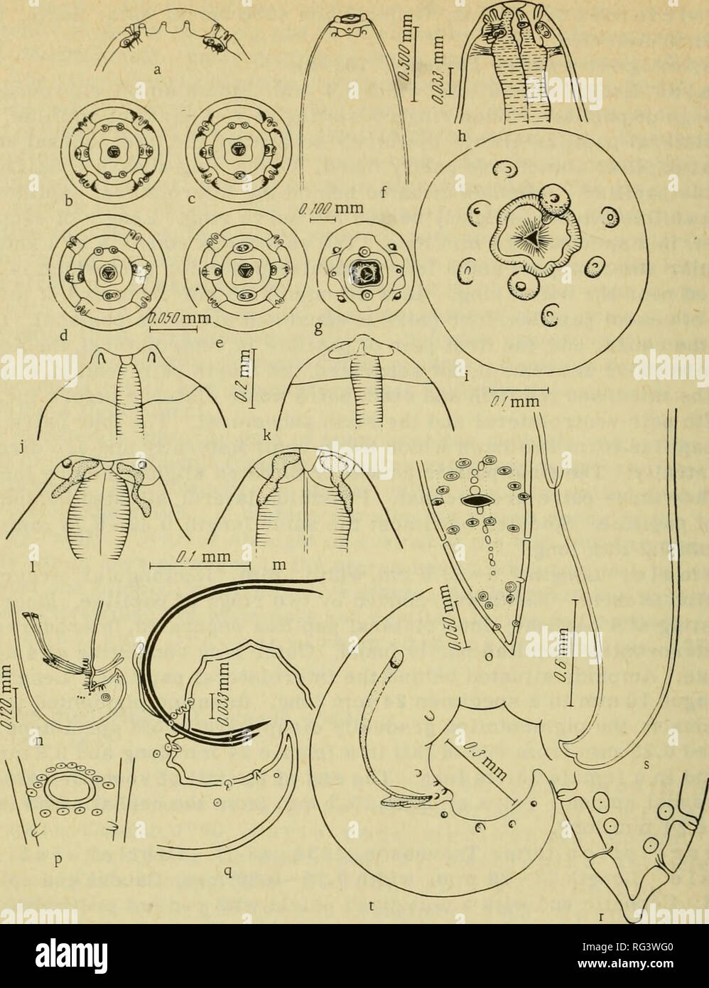 . Camallanata of animals and man and diseases caused by them = Kamallanaty zhivotnykh i cheloveka i vyzyvaemye ime zabolevaniya. Helminths; Worms as carriers of disease. (245). FIGURE 143. Dracunculus medinensis (Linnaeus, 1758). Cephalic ends: a—e — after Moorthy, 1937; f,g — after Skrjabin and Shul'ts, 1931; h — after Chitwood and Chitwood, 1950; i—m — after Travassos, 1934. Caudal ends: n, ? — after Moorthy, 1937; q,r — after Chitwood and Chitwood, 1950; s, t — after Travassos, 1934; p — region of cloaca — after Chitwood and Chitwood, 1950. Female. Length 465 —490 mm, width 1.5 mm. Body cyl Stock Photohttps://www.alamy.com/image-license-details/?v=1https://www.alamy.com/camallanata-of-animals-and-man-and-diseases-caused-by-them-=-kamallanaty-zhivotnykh-i-cheloveka-i-vyzyvaemye-ime-zabolevaniya-helminths-worms-as-carriers-of-disease-245-figure-143-dracunculus-medinensis-linnaeus-1758-cephalic-ends-ae-after-moorthy-1937-fg-after-skrjabin-and-shults-1931-h-after-chitwood-and-chitwood-1950-im-after-travassos-1934-caudal-ends-n-after-moorthy-1937-qr-after-chitwood-and-chitwood-1950-s-t-after-travassos-1934-p-region-of-cloaca-after-chitwood-and-chitwood-1950-female-length-465-490-mm-width-15-mm-body-cyl-image233655184.html
RMRG3WG0–. Camallanata of animals and man and diseases caused by them = Kamallanaty zhivotnykh i cheloveka i vyzyvaemye ime zabolevaniya. Helminths; Worms as carriers of disease. (245). FIGURE 143. Dracunculus medinensis (Linnaeus, 1758). Cephalic ends: a—e — after Moorthy, 1937; f,g — after Skrjabin and Shul'ts, 1931; h — after Chitwood and Chitwood, 1950; i—m — after Travassos, 1934. Caudal ends: n, ? — after Moorthy, 1937; q,r — after Chitwood and Chitwood, 1950; s, t — after Travassos, 1934; p — region of cloaca — after Chitwood and Chitwood, 1950. Female. Length 465 —490 mm, width 1.5 mm. Body cyl
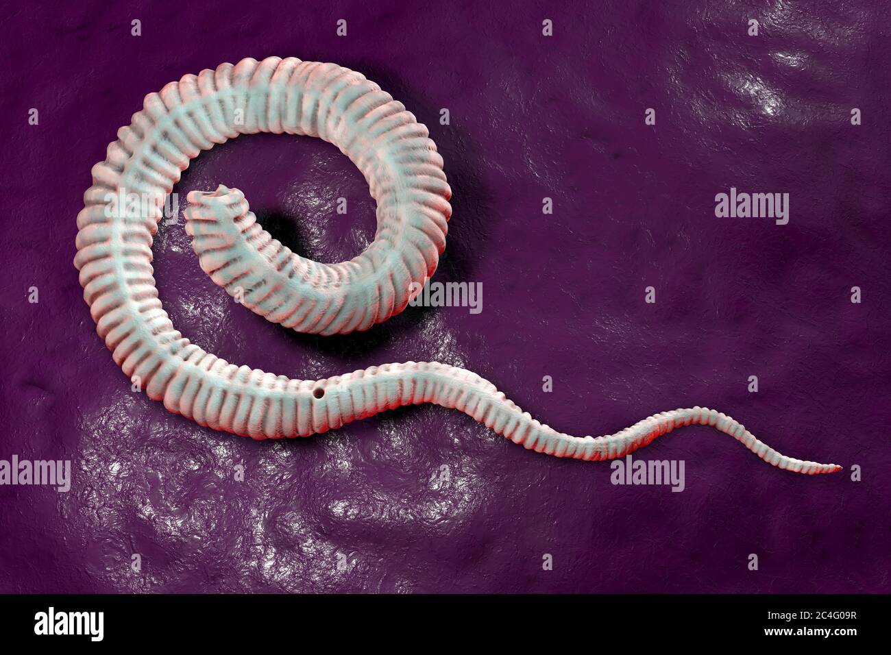 Guinea worm (Dracunculus medinensis) first-stage larva, computer illustration. Larvae are excreted from female worm parasitising under the skin of human extremities in patient's with dracunculiasis. Stock Photohttps://www.alamy.com/image-license-details/?v=1https://www.alamy.com/guinea-worm-dracunculus-medinensis-first-stage-larva-computer-illustration-larvae-are-excreted-from-female-worm-parasitising-under-the-skin-of-human-extremities-in-patients-with-dracunculiasis-image364227859.html
RF2C4G09R–Guinea worm (Dracunculus medinensis) first-stage larva, computer illustration. Larvae are excreted from female worm parasitising under the skin of human extremities in patient's with dracunculiasis.
 Looping Thread around Guinea Worm Emerging from Leg, Near Tera Niger Africa. Stock Photohttps://www.alamy.com/image-license-details/?v=1https://www.alamy.com/looping-thread-around-guinea-worm-emerging-from-leg-near-tera-niger-image6323489.html
RMA4JWP2–Looping Thread around Guinea Worm Emerging from Leg, Near Tera Niger Africa.
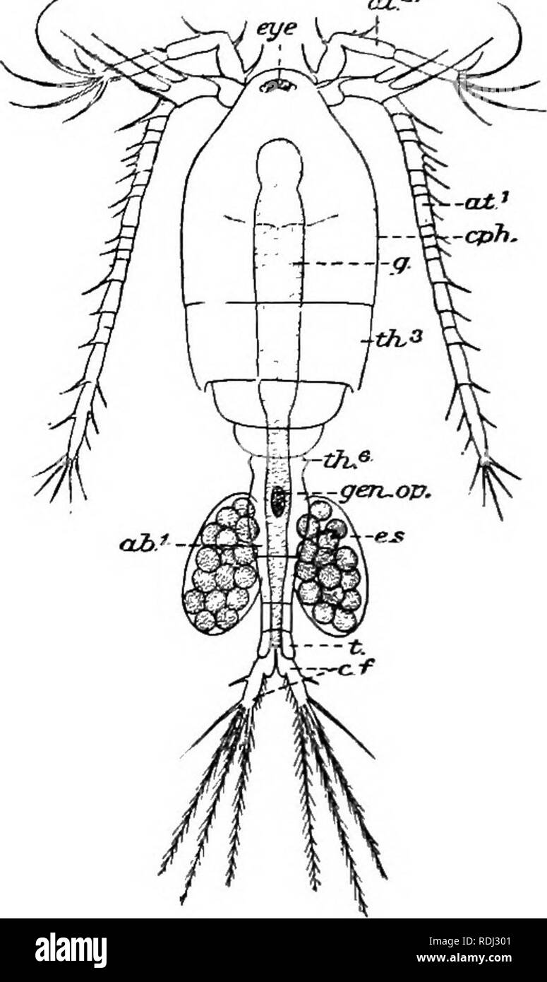 . A manual of elementary zoology . Zoology. Fig. 213.—The Guinea Worm (Dracunculus medinensis). A, Adult female, reduced; J3, larva, much magnified.. Fig. 214.—Cyclops. ad.1, First abdominal segment; a/.1, antennule ; at.^, antenna; c./.% caudal fork; cph,, cephalothorax (fused head and first two thoracic segments) ; c.3-., egg sac; eye (single and median); £"., alimentary canal; gcn.op.y genital opening; t., telson; M.3, th.% third and sixth thoracic segments. In comparing this crustacean with the crayfish, note the absence of proventi iculus, paired eyes, uropods, and carapace, the pr Stock Photohttps://www.alamy.com/image-license-details/?v=1https://www.alamy.com/a-manual-of-elementary-zoology-zoology-fig-213the-guinea-worm-dracunculus-medinensis-a-adult-female-reduced-j3-larva-much-magnified-fig-214cyclops-ad1-first-abdominal-segment-a1-antennule-at-antenna-c-caudal-fork-cph-cephalothorax-fused-head-and-first-two-thoracic-segments-c3-egg-sac-eye-single-and-median-quot-alimentary-canal-gcnopy-genital-opening-t-telson-m3-th-third-and-sixth-thoracic-segments-in-comparing-this-crustacean-with-the-crayfish-note-the-absence-of-proventi-iculus-paired-eyes-uropods-and-carapace-the-pr-image232122801.html
RMRDJ301–. A manual of elementary zoology . Zoology. Fig. 213.—The Guinea Worm (Dracunculus medinensis). A, Adult female, reduced; J3, larva, much magnified.. Fig. 214.—Cyclops. ad.1, First abdominal segment; a/.1, antennule ; at.^, antenna; c./.% caudal fork; cph,, cephalothorax (fused head and first two thoracic segments) ; c.3-., egg sac; eye (single and median); £"., alimentary canal; gcn.op.y genital opening; t., telson; M.3, th.% third and sixth thoracic segments. In comparing this crustacean with the crayfish, note the absence of proventi iculus, paired eyes, uropods, and carapace, the pr
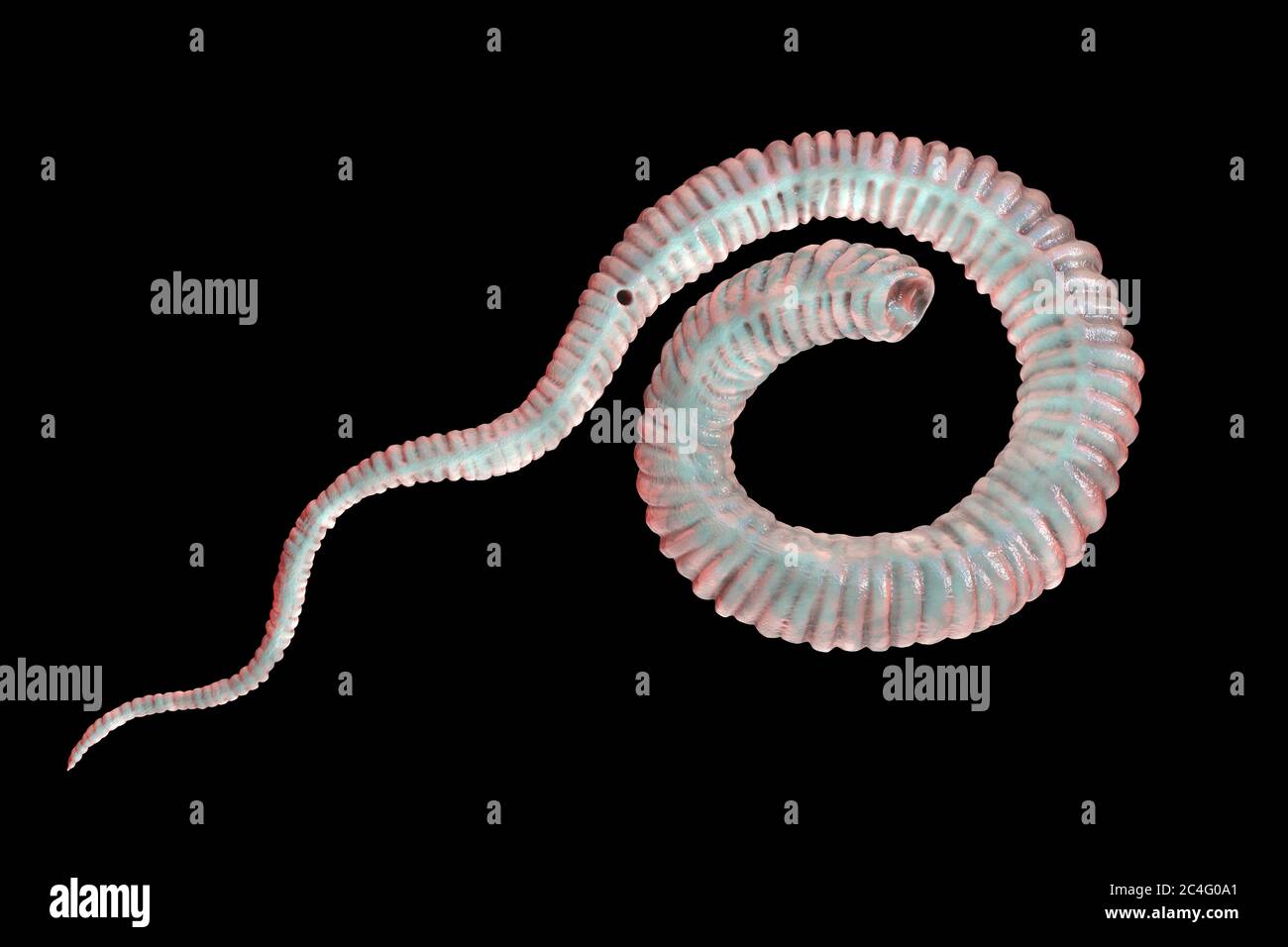 Guinea worm (Dracunculus medinensis) first-stage larva, computer illustration. Larvae are excreted from female worm parasitising under the skin of human extremities in patient's with dracunculiasis. Stock Photohttps://www.alamy.com/image-license-details/?v=1https://www.alamy.com/guinea-worm-dracunculus-medinensis-first-stage-larva-computer-illustration-larvae-are-excreted-from-female-worm-parasitising-under-the-skin-of-human-extremities-in-patients-with-dracunculiasis-image364227865.html
RF2C4G0A1–Guinea worm (Dracunculus medinensis) first-stage larva, computer illustration. Larvae are excreted from female worm parasitising under the skin of human extremities in patient's with dracunculiasis.
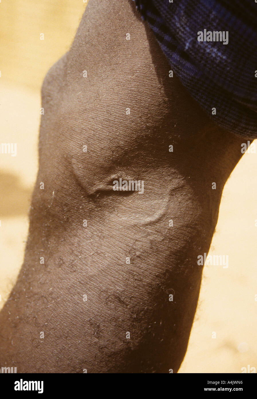 Guinea Worm Under Skin Behind Knee, Near Bankilare, Niger, West Africa. Stock Photohttps://www.alamy.com/image-license-details/?v=1https://www.alamy.com/guinea-worm-under-skin-behind-knee-near-bankilare-niger-west-africa-image6323477.html
RMA4JWN6–Guinea Worm Under Skin Behind Knee, Near Bankilare, Niger, West Africa.
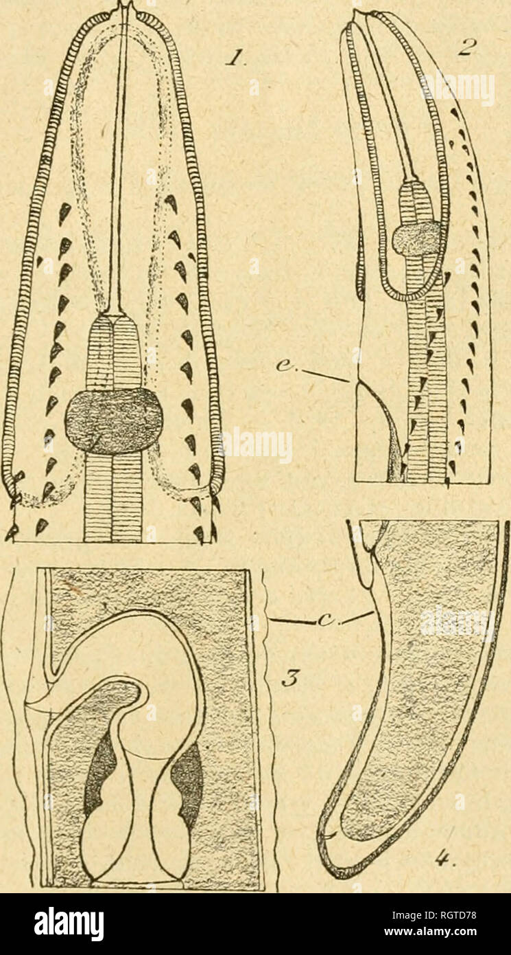 . Bulletin biologique de la France et de la Belgique. Biology; Natural history. FORMES Larvaires des nématodës parasites 371 Dictyocaulu-s Raillikt et Hk.nry (1907). Dictyocaulus viviparus lÎLOGH, 1782. Synon. Strongylus micrurus Mehlis, 1831. -- Lu prétendue larve de ce Strongle figurée par Cobbold (1886) est un Rhabditis libre : Strôse, 1891. Di'acunculus Kniphoff (1759). Dracunculus medinensis (Velsch, 1674). — Larves dans les Cyclops des oasis tunisiennes : Chatton, 1918.. Kig. 1I. - Echinuria pho'iiicopteri Seuhat. ^ Larve du 4' slade, p* la mue. 1 et 2, exirémité antérieure vue par Li l Stock Photohttps://www.alamy.com/image-license-details/?v=1https://www.alamy.com/bulletin-biologique-de-la-france-et-de-la-belgique-biology-natural-history-formes-larvaires-des-nmatods-parasites-371-dictyocaulu-s-raillikt-et-hknry-1907-dictyocaulus-viviparus-llogh-1782-synon-strongylus-micrurus-mehlis-1831-lu-prtendue-larve-de-ce-strongle-figure-par-cobbold-1886-est-un-rhabditis-libre-strse-1891-diacunculus-kniphoff-1759-dracunculus-medinensis-velsch-1674-larves-dans-les-cyclops-des-oasis-tunisiennes-chatton-1918-kig-1i-echinuria-phoiiicopteri-seuhat-larve-du-4-slade-p-la-mue-1-et-2-exirmit-antrieure-vue-par-li-l-image234106524.html
RMRGTD78–. Bulletin biologique de la France et de la Belgique. Biology; Natural history. FORMES Larvaires des nématodës parasites 371 Dictyocaulu-s Raillikt et Hk.nry (1907). Dictyocaulus viviparus lÎLOGH, 1782. Synon. Strongylus micrurus Mehlis, 1831. -- Lu prétendue larve de ce Strongle figurée par Cobbold (1886) est un Rhabditis libre : Strôse, 1891. Di'acunculus Kniphoff (1759). Dracunculus medinensis (Velsch, 1674). — Larves dans les Cyclops des oasis tunisiennes : Chatton, 1918.. Kig. 1I. - Echinuria pho'iiicopteri Seuhat. ^ Larve du 4' slade, p* la mue. 1 et 2, exirémité antérieure vue par Li l
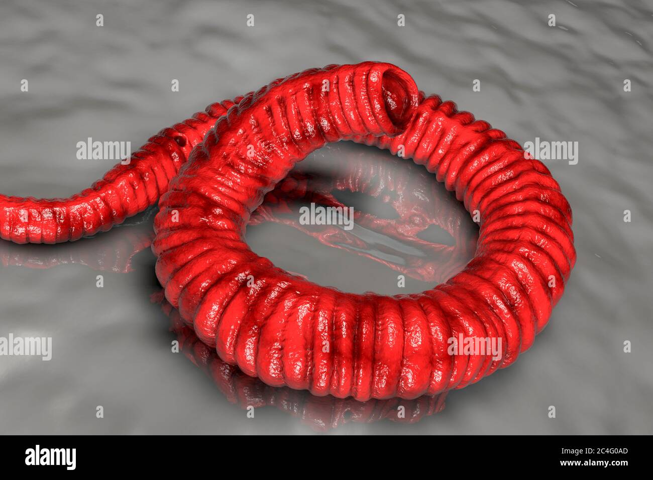 Guinea worm (Dracunculus medinensis) first-stage larva, computer illustration. Larvae are excreted from female worm parasitising under the skin of human extremities in patient's with dracunculiasis. Stock Photohttps://www.alamy.com/image-license-details/?v=1https://www.alamy.com/guinea-worm-dracunculus-medinensis-first-stage-larva-computer-illustration-larvae-are-excreted-from-female-worm-parasitising-under-the-skin-of-human-extremities-in-patients-with-dracunculiasis-image364227877.html
RF2C4G0AD–Guinea worm (Dracunculus medinensis) first-stage larva, computer illustration. Larvae are excreted from female worm parasitising under the skin of human extremities in patient's with dracunculiasis.
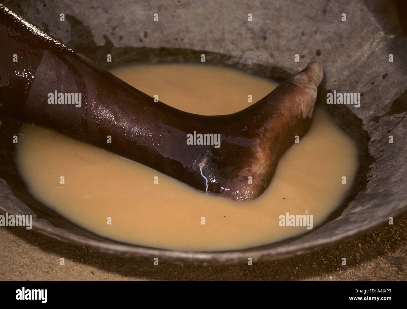 Guinea Worm Emerging. Stock Photohttps://www.alamy.com/image-license-details/?v=1https://www.alamy.com/guinea-worm-emerging-image6323682.html
RMA4JXP3–Guinea Worm Emerging.
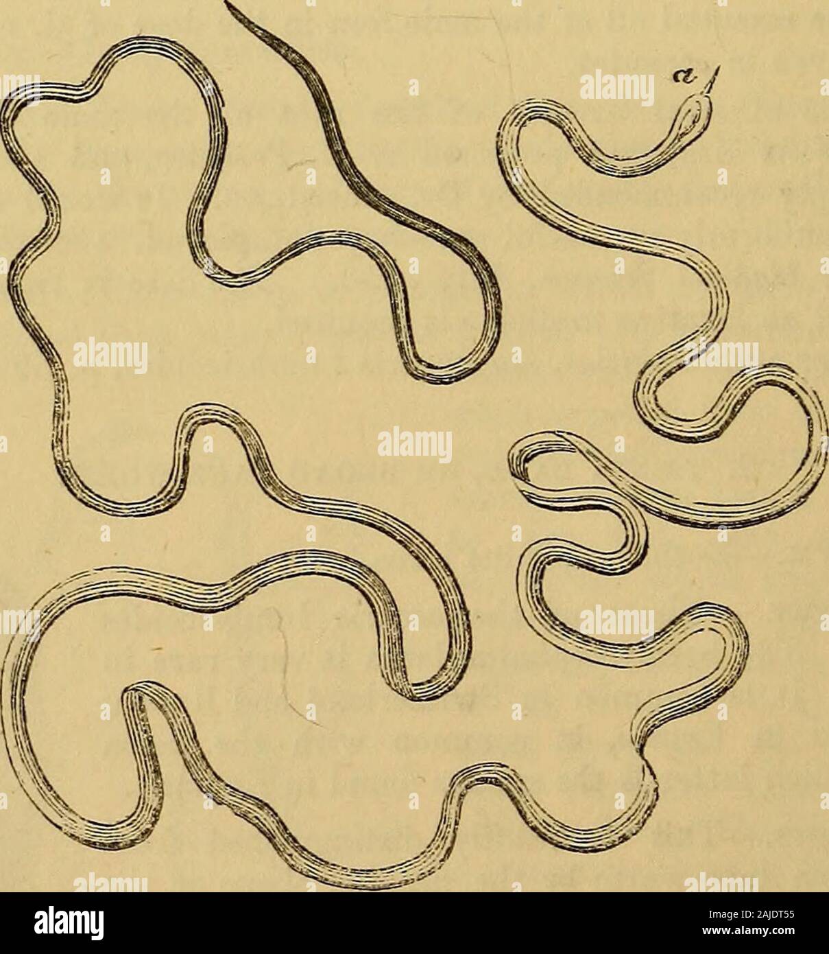 Hooper's physician's vade mecum, or, A manual of the principles and practice of physic . 2 Q 2 596 FILARIA MEDINENSIS. FILARIA MEDINENSIS—GUINEA-WORM. Synonyms.—Dracunculus. Hair-worm. Symptoms.—An itching sensation is first felt in the skin of somepart of the upper or lower extremities ; most frequently in the lowerextremities, and especially in the feet. This itching sensation is soonfollowed by the appearance of a small vesicle, succeeded by an indolentinflamed swelling like a boil, which breaks and discharges its contents,revealing the head of the worm, which gradually protrudes throughthe Stock Photohttps://www.alamy.com/image-license-details/?v=1https://www.alamy.com/hoopers-physicians-vade-mecum-or-a-manual-of-the-principles-and-practice-of-physic-2-q-2-596-filaria-medinensis-filaria-medinensisguinea-worm-synonymsdracunculus-hair-worm-symptomsan-itching-sensation-is-first-felt-in-the-skin-of-somepart-of-the-upper-or-lower-extremities-most-frequently-in-the-lowerextremities-and-especially-in-the-feet-this-itching-sensation-is-soonfollowed-by-the-appearance-of-a-small-vesicle-succeeded-by-an-indolentinflamed-swelling-like-a-boil-which-breaks-and-discharges-its-contentsrevealing-the-head-of-the-worm-which-gradually-protrudes-throughthe-image338365137.html
RM2AJDT55–Hooper's physician's vade mecum, or, A manual of the principles and practice of physic . 2 Q 2 596 FILARIA MEDINENSIS. FILARIA MEDINENSIS—GUINEA-WORM. Synonyms.—Dracunculus. Hair-worm. Symptoms.—An itching sensation is first felt in the skin of somepart of the upper or lower extremities ; most frequently in the lowerextremities, and especially in the feet. This itching sensation is soonfollowed by the appearance of a small vesicle, succeeded by an indolentinflamed swelling like a boil, which breaks and discharges its contents,revealing the head of the worm, which gradually protrudes throughthe
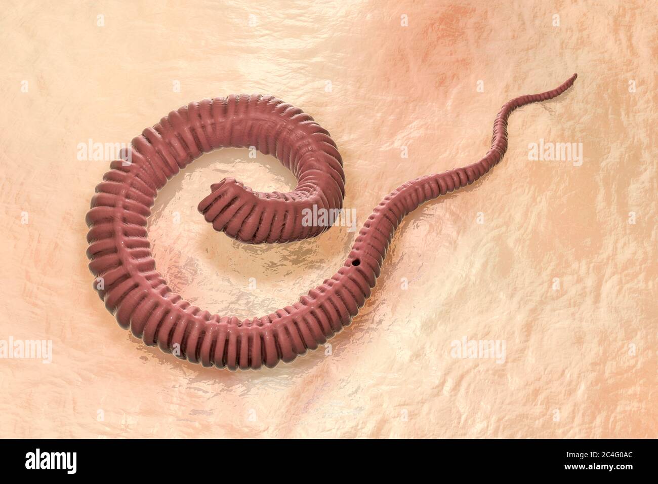 Guinea worm (Dracunculus medinensis) first-stage larva, computer illustration. Larvae are excreted from female worm parasitising under the skin of human extremities in patient's with dracunculiasis. Stock Photohttps://www.alamy.com/image-license-details/?v=1https://www.alamy.com/guinea-worm-dracunculus-medinensis-first-stage-larva-computer-illustration-larvae-are-excreted-from-female-worm-parasitising-under-the-skin-of-human-extremities-in-patients-with-dracunculiasis-image364227876.html
RF2C4G0AC–Guinea worm (Dracunculus medinensis) first-stage larva, computer illustration. Larvae are excreted from female worm parasitising under the skin of human extremities in patient's with dracunculiasis.
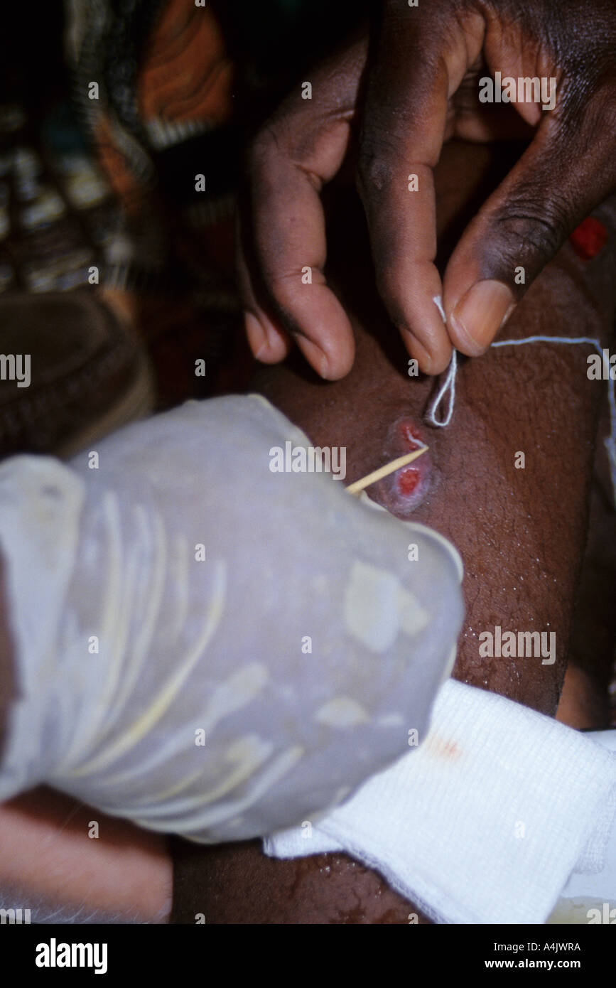 Looping Thread Around Guinea Worm Emerging from Leg, Near Tera, Niger, West Africa. Stock Photohttps://www.alamy.com/image-license-details/?v=1https://www.alamy.com/looping-thread-around-guinea-worm-emerging-from-leg-near-tera-niger-image6323513.html
RMA4JWRA–Looping Thread Around Guinea Worm Emerging from Leg, Near Tera, Niger, West Africa.
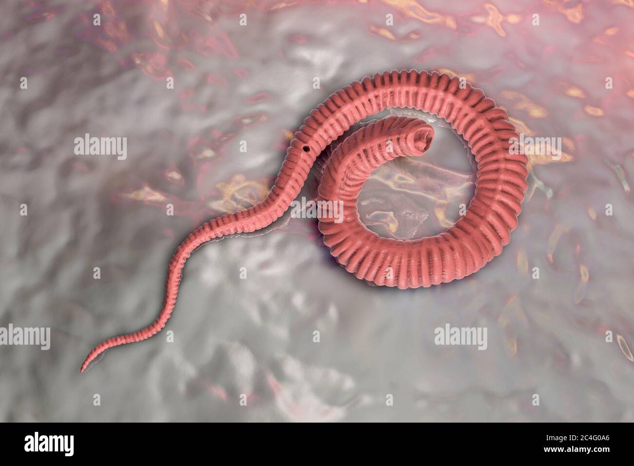 Guinea worm (Dracunculus medinensis) first-stage larva, computer illustration. Larvae are excreted from female worm parasitising under the skin of human extremities in patient's with dracunculiasis. Stock Photohttps://www.alamy.com/image-license-details/?v=1https://www.alamy.com/guinea-worm-dracunculus-medinensis-first-stage-larva-computer-illustration-larvae-are-excreted-from-female-worm-parasitising-under-the-skin-of-human-extremities-in-patients-with-dracunculiasis-image364227870.html
RF2C4G0A6–Guinea worm (Dracunculus medinensis) first-stage larva, computer illustration. Larvae are excreted from female worm parasitising under the skin of human extremities in patient's with dracunculiasis.
 Guinea Worm Emerging. Near Zinder, Niger. Stock Photohttps://www.alamy.com/image-license-details/?v=1https://www.alamy.com/guinea-worm-emerging-near-zinder-niger-image6323639.html
RMA4JXK8–Guinea Worm Emerging. Near Zinder, Niger.
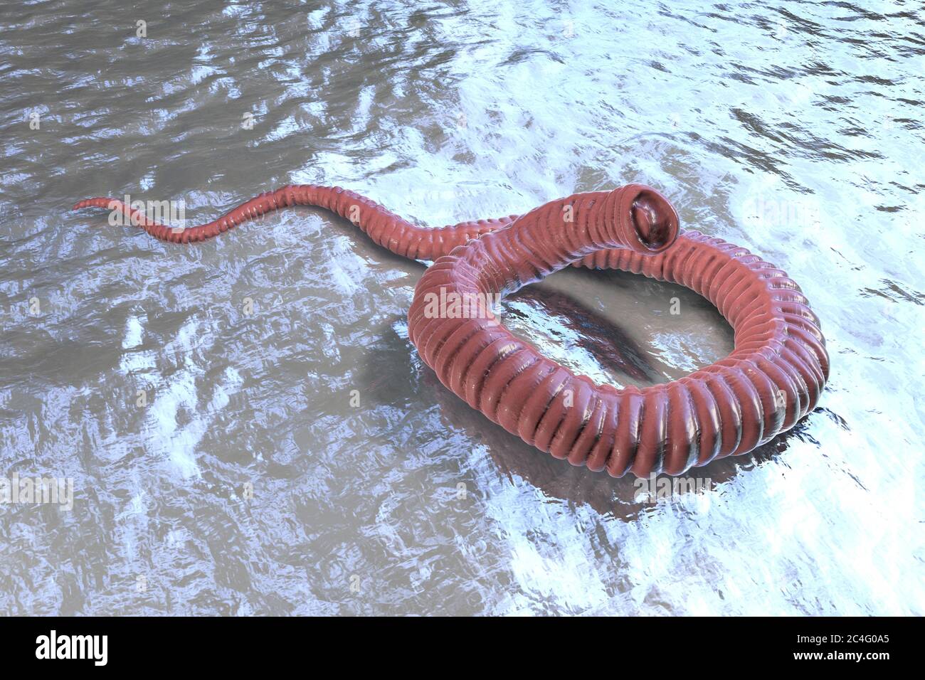 Guinea worm (Dracunculus medinensis) first-stage larva, computer illustration. Larvae are excreted from female worm parasitising under the skin of human extremities in patient's with dracunculiasis. Stock Photohttps://www.alamy.com/image-license-details/?v=1https://www.alamy.com/guinea-worm-dracunculus-medinensis-first-stage-larva-computer-illustration-larvae-are-excreted-from-female-worm-parasitising-under-the-skin-of-human-extremities-in-patients-with-dracunculiasis-image364227869.html
RF2C4G0A5–Guinea worm (Dracunculus medinensis) first-stage larva, computer illustration. Larvae are excreted from female worm parasitising under the skin of human extremities in patient's with dracunculiasis.
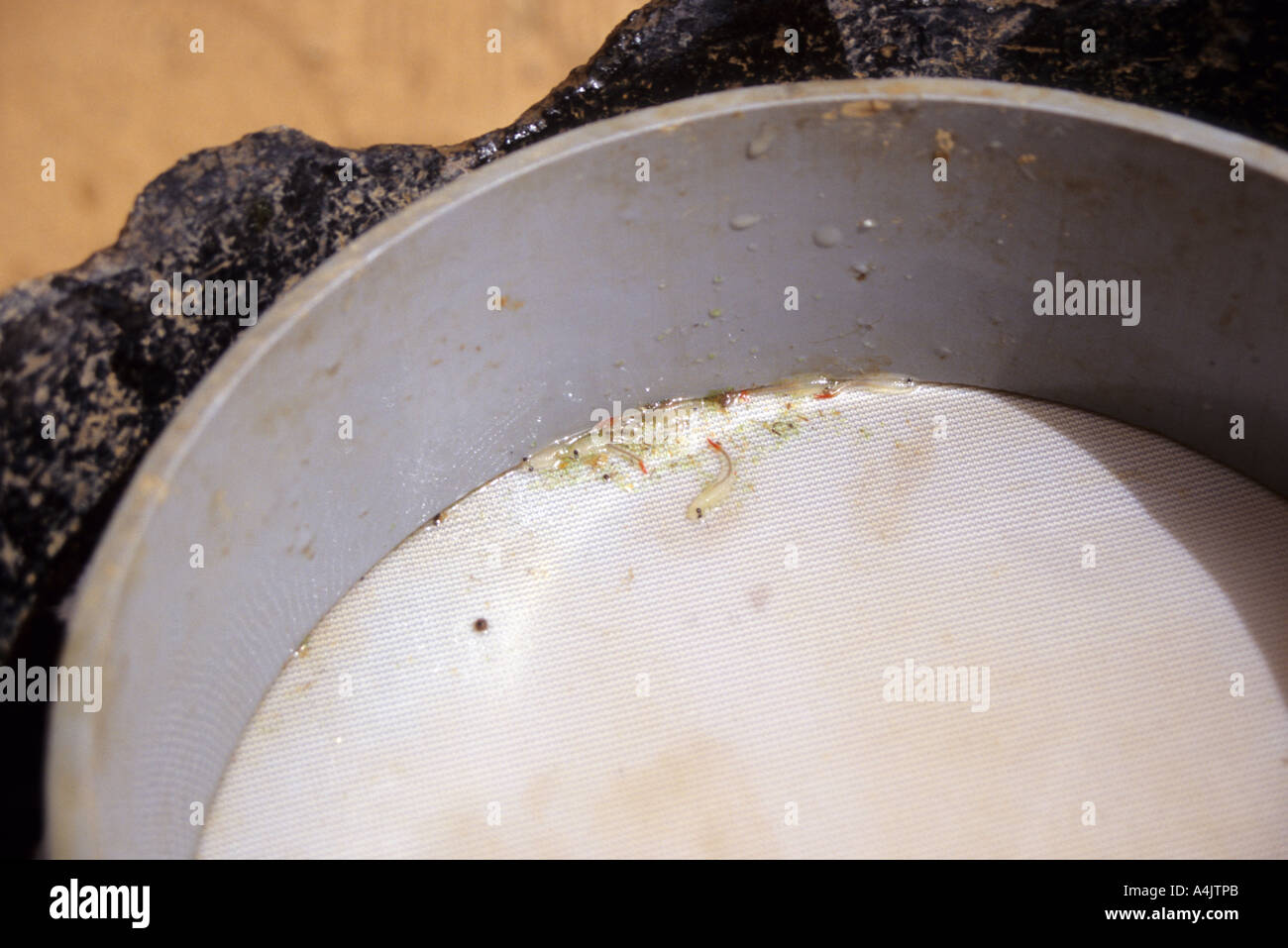 Residue in Water Filter, Niger Stock Photohttps://www.alamy.com/image-license-details/?v=1https://www.alamy.com/residue-in-water-filter-niger-image6323306.html
RMA4JTPB–Residue in Water Filter, Niger
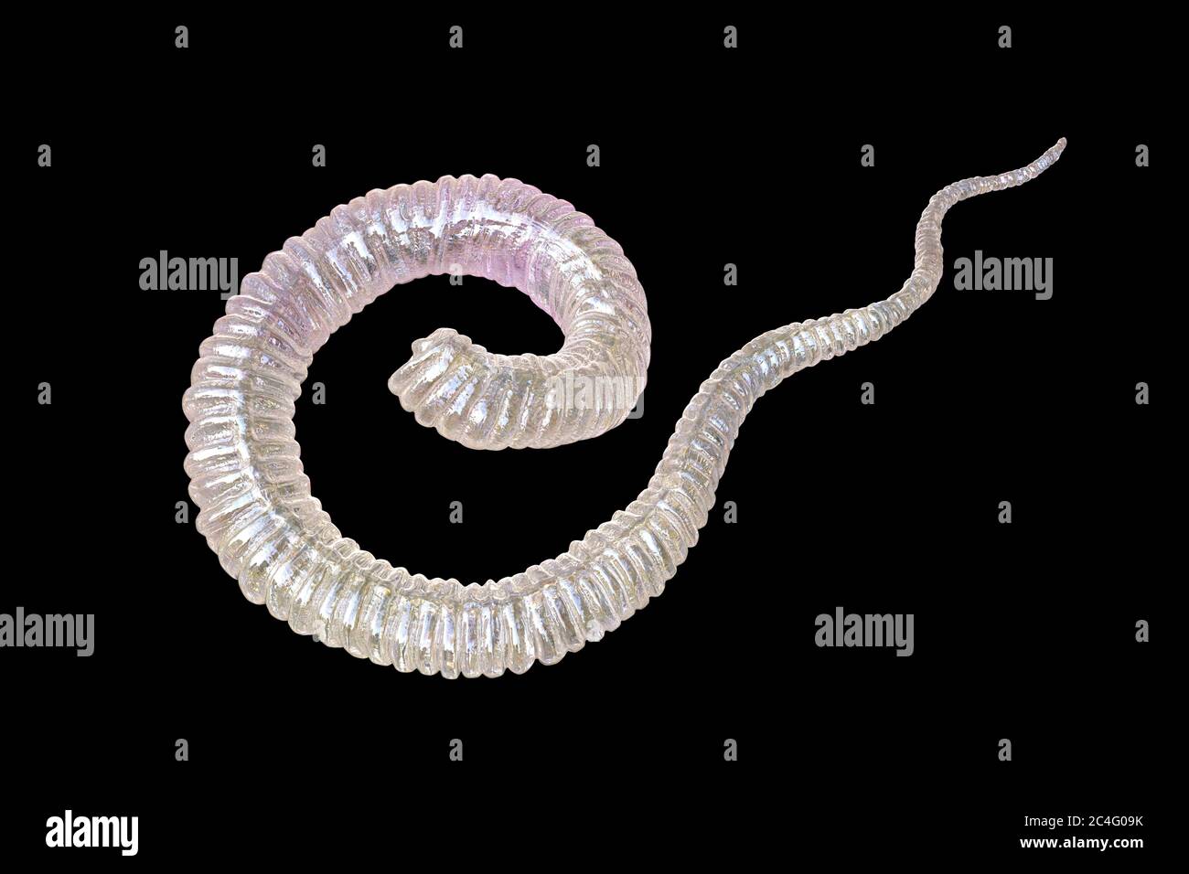 Guinea worm (Dracunculus medinensis) first-stage larva, computer illustration. Larvae are excreted from female worm parasitising under the skin of human extremities in patient's with dracunculiasis. Stock Photohttps://www.alamy.com/image-license-details/?v=1https://www.alamy.com/guinea-worm-dracunculus-medinensis-first-stage-larva-computer-illustration-larvae-are-excreted-from-female-worm-parasitising-under-the-skin-of-human-extremities-in-patients-with-dracunculiasis-image364227855.html
RF2C4G09K–Guinea worm (Dracunculus medinensis) first-stage larva, computer illustration. Larvae are excreted from female worm parasitising under the skin of human extremities in patient's with dracunculiasis.
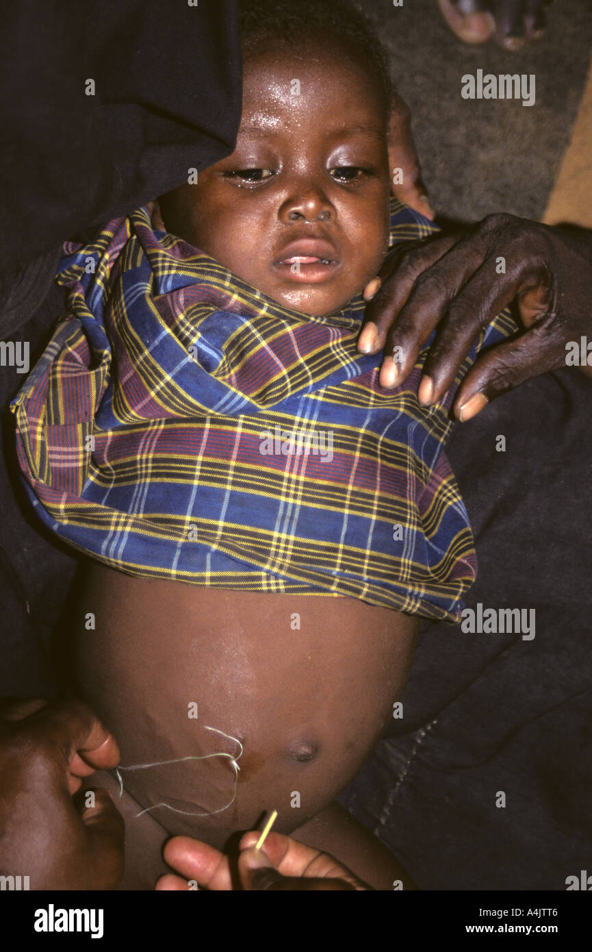 Doctors Tying Thread Around Emerging Guinea Worm, Niger. Stock Photohttps://www.alamy.com/image-license-details/?v=1https://www.alamy.com/doctors-tying-thread-around-emerging-guinea-worm-niger-image6323333.html
RMA4JTT6–Doctors Tying Thread Around Emerging Guinea Worm, Niger.
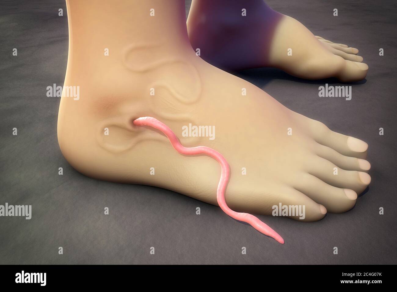 Illustration of a guinea worm (Dracunculus medinensis) emerging from an infected foot. The white thread-like worm is seen at centre; the female worm i Stock Photohttps://www.alamy.com/image-license-details/?v=1https://www.alamy.com/illustration-of-a-guinea-worm-dracunculus-medinensis-emerging-from-an-infected-foot-the-white-thread-like-worm-is-seen-at-centre-the-female-worm-i-image364227799.html
RF2C4G07K–Illustration of a guinea worm (Dracunculus medinensis) emerging from an infected foot. The white thread-like worm is seen at centre; the female worm i
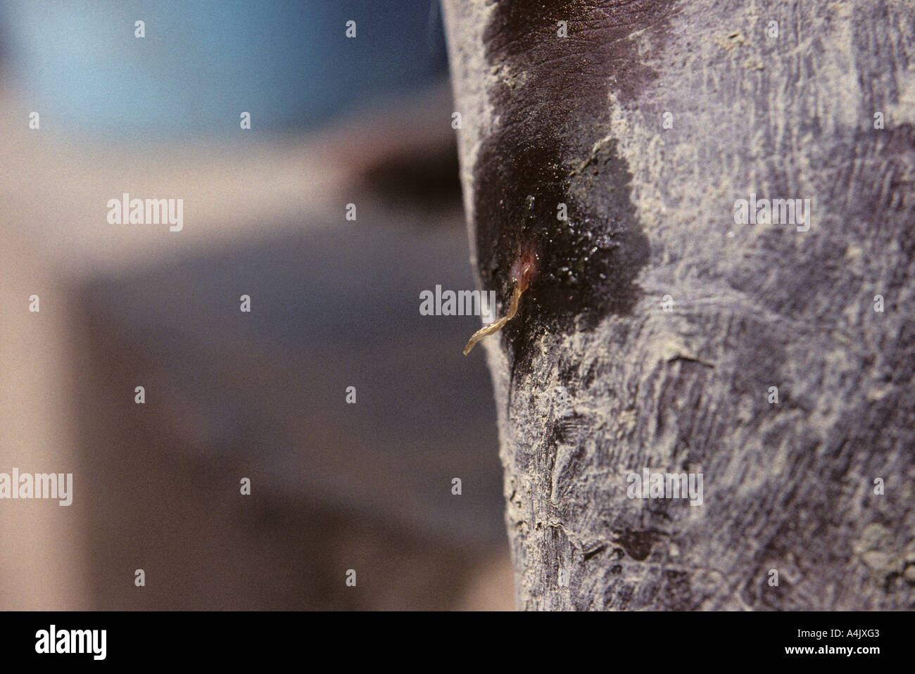 Guinea Worm Emerging. Ivory Coast, Cote d'Ivoire. Stock Photohttps://www.alamy.com/image-license-details/?v=1https://www.alamy.com/guinea-worm-emerging-ivory-coast-cote-divoire-image6323586.html
RMA4JXG3–Guinea Worm Emerging. Ivory Coast, Cote d'Ivoire.
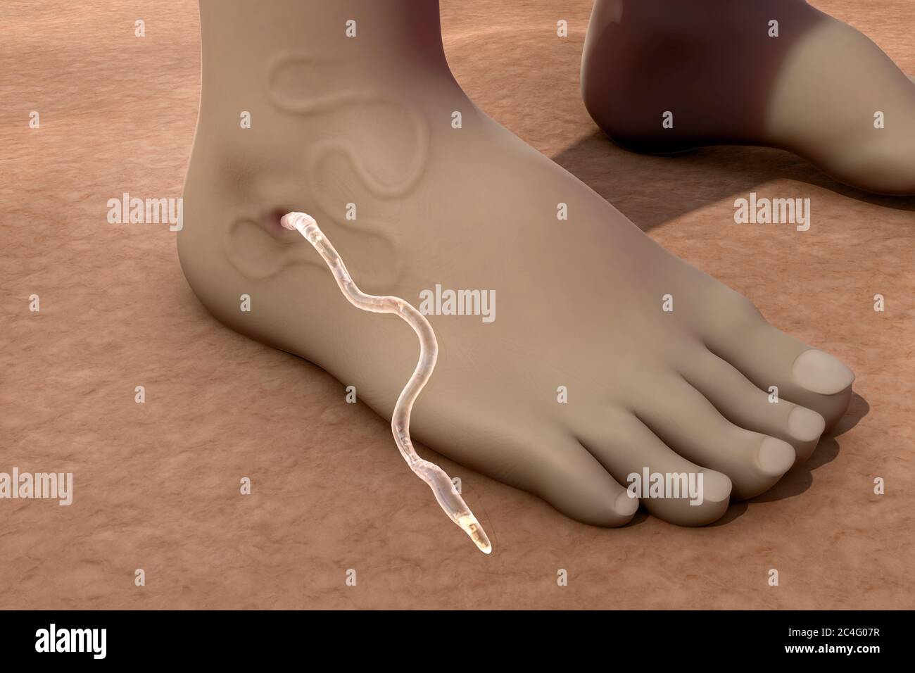 Illustration of a guinea worm (Dracunculus medinensis) emerging from an infected foot. The white thread-like worm is seen at centre; the female worm i Stock Photohttps://www.alamy.com/image-license-details/?v=1https://www.alamy.com/illustration-of-a-guinea-worm-dracunculus-medinensis-emerging-from-an-infected-foot-the-white-thread-like-worm-is-seen-at-centre-the-female-worm-i-image364227803.html
RF2C4G07R–Illustration of a guinea worm (Dracunculus medinensis) emerging from an infected foot. The white thread-like worm is seen at centre; the female worm i
 Guinea Worm Breaking through Skin of Young Victim, Niger. Stock Photohttps://www.alamy.com/image-license-details/?v=1https://www.alamy.com/guinea-worm-breaking-through-skin-of-young-victim-niger-image6323323.html
RMA4JTRC–Guinea Worm Breaking through Skin of Young Victim, Niger.
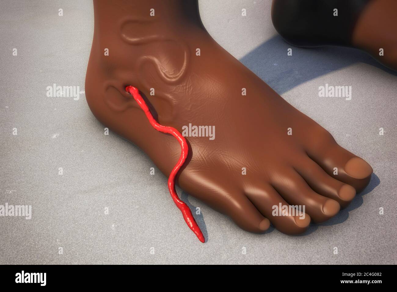 Illustration of a guinea worm (Dracunculus medinensis) emerging from an infected foot. The white thread-like worm is seen at centre; the female worm i Stock Photohttps://www.alamy.com/image-license-details/?v=1https://www.alamy.com/illustration-of-a-guinea-worm-dracunculus-medinensis-emerging-from-an-infected-foot-the-white-thread-like-worm-is-seen-at-centre-the-female-worm-i-image364227810.html
RF2C4G082–Illustration of a guinea worm (Dracunculus medinensis) emerging from an infected foot. The white thread-like worm is seen at centre; the female worm i
 Water Source, Pond, Girl, Cow, Ivory Coast, Cote d'Ivoire. Stock Photohttps://www.alamy.com/image-license-details/?v=1https://www.alamy.com/water-source-pond-girl-cow-ivory-coast-cote-divoire-image6323194.html
RMA4JRYB–Water Source, Pond, Girl, Cow, Ivory Coast, Cote d'Ivoire.
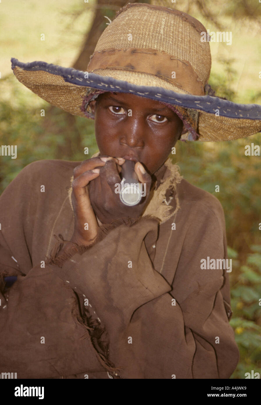 Boy with Drinking Tube Filter to Avoid Ingesting Guinea Worm, Niger. Stock Photohttps://www.alamy.com/image-license-details/?v=1https://www.alamy.com/boy-with-drinking-tube-filter-to-avoid-ingesting-guinea-worm-niger-image6323448.html
RMA4JWK9–Boy with Drinking Tube Filter to Avoid Ingesting Guinea Worm, Niger.
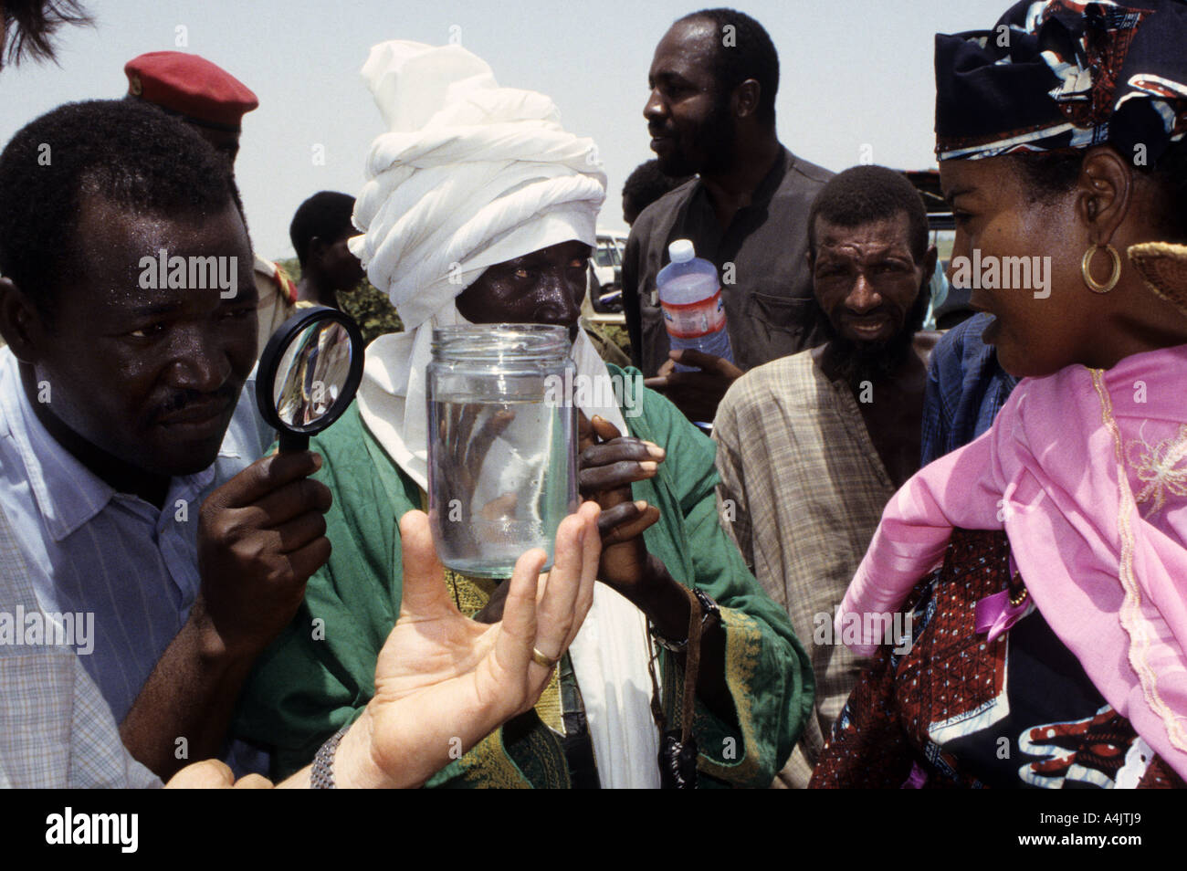 Health Official Examines Water for Guinea Worm, Niger. Stock Photohttps://www.alamy.com/image-license-details/?v=1https://www.alamy.com/health-official-examines-water-for-guinea-worm-niger-image6323240.html
RMA4JTJ9–Health Official Examines Water for Guinea Worm, Niger.
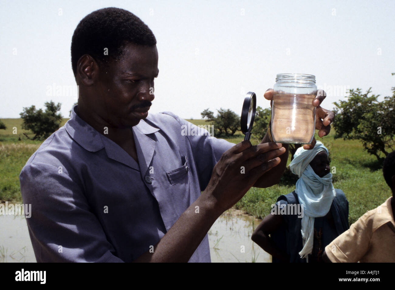 Nigerien Doctor Examines Water for Guinea Worm Transmitters, Niger. Stock Photohttps://www.alamy.com/image-license-details/?v=1https://www.alamy.com/nigerien-doctor-examines-water-for-guinea-worm-transmitters-niger-image6323232.html
RMA4JTJ1–Nigerien Doctor Examines Water for Guinea Worm Transmitters, Niger.
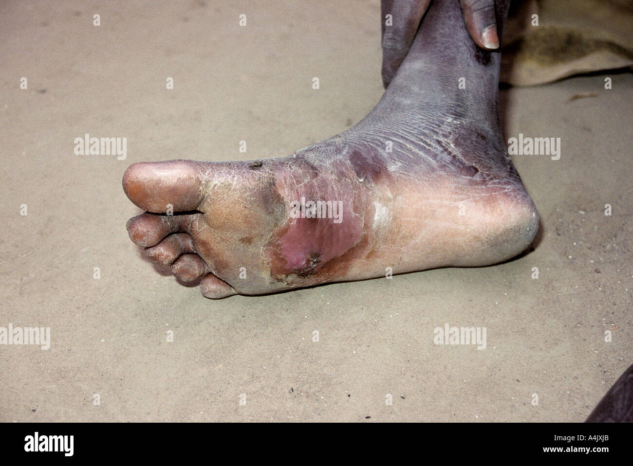 Healing Secondary Infection Where Guinea Worm has Exited. Stock Photohttps://www.alamy.com/image-license-details/?v=1https://www.alamy.com/healing-secondary-infection-where-guinea-worm-has-exited-image6323626.html
RMA4JXJB–Healing Secondary Infection Where Guinea Worm has Exited.
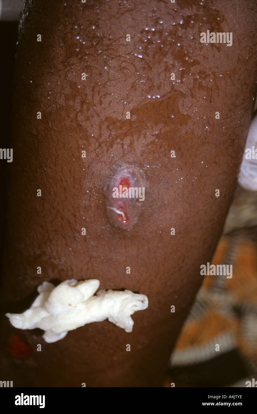 Coaxing Guinea Worm to Emerge, Niger. Stock Photohttps://www.alamy.com/image-license-details/?v=1https://www.alamy.com/coaxing-guinea-worm-to-emerge-niger-image6323389.html
RMA4JTYE–Coaxing Guinea Worm to Emerge, Niger.
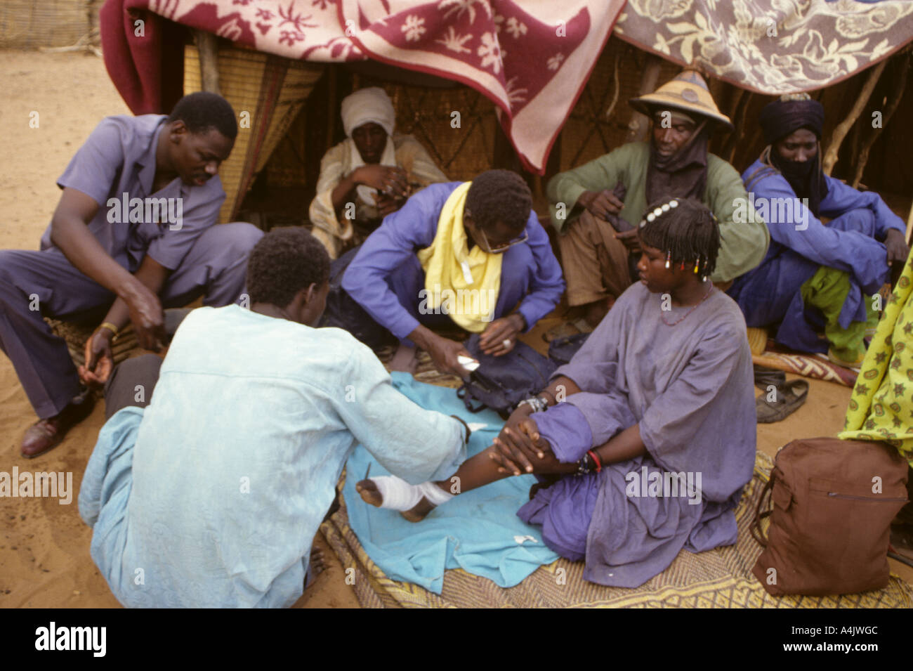 Bankilare, Niger, Africa. Examining Bella woman with Guinea worm. Stock Photohttps://www.alamy.com/image-license-details/?v=1https://www.alamy.com/bankilare-niger-africa-examining-bella-woman-with-guinea-worm-image6323403.html
RMA4JWGC–Bankilare, Niger, Africa. Examining Bella woman with Guinea worm.
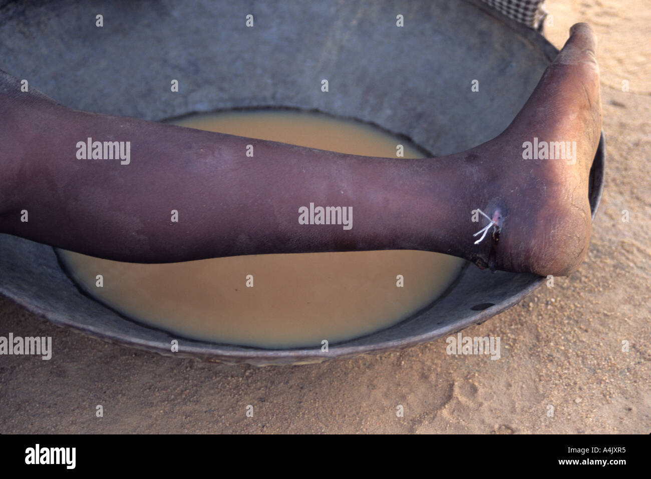 Guinea Worm Emerging. Near Zinder, Niger. Stock Photohttps://www.alamy.com/image-license-details/?v=1https://www.alamy.com/guinea-worm-emerging-near-zinder-niger-image6323700.html
RMA4JXR5–Guinea Worm Emerging. Near Zinder, Niger.
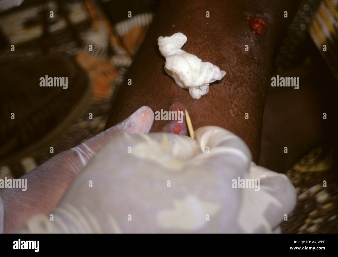 Looping Thread around Guinea Worm Emerging from Leg, Near Tera, Niger, West Africa. Stock Photohttps://www.alamy.com/image-license-details/?v=1https://www.alamy.com/looping-thread-around-guinea-worm-emerging-from-leg-near-tera-niger-image6323501.html
RMA4JWPE–Looping Thread around Guinea Worm Emerging from Leg, Near Tera, Niger, West Africa.
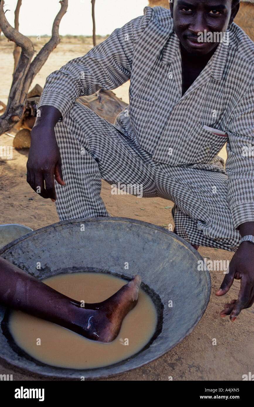 Guinea Worm Emerging. Near Zinder, Niger. Stock Photohttps://www.alamy.com/image-license-details/?v=1https://www.alamy.com/guinea-worm-emerging-near-zinder-niger-image6323668.html
RMA4JXN5–Guinea Worm Emerging. Near Zinder, Niger.
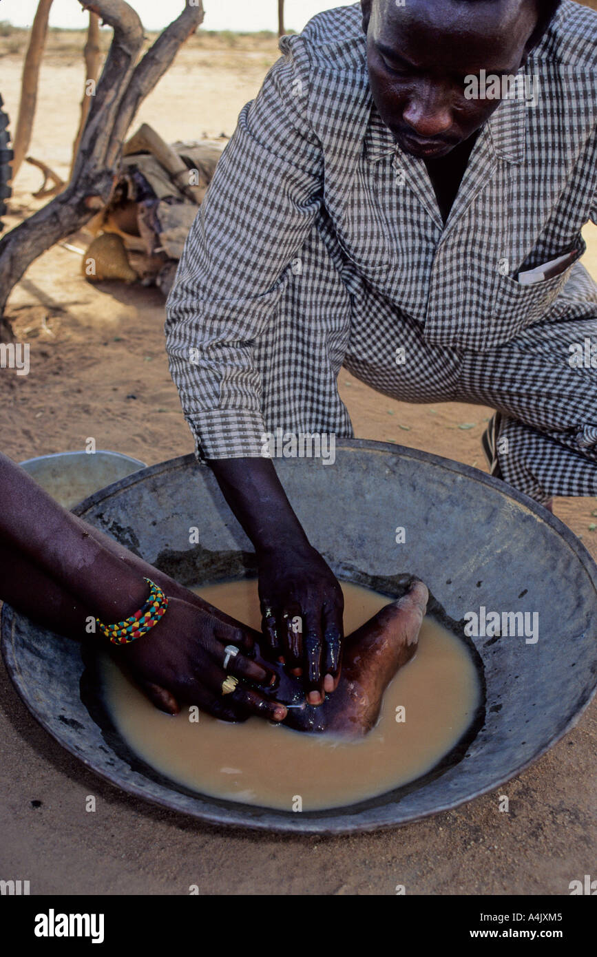 Guinea Worm Emerging. Near Zinder, Niger. Stock Photohttps://www.alamy.com/image-license-details/?v=1https://www.alamy.com/guinea-worm-emerging-near-zinder-niger-image6323652.html
RMA4JXM5–Guinea Worm Emerging. Near Zinder, Niger.
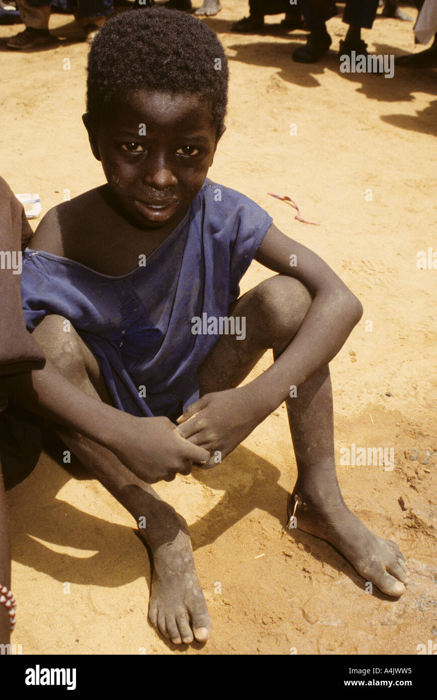 Guinea Worm Emerging from Boy's Ankle, Niger. Stock Photohttps://www.alamy.com/image-license-details/?v=1https://www.alamy.com/guinea-worm-emerging-from-boys-ankle-niger-image6323540.html
RMA4JWW5–Guinea Worm Emerging from Boy's Ankle, Niger.
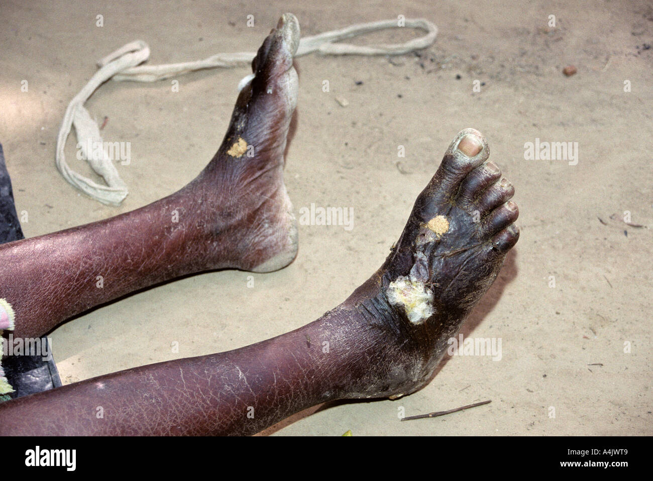 Yorobodi, Ivory Coast, Cote d'Ivoire, Africa. Sores healing after Guinea worm extraction. 208.11 Stock Photohttps://www.alamy.com/image-license-details/?v=1https://www.alamy.com/yorobodi-ivory-coast-cote-divoire-africa-sores-healing-after-guinea-image6323528.html
RMA4JWT9–Yorobodi, Ivory Coast, Cote d'Ivoire, Africa. Sores healing after Guinea worm extraction. 208.11
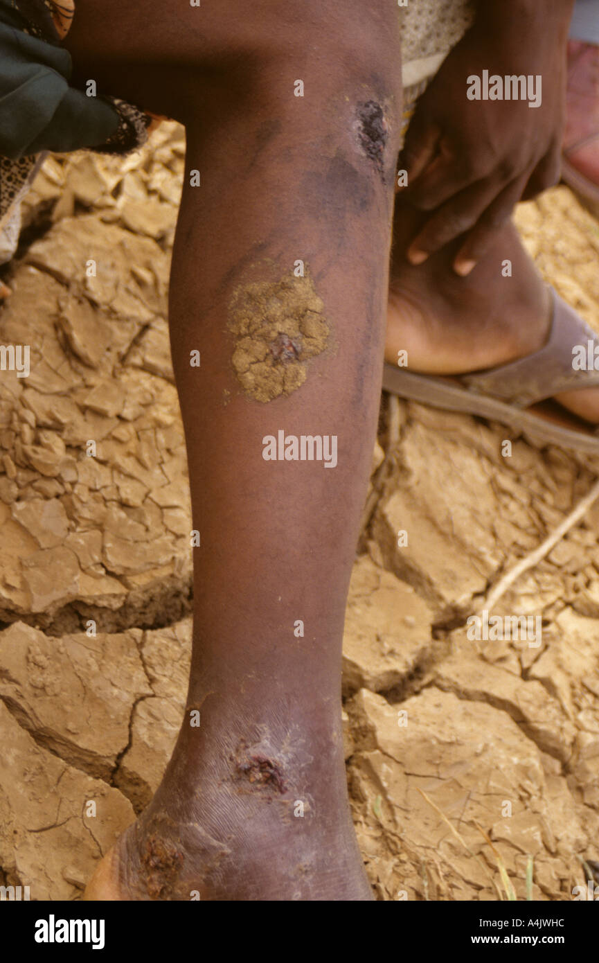 Bankilare, Niger, West Africa. Cow dung covers open sores caused by Guinea worm, Niger. Stock Photohttps://www.alamy.com/image-license-details/?v=1https://www.alamy.com/bankilare-niger-west-africa-cow-dung-covers-open-sores-caused-by-guinea-image6323419.html
RMA4JWHC–Bankilare, Niger, West Africa. Cow dung covers open sores caused by Guinea worm, Niger.
 Filtering Water to Eliminate Cyclopods, Guinea Worm Vectors, Niger Stock Photohttps://www.alamy.com/image-license-details/?v=1https://www.alamy.com/filtering-water-to-eliminate-cyclopods-guinea-worm-vectors-niger-image6323283.html
RMA4JTN4–Filtering Water to Eliminate Cyclopods, Guinea Worm Vectors, Niger
 Woman's Fabric Carries Guinea Worm Eradication Message, Niger. Stock Photohttps://www.alamy.com/image-license-details/?v=1https://www.alamy.com/womans-fabric-carries-guinea-worm-eradication-message-niger-image6323433.html
RMA4JWJA–Woman's Fabric Carries Guinea Worm Eradication Message, Niger.
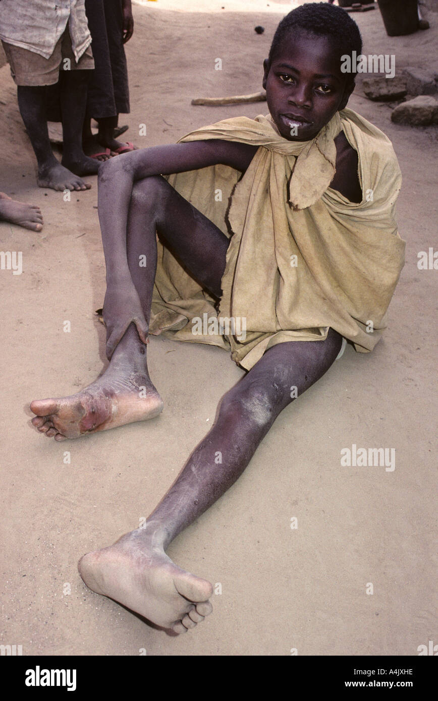 Boy Showing Healing Foot where Guinea Worm has Exited the Foot. Ivory Coast, Cote d'Ivoire. Stock Photohttps://www.alamy.com/image-license-details/?v=1https://www.alamy.com/boy-showing-healing-foot-where-guinea-worm-has-exited-the-foot-ivory-image6323613.html
RMA4JXHE–Boy Showing Healing Foot where Guinea Worm has Exited the Foot. Ivory Coast, Cote d'Ivoire.
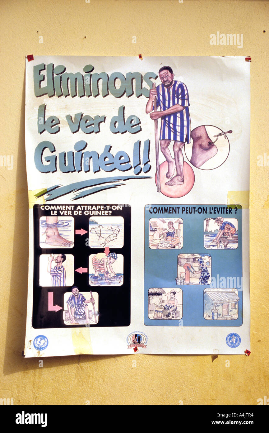 Guinea Worm Education Poster, Ivory Coast, Cote d'Ivoire. Stock Photohttps://www.alamy.com/image-license-details/?v=1https://www.alamy.com/guinea-worm-education-poster-ivory-coast-cote-divoire-image6323315.html
RMA4JTR4–Guinea Worm Education Poster, Ivory Coast, Cote d'Ivoire.
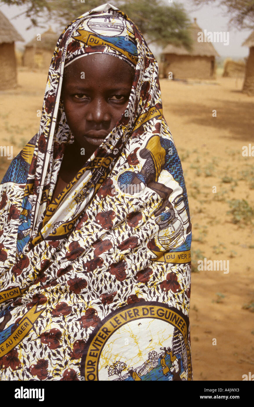 Guinea Worm Eradication Message on Clothing Fabric, Niger. Stock Photohttps://www.alamy.com/image-license-details/?v=1https://www.alamy.com/guinea-worm-eradication-message-on-clothing-fabric-niger-image6323554.html
RMA4JWX3–Guinea Worm Eradication Message on Clothing Fabric, Niger.
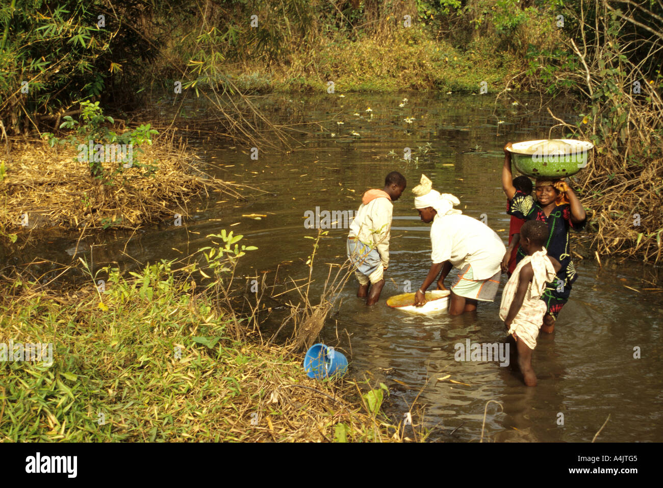 Water Source, Girls Getting Water from Pond, Ivory Coast., Cote d'Ivoire. Stock Photohttps://www.alamy.com/image-license-details/?v=1https://www.alamy.com/water-source-girls-getting-water-from-pond-ivory-coast-cote-divoire-image6323204.html
RMA4JTG5–Water Source, Girls Getting Water from Pond, Ivory Coast., Cote d'Ivoire.
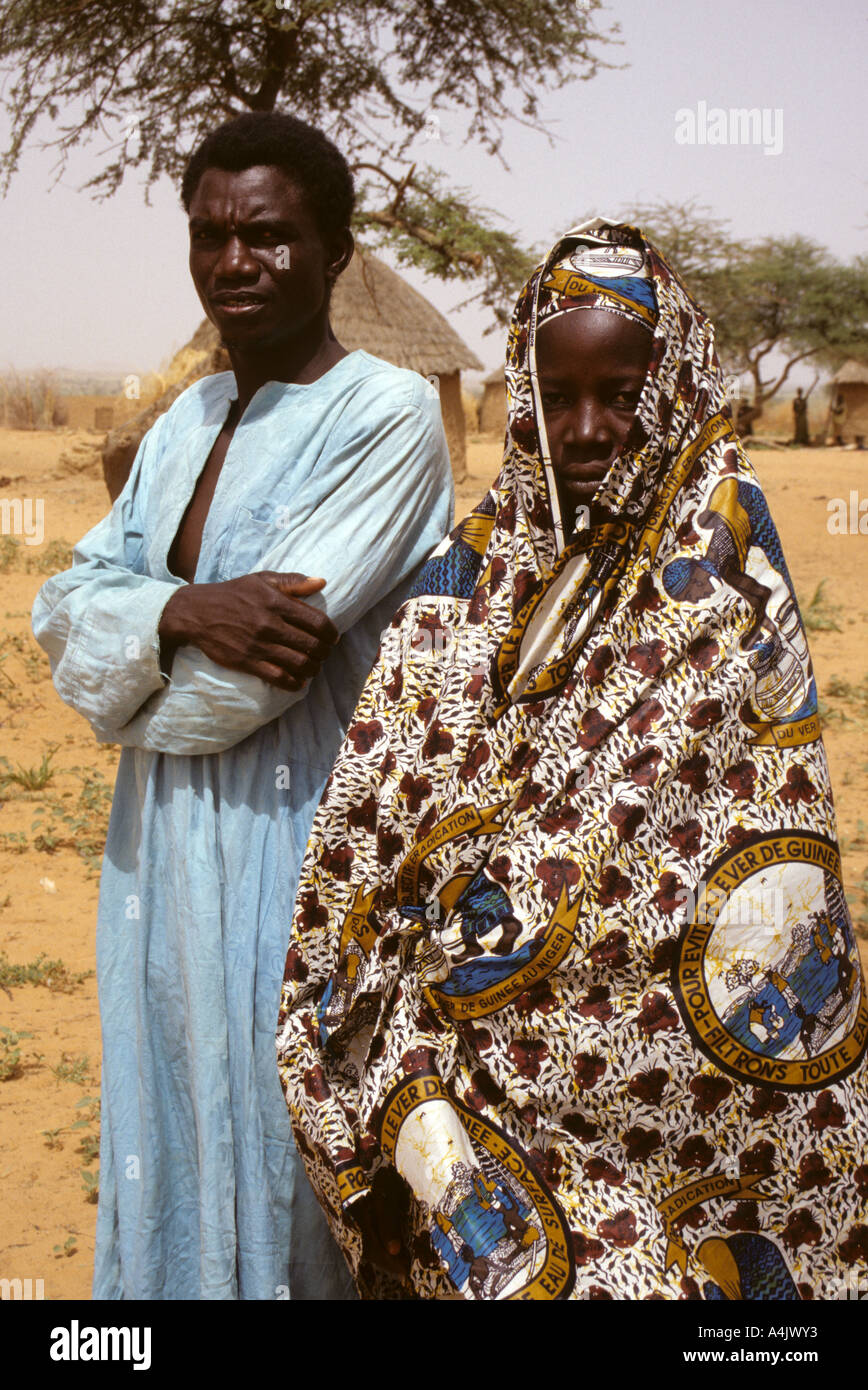 Guinea Worm Eradication Message on Woman's Fabric, Niger. Stock Photohttps://www.alamy.com/image-license-details/?v=1https://www.alamy.com/guinea-worm-eradication-message-on-womans-fabric-niger-image6323570.html
RMA4JWY3–Guinea Worm Eradication Message on Woman's Fabric, Niger.
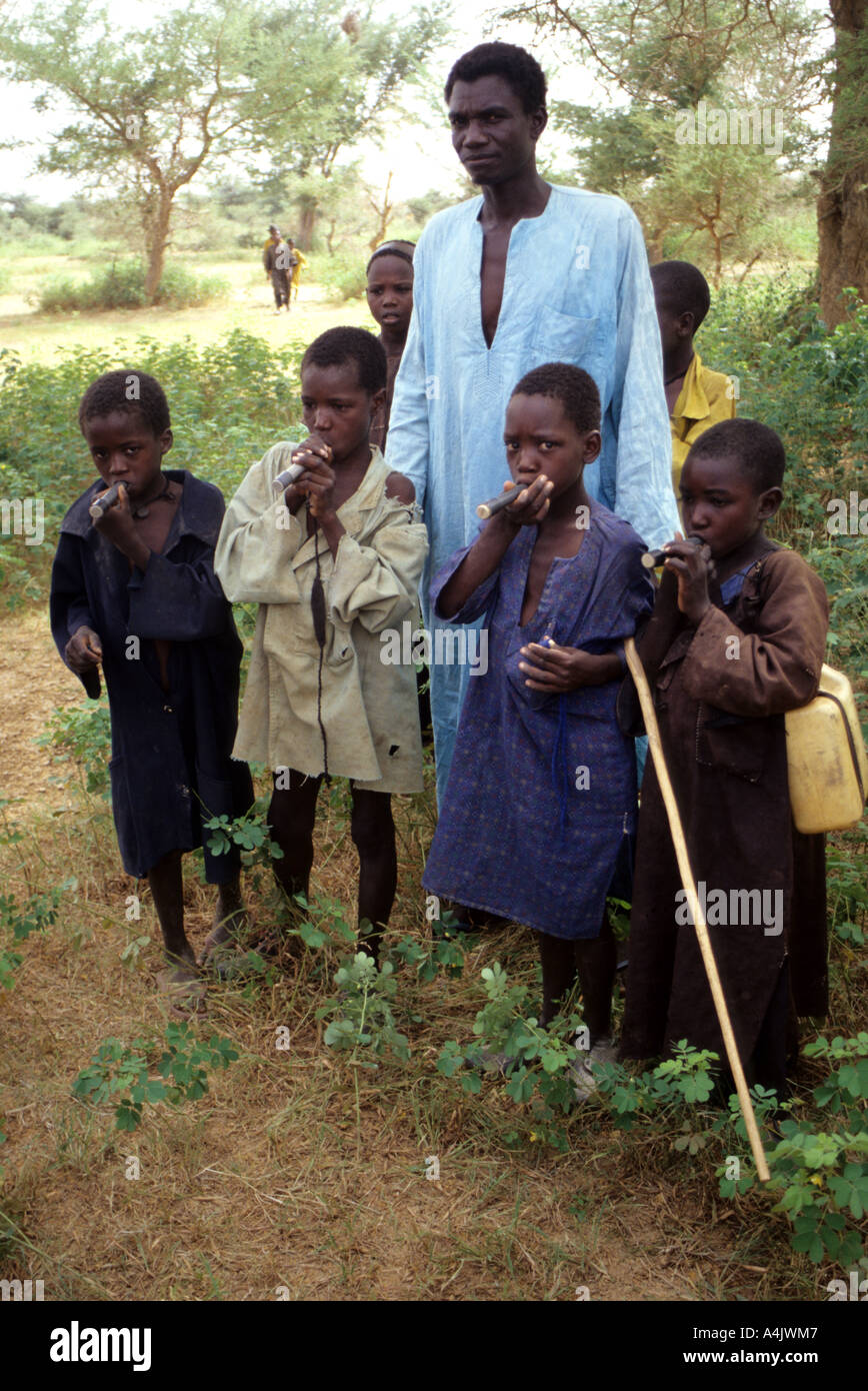 Boys with Drinking Tube Filters to Avoid Ingesting Guinea Worm, Niger. Stock Photohttps://www.alamy.com/image-license-details/?v=1https://www.alamy.com/boys-with-drinking-tube-filters-to-avoid-ingesting-guinea-worm-niger-image6323462.html
RMA4JWM7–Boys with Drinking Tube Filters to Avoid Ingesting Guinea Worm, Niger.
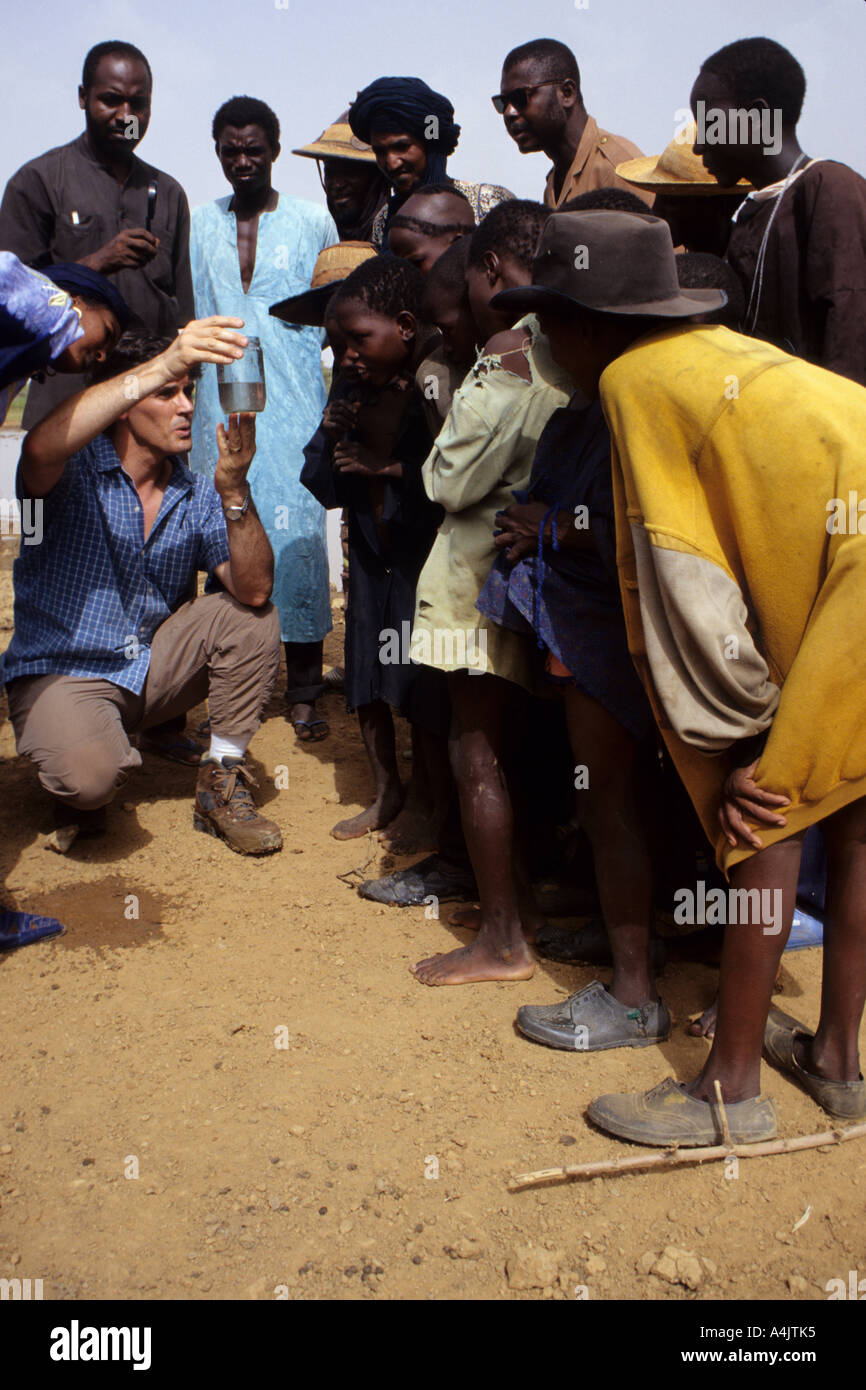 Guinea Worm Eradication Program, Niger. Doctor showing cyclopods in water. Stock Photohttps://www.alamy.com/image-license-details/?v=1https://www.alamy.com/guinea-worm-eradication-program-niger-doctor-showing-cyclopods-in-image6323252.html
RMA4JTK5–Guinea Worm Eradication Program, Niger. Doctor showing cyclopods in water.
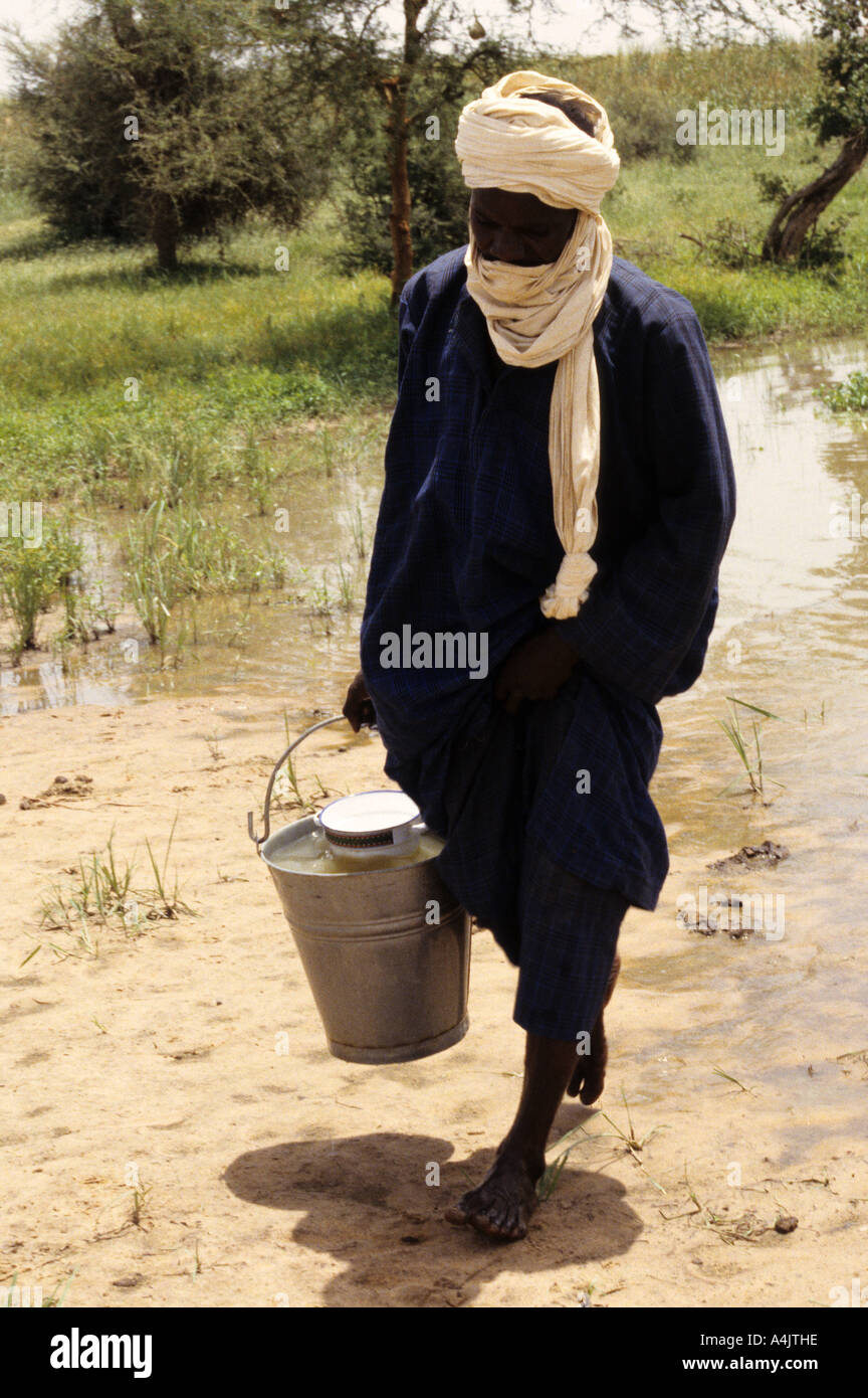 Carrying Water from Guinea Worm-infested Pond, Niger. Stock Photohttps://www.alamy.com/image-license-details/?v=1https://www.alamy.com/carrying-water-from-guinea-worm-infested-pond-niger-image6323229.html
RMA4JTHE–Carrying Water from Guinea Worm-infested Pond, Niger.
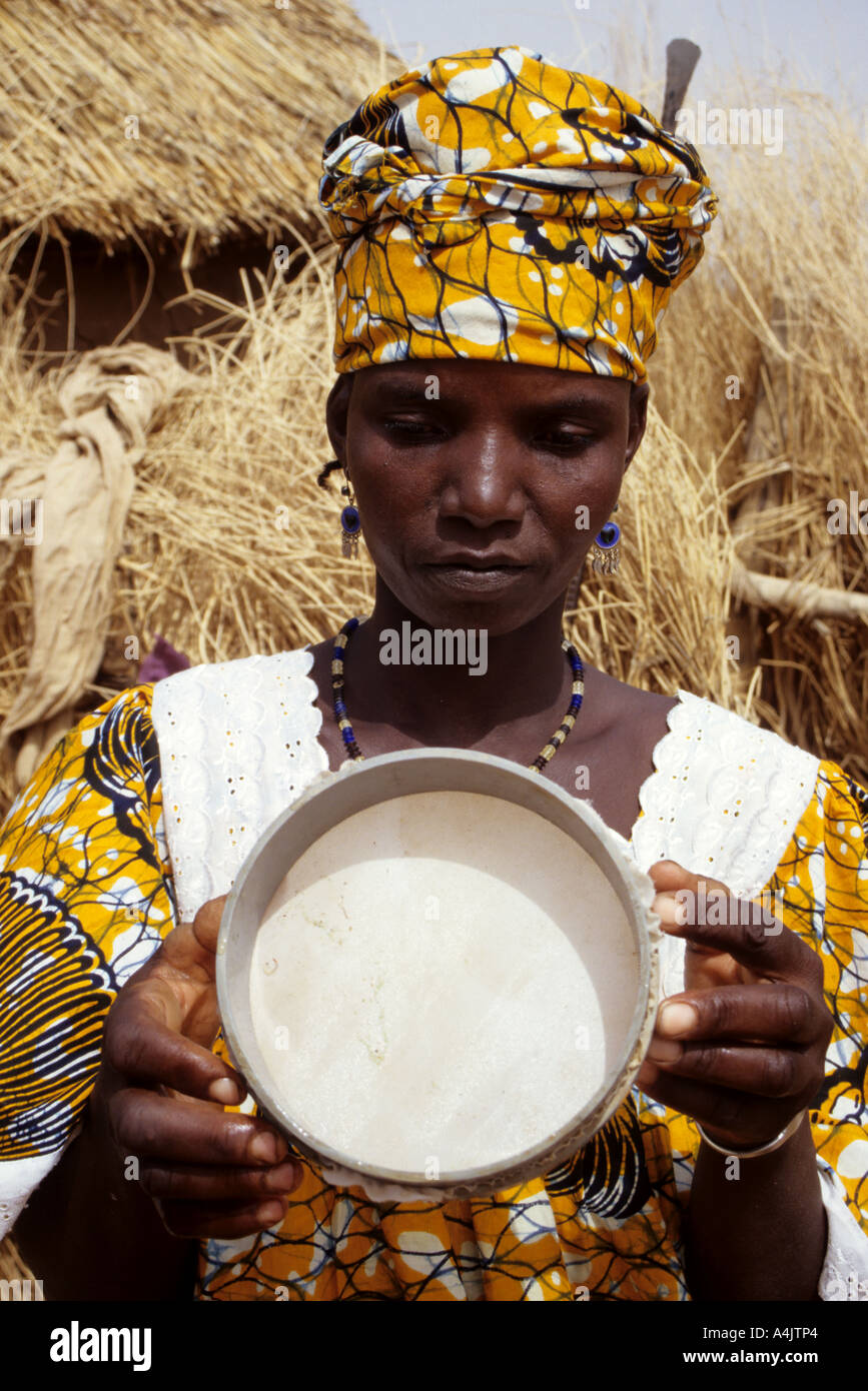 Showing Water Filter, Niger. Stock Photohttps://www.alamy.com/image-license-details/?v=1https://www.alamy.com/showing-water-filter-niger-image6323299.html
RMA4JTP4–Showing Water Filter, Niger.
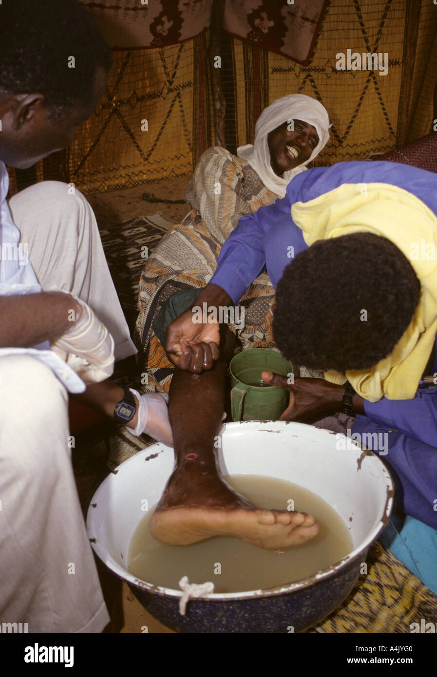 Guinea Worm Extraction, Niger Stock Photohttps://www.alamy.com/image-license-details/?v=1https://www.alamy.com/guinea-worm-extraction-niger-image6323775.html
RMA4JYG0–Guinea Worm Extraction, Niger
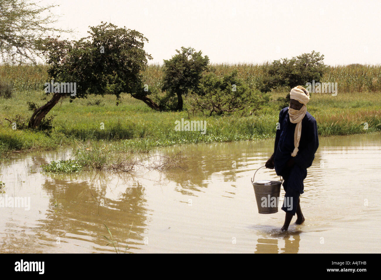 Carrying Water from Guinea Worm-infested Pond, Niger. Stock Photohttps://www.alamy.com/image-license-details/?v=1https://www.alamy.com/carrying-water-from-guinea-worm-infested-pond-niger-image6323226.html
RMA4JTHB–Carrying Water from Guinea Worm-infested Pond, Niger.
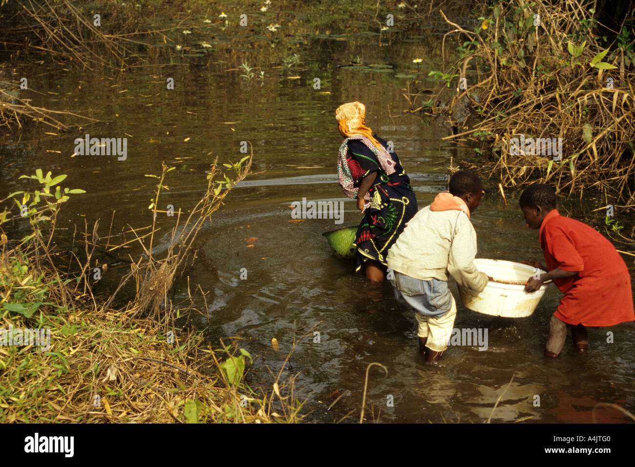 Water Source, Children Getting Water from Pond, Ivory Coast, Cote d'Ivoire. Stock Photohttps://www.alamy.com/image-license-details/?v=1https://www.alamy.com/water-source-children-getting-water-from-pond-ivory-coast-cote-divoire-image6323199.html
RMA4JTG0–Water Source, Children Getting Water from Pond, Ivory Coast, Cote d'Ivoire.
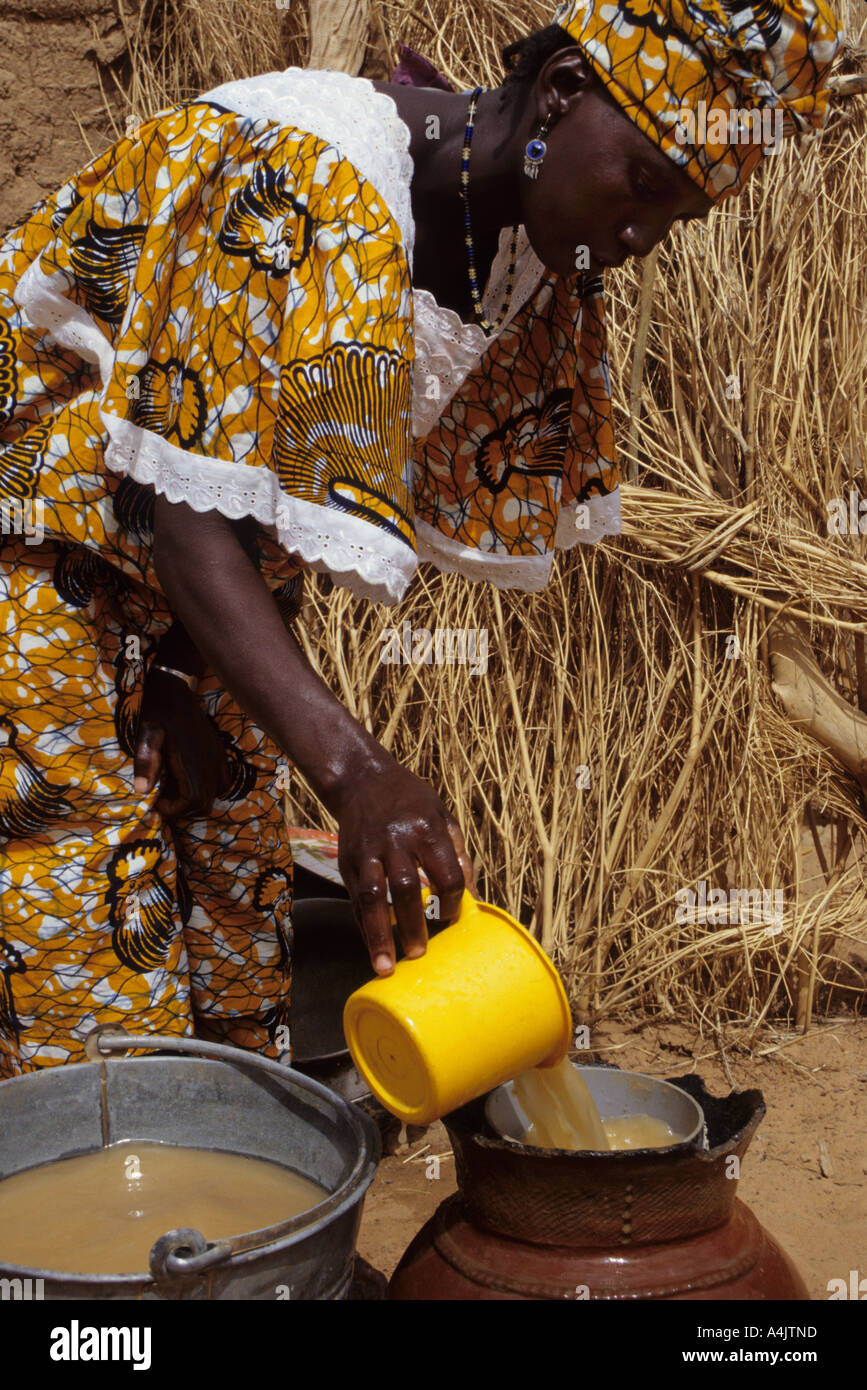 Filtering Water to Eliminate Cyclopods, Niger. Stock Photohttps://www.alamy.com/image-license-details/?v=1https://www.alamy.com/filtering-water-to-eliminate-cyclopods-niger-image6323292.html
RMA4JTND–Filtering Water to Eliminate Cyclopods, Niger.
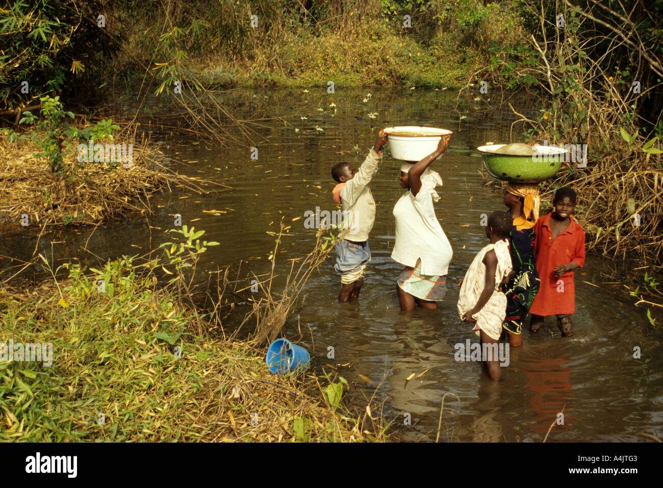 Water Source, Children Getting Water from Pond, Ivory Coast, Cote d'Ivoire. Stock Photohttps://www.alamy.com/image-license-details/?v=1https://www.alamy.com/water-source-children-getting-water-from-pond-ivory-coast-cote-divoire-image6323202.html
RMA4JTG3–Water Source, Children Getting Water from Pond, Ivory Coast, Cote d'Ivoire.
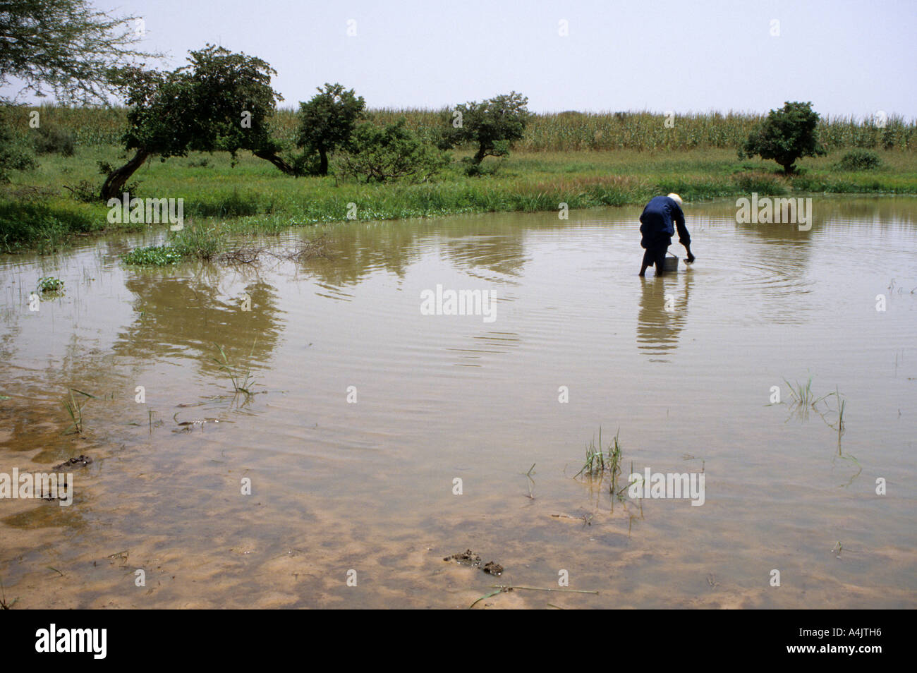 Collecting Water in Guinea Worm-infested Pond, Niger. Stock Photohttps://www.alamy.com/image-license-details/?v=1https://www.alamy.com/collecting-water-in-guinea-worm-infested-pond-niger-image6323221.html
RMA4JTH6–Collecting Water in Guinea Worm-infested Pond, Niger.
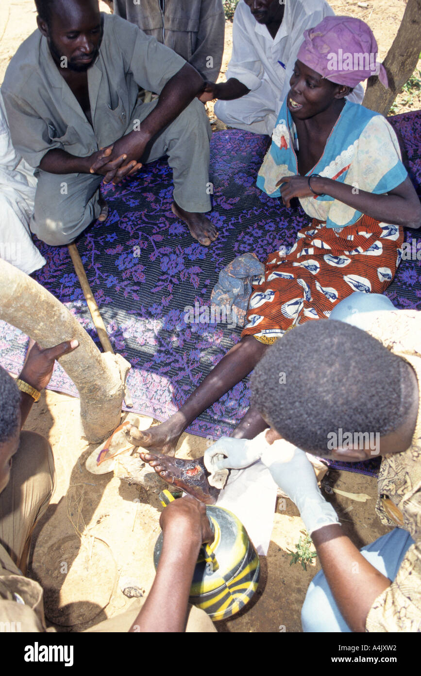 Treating Secondary Infection of Victim of Guinea Worm Infestation, Niger Stock Photohttps://www.alamy.com/image-license-details/?v=1https://www.alamy.com/treating-secondary-infection-of-victim-of-guinea-worm-infestation-image6323729.html
RMA4JXW2–Treating Secondary Infection of Victim of Guinea Worm Infestation, Niger
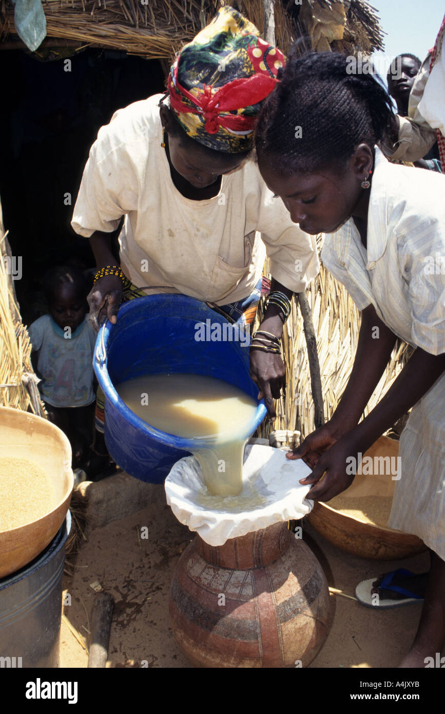 Filtering water to remove Guinea Worm parasite transmitters. Near Tera, Niger. Stock Photohttps://www.alamy.com/image-license-details/?v=1https://www.alamy.com/filtering-water-to-remove-guinea-worm-parasite-transmitters-near-tera-image6323770.html
RMA4JXYB–Filtering water to remove Guinea Worm parasite transmitters. Near Tera, Niger.
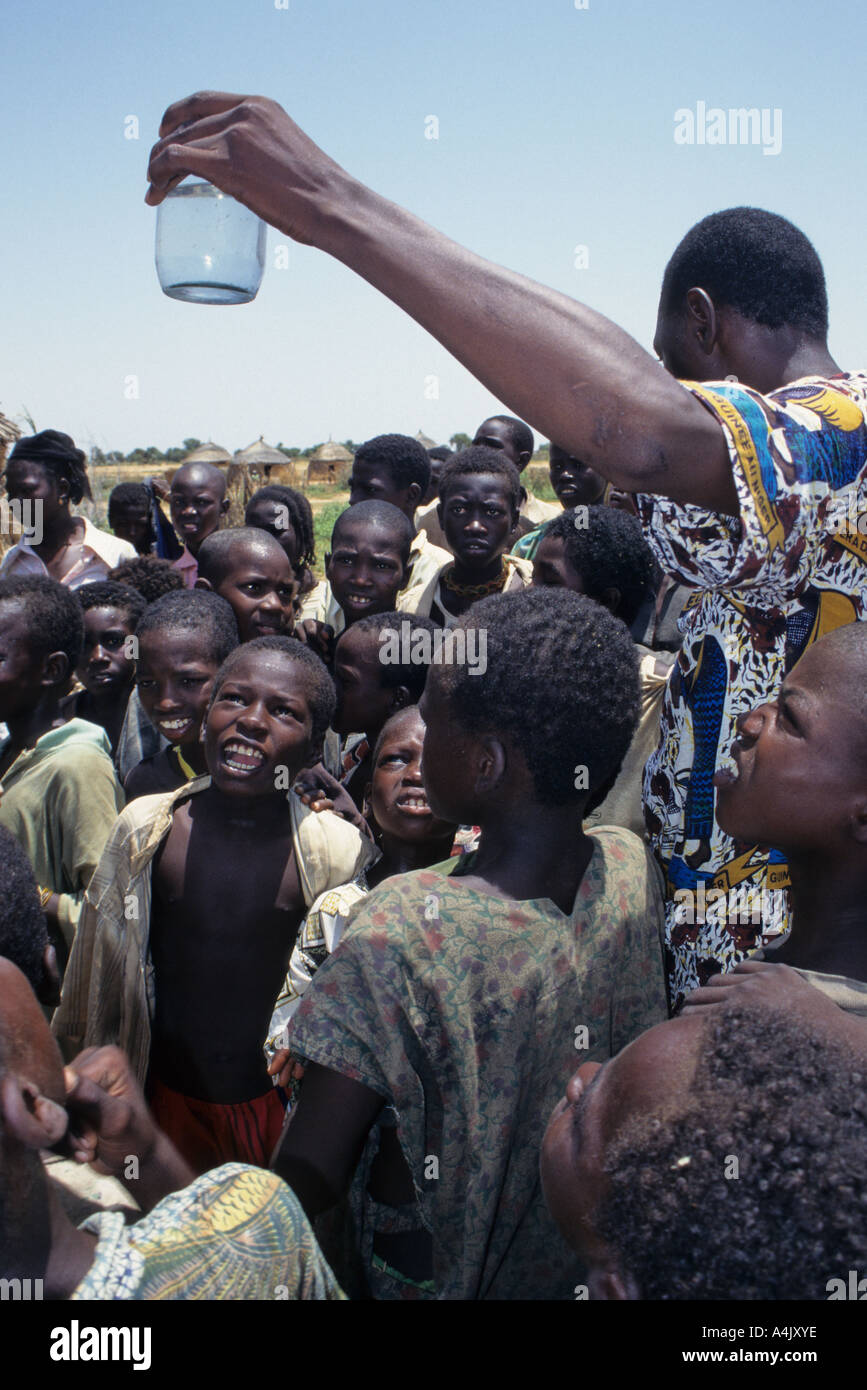 Guinea Worm Education and Eradication, Niger. Doctor showing copepods in water. Stock Photohttps://www.alamy.com/image-license-details/?v=1https://www.alamy.com/guinea-worm-education-and-eradication-niger-doctor-showing-copepods-image6323773.html
RMA4JXYE–Guinea Worm Education and Eradication, Niger. Doctor showing copepods in water.
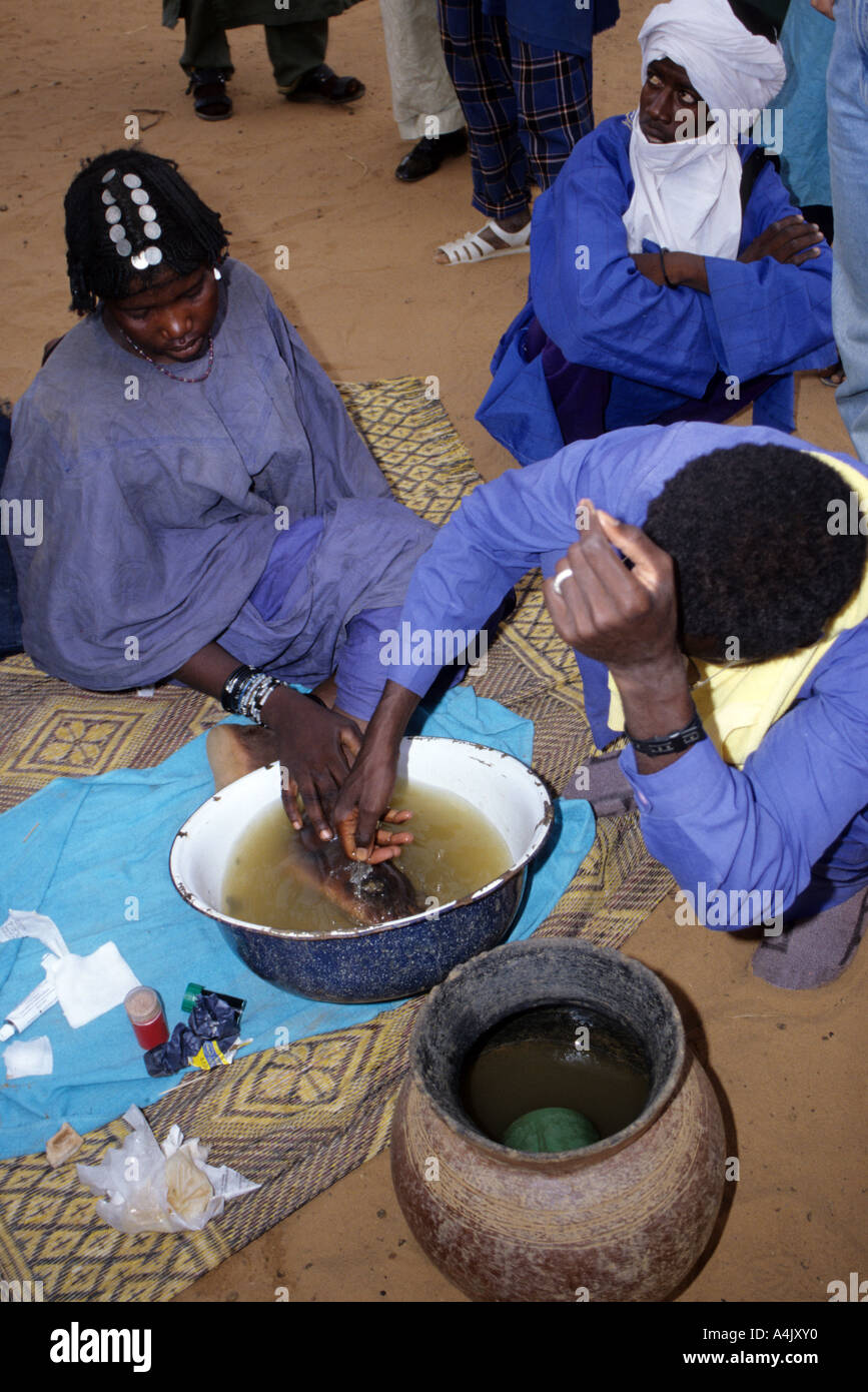 Extracting Guinea Worm from Bella Woman, Niger Stock Photohttps://www.alamy.com/image-license-details/?v=1https://www.alamy.com/extracting-guinea-worm-from-bella-woman-niger-image6323759.html
RMA4JXY0–Extracting Guinea Worm from Bella Woman, Niger
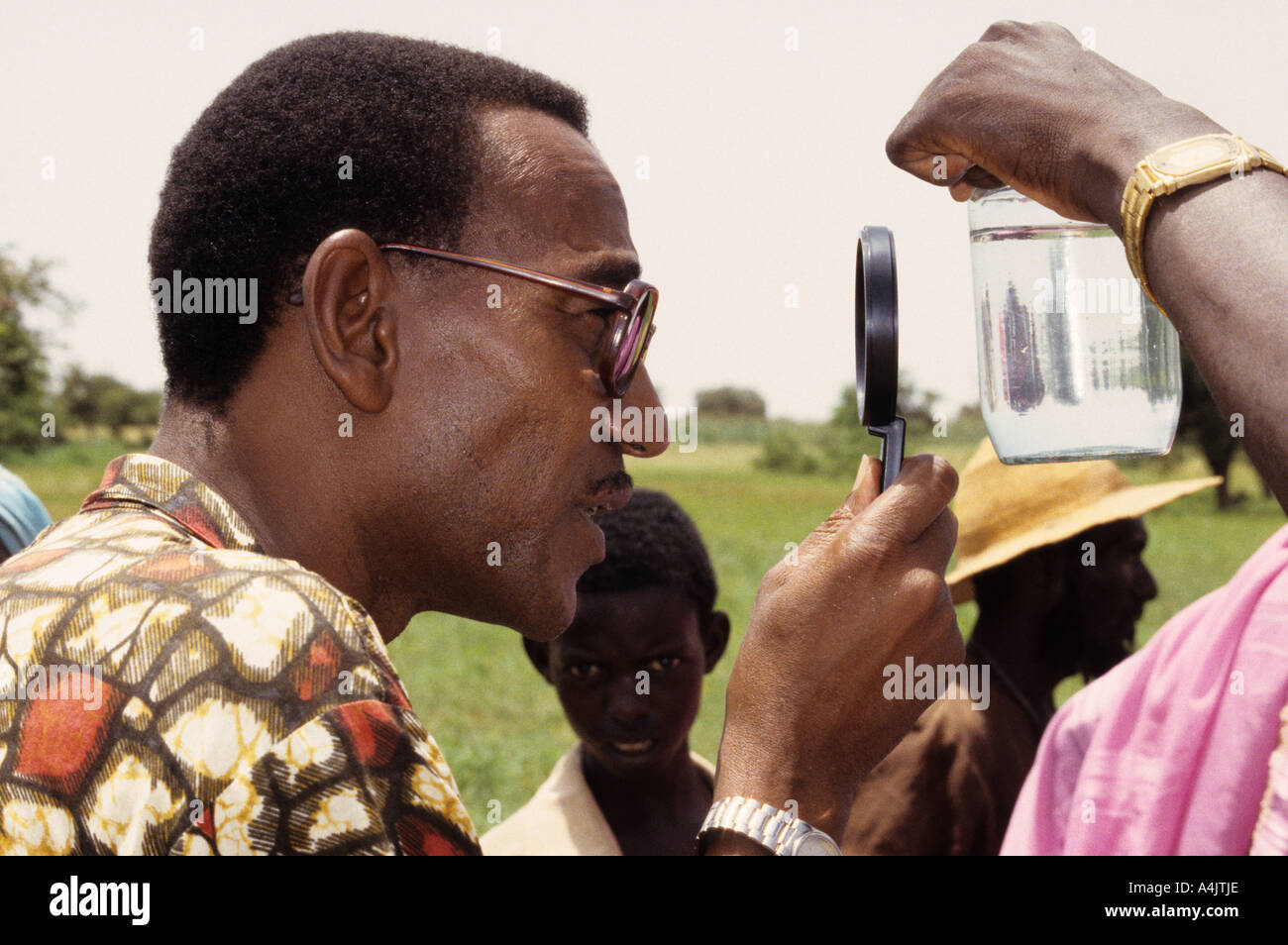 Journalist Examines Water for Guinea Worm Cyclopods, Niger. Stock Photohttps://www.alamy.com/image-license-details/?v=1https://www.alamy.com/journalist-examines-water-for-guinea-worm-cyclopods-niger-image6323245.html
RMA4JTJE–Journalist Examines Water for Guinea Worm Cyclopods, Niger.
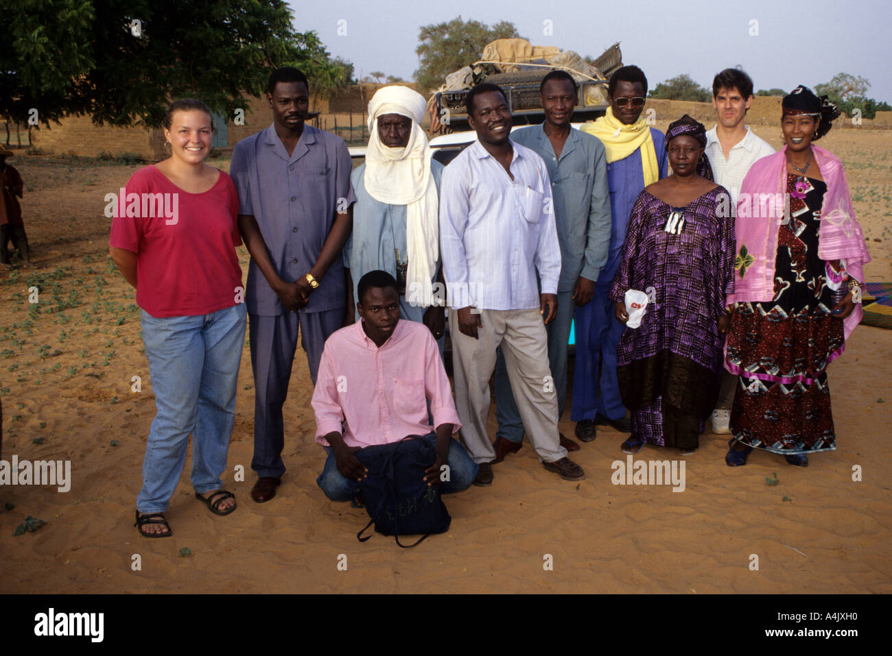 Niger. Peace Corps Volunteer and Guinea Worm Education Team. Stock Photohttps://www.alamy.com/image-license-details/?v=1https://www.alamy.com/niger-peace-corps-volunteer-and-guinea-worm-education-team-image6323599.html
RMA4JXH0–Niger. Peace Corps Volunteer and Guinea Worm Education Team.
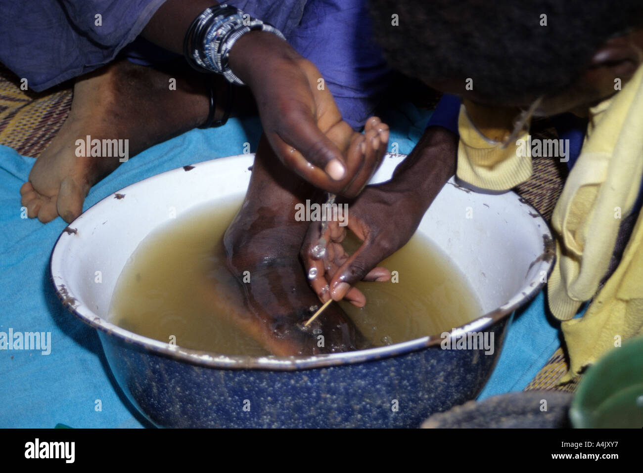 Extracting Guinea Worm from Bella Woman, Niger Stock Photohttps://www.alamy.com/image-license-details/?v=1https://www.alamy.com/extracting-guinea-worm-from-bella-woman-niger-image6323766.html
RMA4JXY7–Extracting Guinea Worm from Bella Woman, Niger
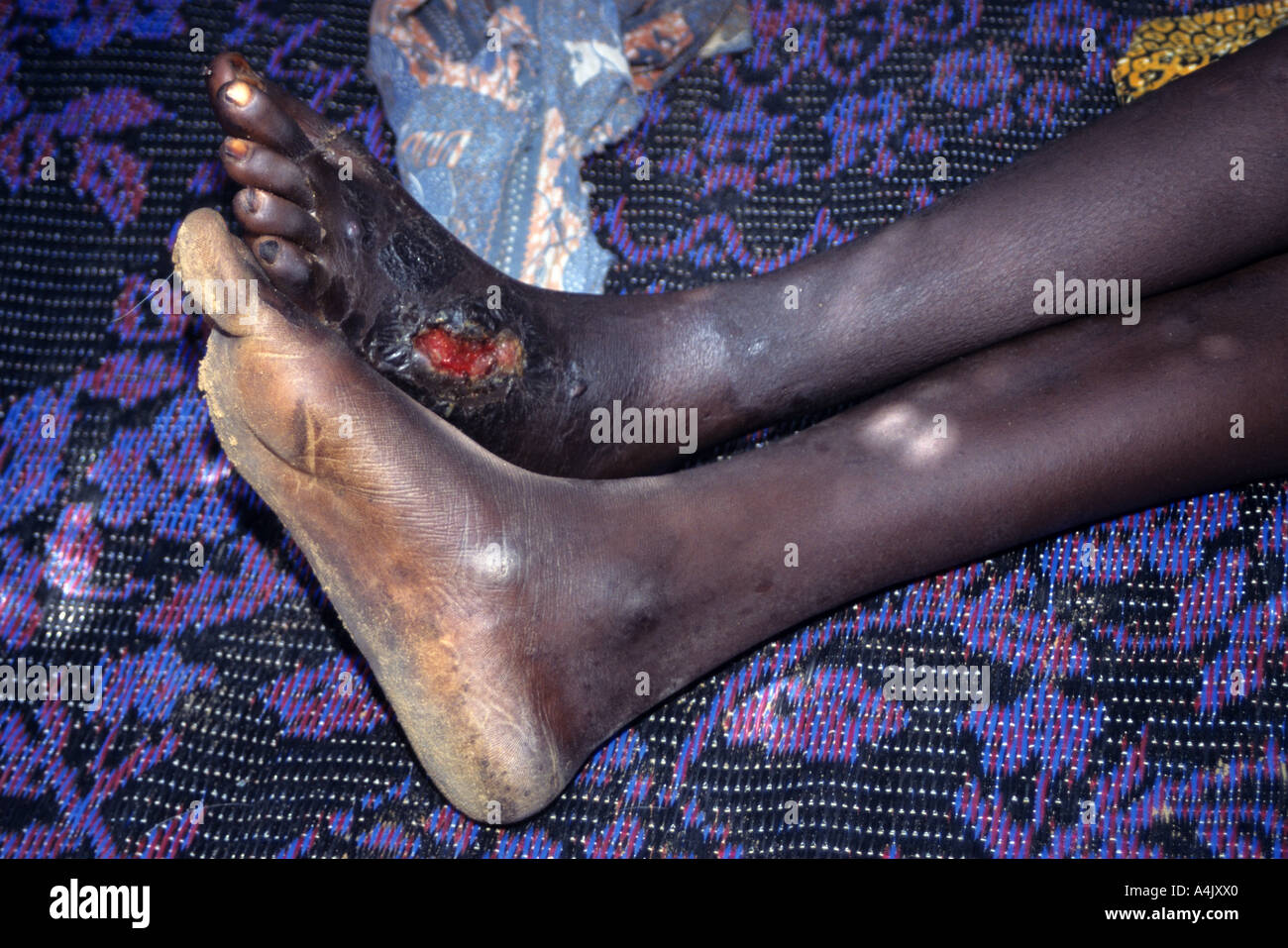 Secondary Infection of Wound Caused by Guinea Worm. Niger Stock Photohttps://www.alamy.com/image-license-details/?v=1https://www.alamy.com/secondary-infection-of-wound-caused-by-guinea-worm-niger-image6323743.html
RMA4JXX0–Secondary Infection of Wound Caused by Guinea Worm. Niger
 Guinea Worm Education Program, Niger Stock Photohttps://www.alamy.com/image-license-details/?v=1https://www.alamy.com/guinea-worm-education-program-niger-image6323265.html
RMA4JTM2–Guinea Worm Education Program, Niger
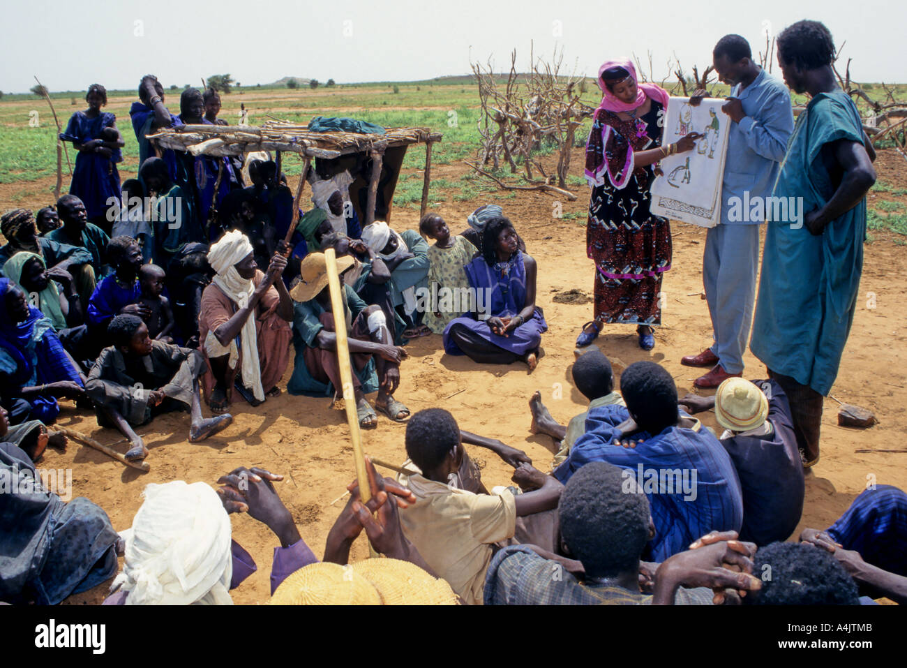 Guinea Worm Education Program, Niger. Stock Photohttps://www.alamy.com/image-license-details/?v=1https://www.alamy.com/guinea-worm-education-program-niger-image6323274.html
RMA4JTMB–Guinea Worm Education Program, Niger.
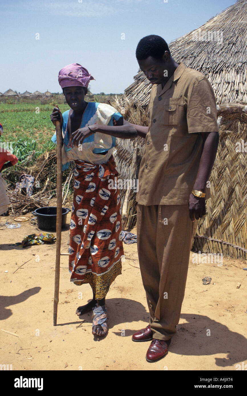 Secondary Infections from Guinea Worm Infestation Cause Great Pain, Niger Stock Photohttps://www.alamy.com/image-license-details/?v=1https://www.alamy.com/secondary-infections-from-guinea-worm-infestation-cause-great-pain-image6323715.html
RMA4JXT4–Secondary Infections from Guinea Worm Infestation Cause Great Pain, Niger