Quick filters:
Gross pathology Stock Photos and Images
 Gross pathology of fixed, cut brain showing haemorrhagic meningitis due to anthrax Stock Photohttps://www.alamy.com/image-license-details/?v=1https://www.alamy.com/stock-photo-gross-pathology-of-fixed-cut-brain-showing-haemorrhagic-meningitis-84973628.html
RMEX6TMC–Gross pathology of fixed, cut brain showing haemorrhagic meningitis due to anthrax
 Gross pathology of amebiasis, intestine. Image courtesy CDC/Dr. Mae Melvin: Dr. E. West of Mobile, AL, 1962. Stock Photohttps://www.alamy.com/image-license-details/?v=1https://www.alamy.com/gross-pathology-of-amebiasis-intestine-image-courtesy-cdcdr-mae-melvin-image155846575.html
RMK1FBW3–Gross pathology of amebiasis, intestine. Image courtesy CDC/Dr. Mae Melvin: Dr. E. West of Mobile, AL, 1962.
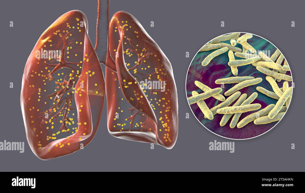 Lungs affected by miliary tuberculosis, illustration Stock Photohttps://www.alamy.com/image-license-details/?v=1https://www.alamy.com/lungs-affected-by-miliary-tuberculosis-illustration-image571248825.html
RF2T5AHKN–Lungs affected by miliary tuberculosis, illustration
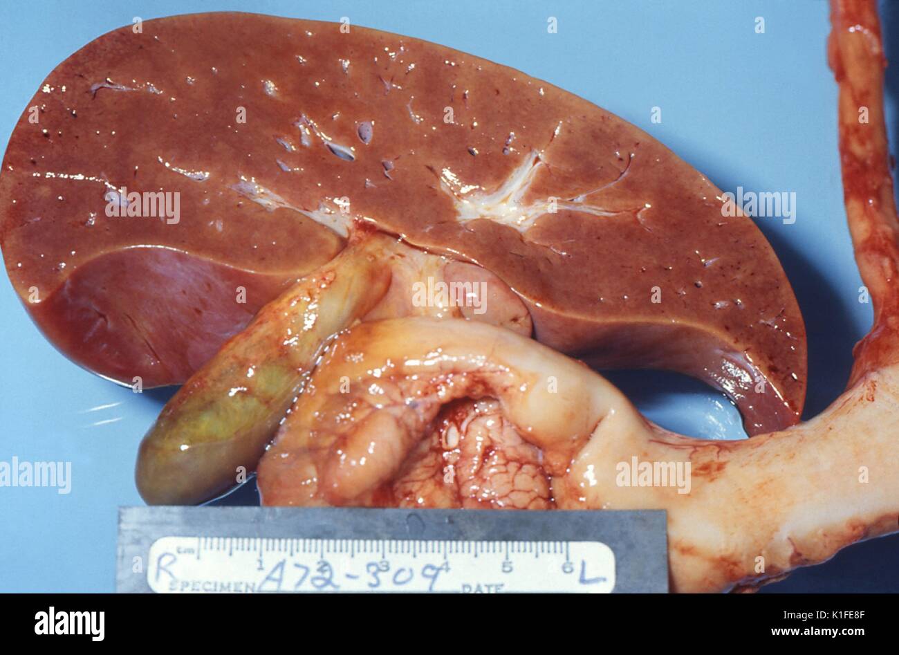 Gross pathology of liver in fatal Reye's syndrome, The cut surface of this gross autopsy specimen of a liver from a child who died of Reye's syndrome, displays a slight pallor, which was due to fat accumulation in the liver cells, also know as histiocytes. Image courtesy CDC/Dr. Edwin P. Ewing, Jr., 1972. Stock Photohttps://www.alamy.com/image-license-details/?v=1https://www.alamy.com/gross-pathology-of-liver-in-fatal-reyes-syndrome-the-cut-surface-of-image155848463.html
RMK1FE8F–Gross pathology of liver in fatal Reye's syndrome, The cut surface of this gross autopsy specimen of a liver from a child who died of Reye's syndrome, displays a slight pallor, which was due to fat accumulation in the liver cells, also know as histiocytes. Image courtesy CDC/Dr. Edwin P. Ewing, Jr., 1972.
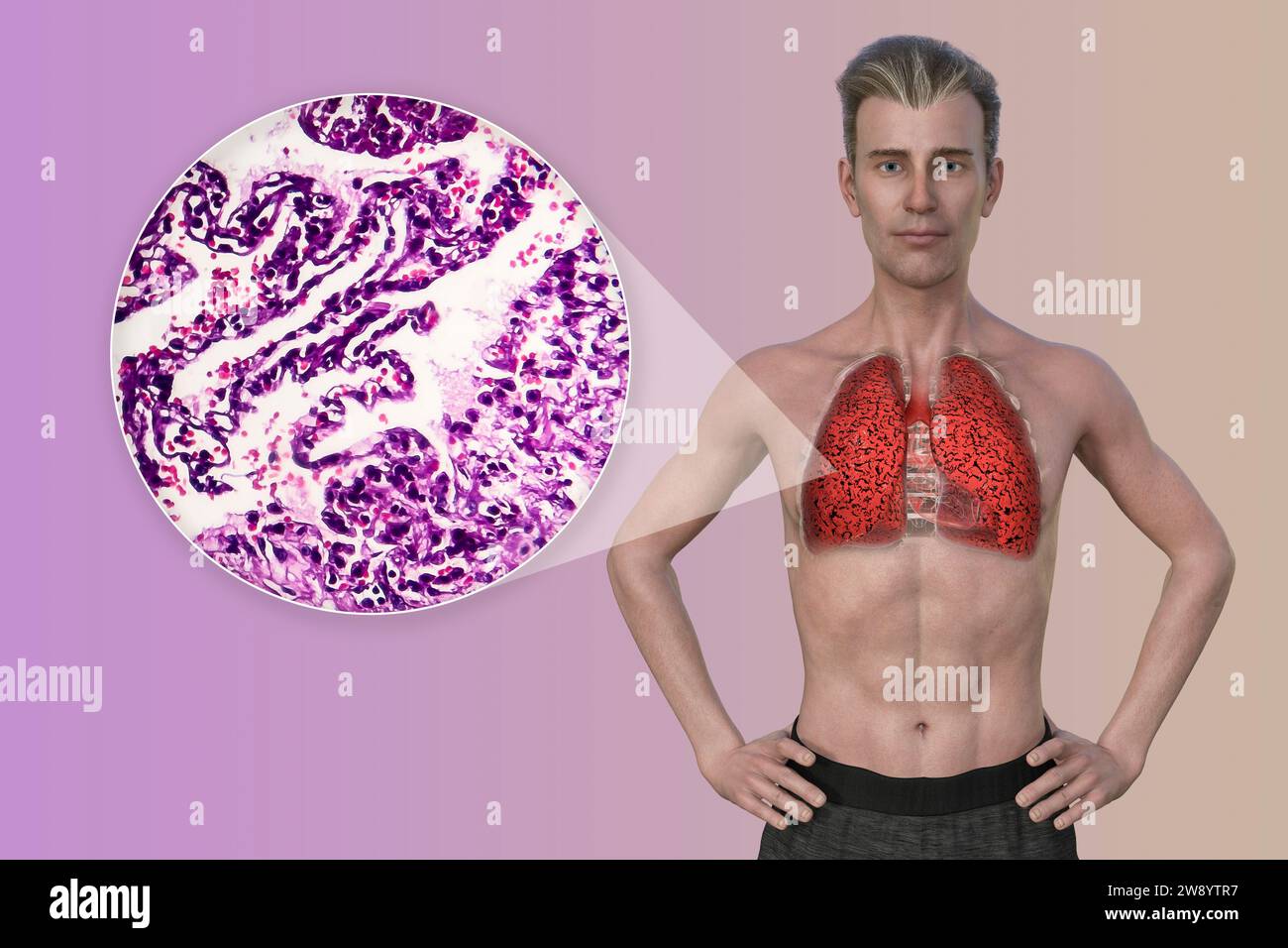 Man with smoker's lungs, illustration Stock Photohttps://www.alamy.com/image-license-details/?v=1https://www.alamy.com/man-with-smokers-lungs-illustration-image590681931.html
RF2W8YTR7–Man with smoker's lungs, illustration
 Gross pathology of intestinal ulcers due to amebiasis. Image courtesy CDC/Dr. Mae Melvin, Dr. E. West of Mobile, AL, 1962. Stock Photohttps://www.alamy.com/image-license-details/?v=1https://www.alamy.com/gross-pathology-of-intestinal-ulcers-due-to-amebiasis-image-courtesy-image155846436.html
RMK1FBM4–Gross pathology of intestinal ulcers due to amebiasis. Image courtesy CDC/Dr. Mae Melvin, Dr. E. West of Mobile, AL, 1962.
 Multiple granulomas, liver Stock Photohttps://www.alamy.com/image-license-details/?v=1https://www.alamy.com/multiple-granulomas-liver-image352788103.html
RM2BDXTR3–Multiple granulomas, liver
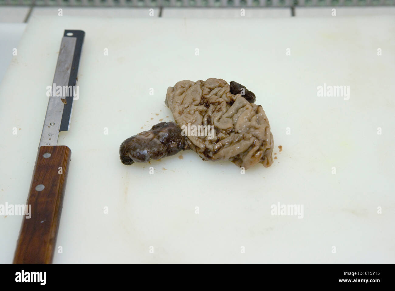 ANATOMIC PATHOLOGY Stock Photohttps://www.alamy.com/image-license-details/?v=1https://www.alamy.com/stock-photo-anatomic-pathology-49304085.html
RMCT5YT5–ANATOMIC PATHOLOGY
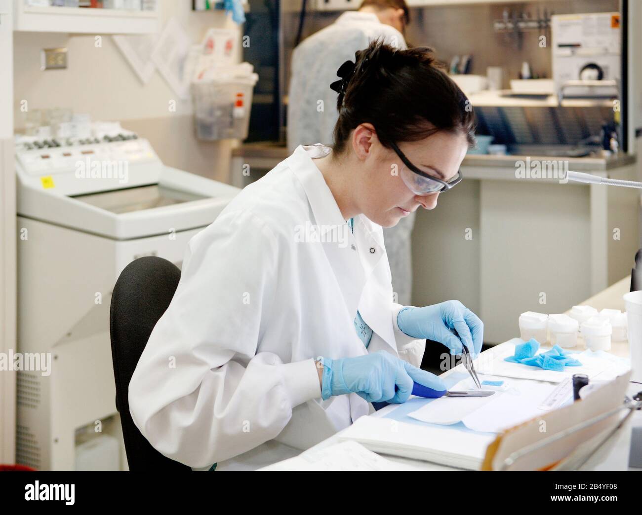 Idaho, USA Jul. 16, 2009 An interior image of the gross room on a modern pathology office. The tecnician is dissecting colon polyps and preparng the Stock Photohttps://www.alamy.com/image-license-details/?v=1https://www.alamy.com/idaho-usa-jul-16-2009-an-interior-image-of-the-gross-room-on-a-modern-pathology-office-the-tecnician-is-dissecting-colon-polyps-and-preparng-the-image347270456.html
RM2B4YF08–Idaho, USA Jul. 16, 2009 An interior image of the gross room on a modern pathology office. The tecnician is dissecting colon polyps and preparng the
 Pathologist Examining a Tumor in a Hospital Laboratory Stock Photohttps://www.alamy.com/image-license-details/?v=1https://www.alamy.com/pathologist-examining-a-tumor-in-a-hospital-laboratory-image184637485.html
RMMMAXYW–Pathologist Examining a Tumor in a Hospital Laboratory
 Pathologist Preparing a Sample Removed from a Patient for Microscopic Examination in a Hospital Laboratory Stock Photohttps://www.alamy.com/image-license-details/?v=1https://www.alamy.com/pathologist-preparing-a-sample-removed-from-a-patient-for-microscopic-examination-in-a-hospital-laboratory-image184601433.html
RMMM9909–Pathologist Preparing a Sample Removed from a Patient for Microscopic Examination in a Hospital Laboratory
 Gross pathology of hydatid cyst from human lung. Image courtesy CDC/Dr. I. Kagan, 1961. Stock Photohttps://www.alamy.com/image-license-details/?v=1https://www.alamy.com/gross-pathology-of-hydatid-cyst-from-human-lung-image-courtesy-cdcdr-image155846674.html
RMK1FC0J–Gross pathology of hydatid cyst from human lung. Image courtesy CDC/Dr. I. Kagan, 1961.
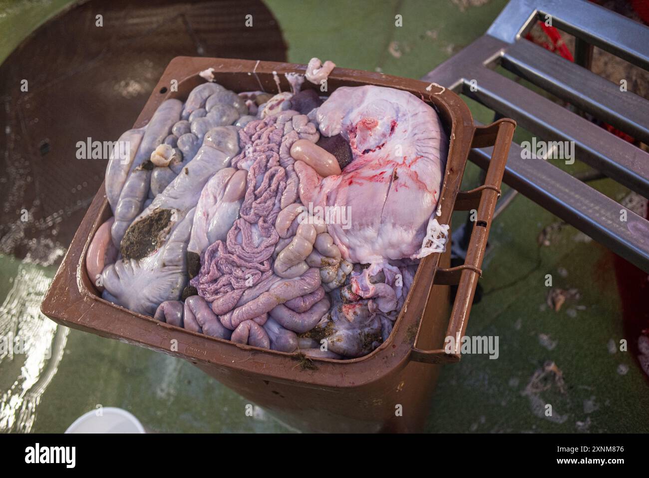 Image of various animal organs placed in a brown container, potentially for medical research or the food industry. The photo showcases a detailed view of raw, freshly retrieved organs. Stock Photohttps://www.alamy.com/image-license-details/?v=1https://www.alamy.com/image-of-various-animal-organs-placed-in-a-brown-container-potentially-for-medical-research-or-the-food-industry-the-photo-showcases-a-detailed-view-of-raw-freshly-retrieved-organs-image615716170.html
RF2XNM876–Image of various animal organs placed in a brown container, potentially for medical research or the food industry. The photo showcases a detailed view of raw, freshly retrieved organs.
 Gross specimen showing two brain slices of the frontal lobe showing an intraparenchymal haemorrhage. Hypertension is the most frequent cause of this t Stock Photohttps://www.alamy.com/image-license-details/?v=1https://www.alamy.com/gross-specimen-showing-two-brain-slices-of-the-frontal-lobe-showing-an-intraparenchymal-haemorrhage-hypertension-is-the-most-frequent-cause-of-this-t-image501929158.html
RF2M4GRNA–Gross specimen showing two brain slices of the frontal lobe showing an intraparenchymal haemorrhage. Hypertension is the most frequent cause of this t
 Alzheimer's Disease Progression Stock Photohttps://www.alamy.com/image-license-details/?v=1https://www.alamy.com/alzheimers-disease-progression-image353184458.html
RM2BEGXAJ–Alzheimer's Disease Progression
 Villous adenoma of the sigmoid colon, gross pathology Stock Photohttps://www.alamy.com/image-license-details/?v=1https://www.alamy.com/stock-photo-villous-adenoma-of-the-sigmoid-colon-gross-pathology-171680867.html
RMKY8MKF–Villous adenoma of the sigmoid colon, gross pathology
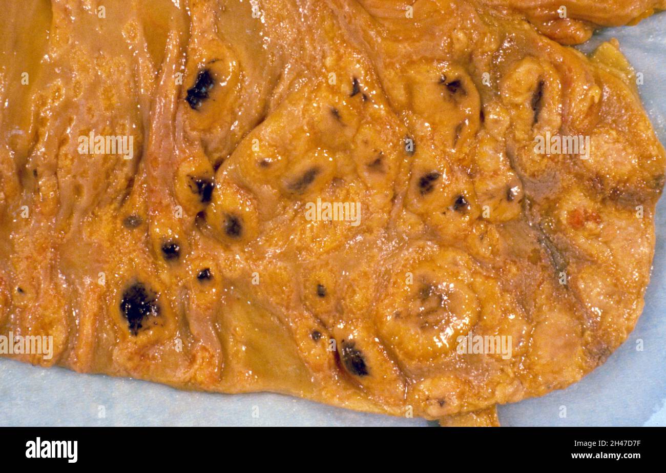 Gross pathology of the caecum and appendix Stock Photohttps://www.alamy.com/image-license-details/?v=1https://www.alamy.com/gross-pathology-of-the-caecum-and-appendix-image450092259.html
RM2H47D7F–Gross pathology of the caecum and appendix
 Bilateral ovarian serous carcinomas, gross pathology Stock Photohttps://www.alamy.com/image-license-details/?v=1https://www.alamy.com/stock-photo-bilateral-ovarian-serous-carcinomas-gross-pathology-171329527.html
RMKXMMFK–Bilateral ovarian serous carcinomas, gross pathology
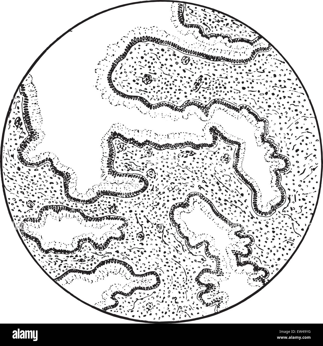 Adenoma of the cervix uteri, vintage engraved illustration. Stock Vectorhttps://www.alamy.com/image-license-details/?v=1https://www.alamy.com/stock-photo-adenoma-of-the-cervix-uteri-vintage-engraved-illustration-84303508.html
RFEW49YG–Adenoma of the cervix uteri, vintage engraved illustration.
 Doctor using surgical knife cut brain specimen after remove abnormal vascular or cerebral cavernous malformation from the brain Stock Photohttps://www.alamy.com/image-license-details/?v=1https://www.alamy.com/doctor-using-surgical-knife-cut-brain-specimen-after-remove-abnormal-vascular-or-cerebral-cavernous-malformation-from-the-brain-image367534848.html
RF2C9XJCG–Doctor using surgical knife cut brain specimen after remove abnormal vascular or cerebral cavernous malformation from the brain
 General and dental pathology with special reference to etiology and pathologic anatomy; a treatise for students and practitioners . ttychange. Results.—Growth is slow and the tumors are benign, althoughsarcomatous change may add a malignant character. ilk I **% £*?:•« *- Fig. 45.—Glioma of the brain. (Delafield and Prudden.) Glioma Glioma is a tumor composed of neuroglia. The favorite seats are the brain and cord, optic tract, nerveand retina, and parts of the olfactory tract. Glioma of the retinaoccurs usually between the second and fourth year of life. (Fig.45.) Gross Pathology.—Gliomata are Stock Photohttps://www.alamy.com/image-license-details/?v=1https://www.alamy.com/general-and-dental-pathology-with-special-reference-to-etiology-and-pathologic-anatomy-a-treatise-for-students-and-practitioners-ttychange-resultsgrowth-is-slow-and-the-tumors-are-benign-althoughsarcomatous-change-may-add-a-malignant-character-ilk-i-fig-45glioma-of-the-brain-delafield-and-prudden-glioma-glioma-is-a-tumor-composed-of-neuroglia-the-favorite-seats-are-the-brain-and-cord-optic-tract-nerveand-retina-and-parts-of-the-olfactory-tract-glioma-of-the-retinaoccurs-usually-between-the-second-and-fourth-year-of-life-fig45-gross-pathologygliomata-are-image339123168.html
RM2AKMB1M–General and dental pathology with special reference to etiology and pathologic anatomy; a treatise for students and practitioners . ttychange. Results.—Growth is slow and the tumors are benign, althoughsarcomatous change may add a malignant character. ilk I **% £*?:•« *- Fig. 45.—Glioma of the brain. (Delafield and Prudden.) Glioma Glioma is a tumor composed of neuroglia. The favorite seats are the brain and cord, optic tract, nerveand retina, and parts of the olfactory tract. Glioma of the retinaoccurs usually between the second and fourth year of life. (Fig.45.) Gross Pathology.—Gliomata are
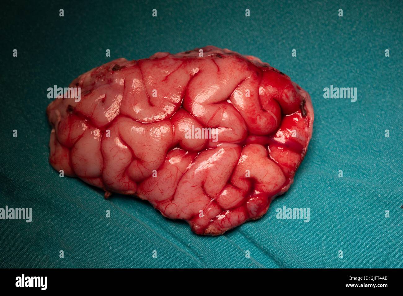 Surgical specimen of brain at frontal lobe causing seizure on green table Stock Photohttps://www.alamy.com/image-license-details/?v=1https://www.alamy.com/surgical-specimen-of-brain-at-frontal-lobe-causing-seizure-on-green-table-image474430051.html
RF2JFT4AB–Surgical specimen of brain at frontal lobe causing seizure on green table
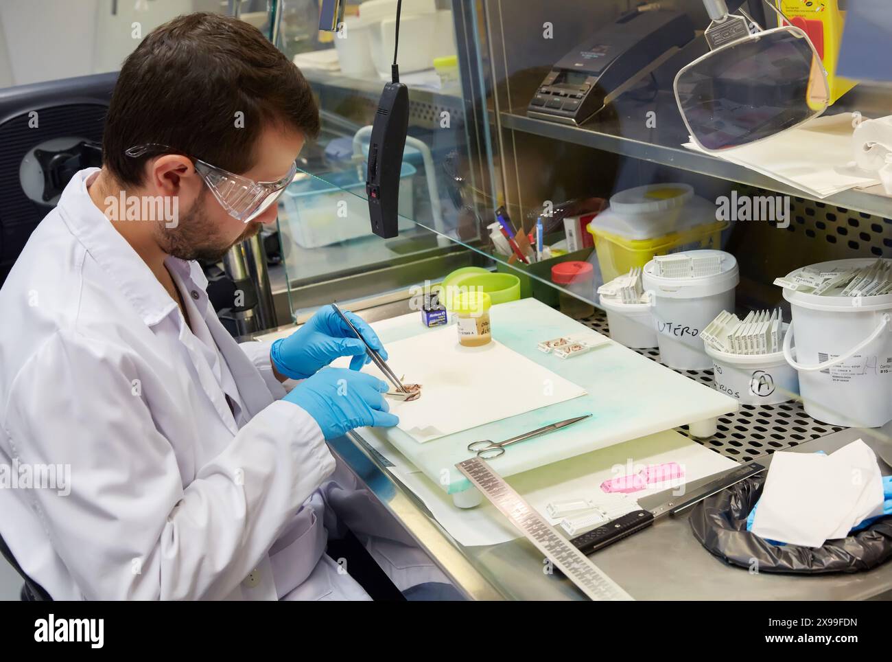 Selecting tissue sample, Anatomic Pathology, Hospital Donostia, San Sebastian, Gipuzkoa, Basque Country, Spain. Stock Photohttps://www.alamy.com/image-license-details/?v=1https://www.alamy.com/selecting-tissue-sample-anatomic-pathology-hospital-donostia-san-sebastian-gipuzkoa-basque-country-spain-image608104497.html
RM2X99FDN–Selecting tissue sample, Anatomic Pathology, Hospital Donostia, San Sebastian, Gipuzkoa, Basque Country, Spain.
 Man with smoker's lungs, illustration Stock Photohttps://www.alamy.com/image-license-details/?v=1https://www.alamy.com/man-with-smokers-lungs-illustration-image590681930.html
RF2W8YTR6–Man with smoker's lungs, illustration
 Unseen old woman rubs, feels tender hand with stiff knot under ring finger. Dupuytren contracture Stock Photohttps://www.alamy.com/image-license-details/?v=1https://www.alamy.com/unseen-old-woman-rubs-feels-tender-hand-with-stiff-knot-under-ring-finger-dupuytren-contracture-image619826045.html
RF2Y0BECD–Unseen old woman rubs, feels tender hand with stiff knot under ring finger. Dupuytren contracture
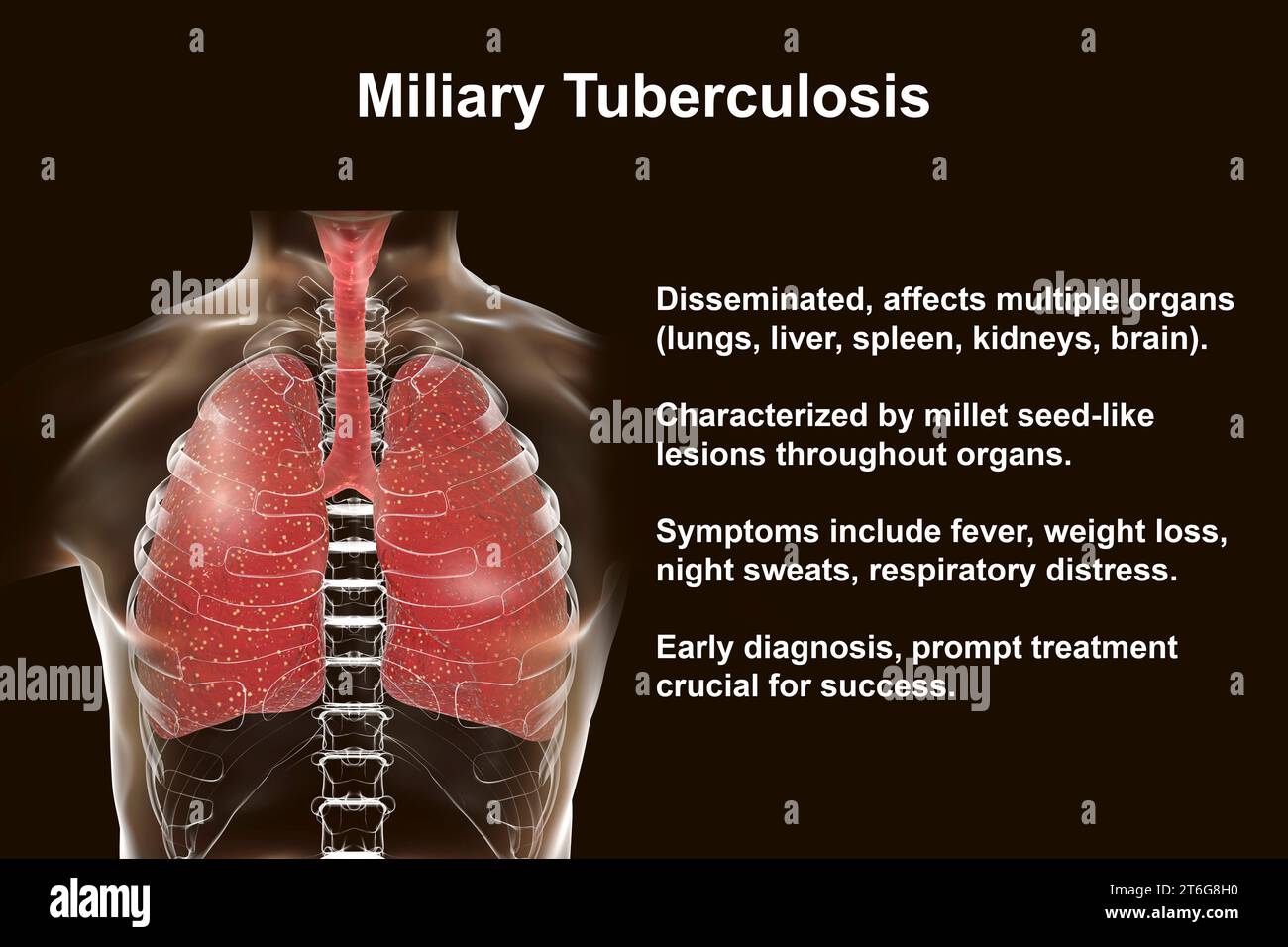 A detailed 3D photorealistic illustration showcasing human lungs affected by miliary tuberculosis Stock Photohttps://www.alamy.com/image-license-details/?v=1https://www.alamy.com/a-detailed-3d-photorealistic-illustration-showcasing-human-lungs-affected-by-miliary-tuberculosis-image571988060.html
RF2T6G8H0–A detailed 3D photorealistic illustration showcasing human lungs affected by miliary tuberculosis
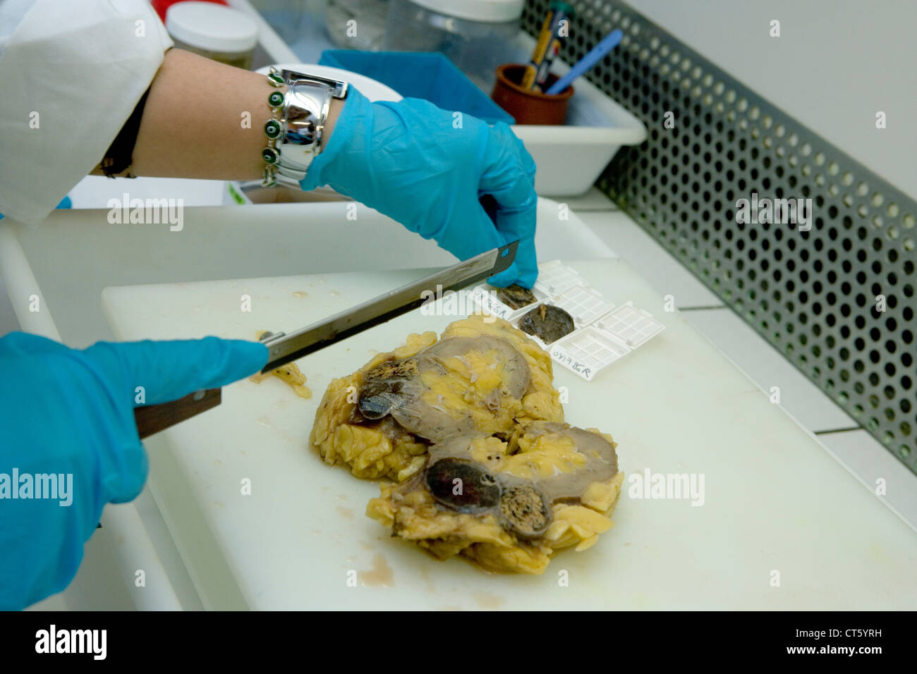 ANATOMIC PATHOLOGY Stock Photohttps://www.alamy.com/image-license-details/?v=1https://www.alamy.com/stock-photo-anatomic-pathology-49304069.html
RMCT5YRH–ANATOMIC PATHOLOGY
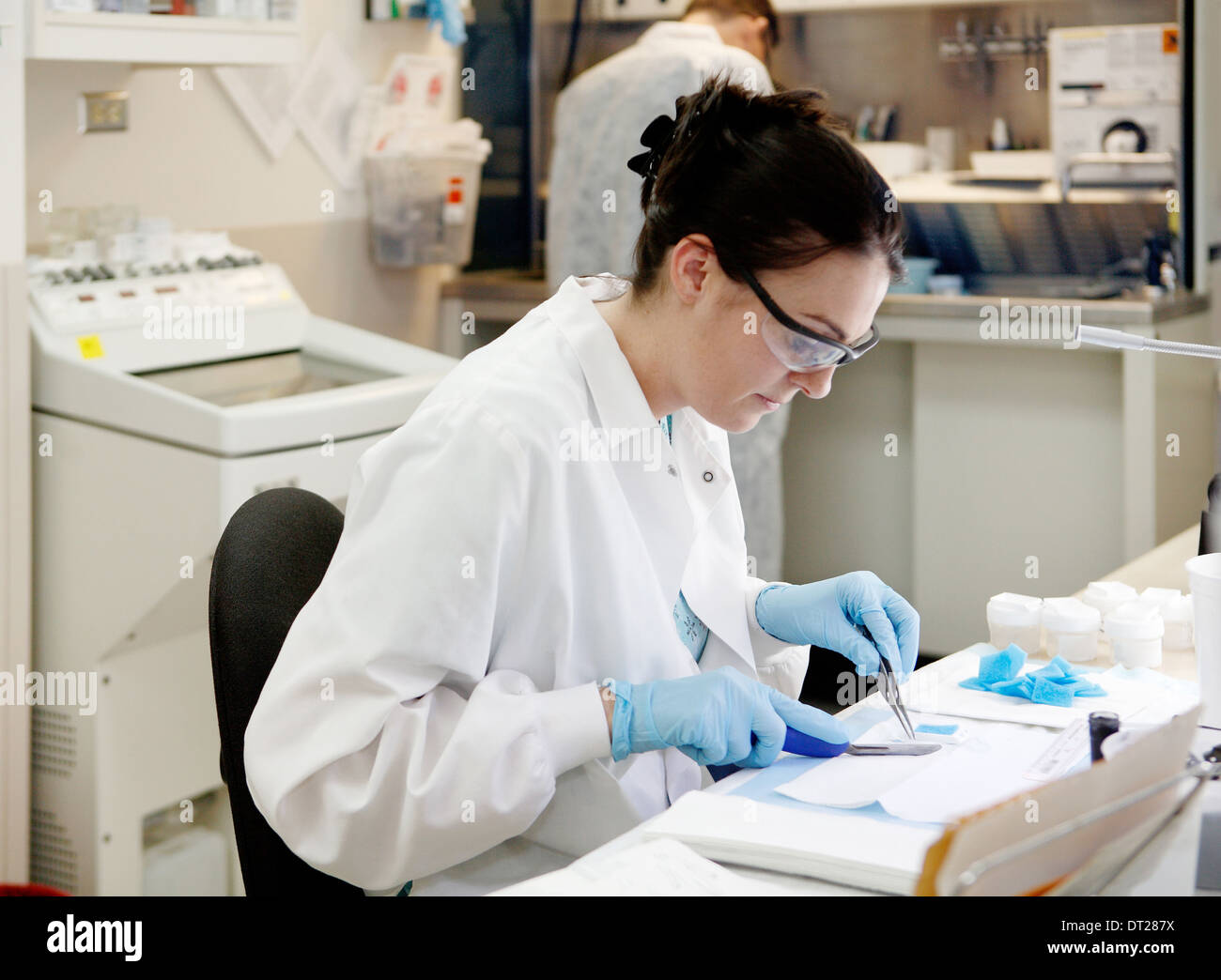 An interior image of the gross room on a modern pathology office. Stock Photohttps://www.alamy.com/image-license-details/?v=1https://www.alamy.com/an-interior-image-of-the-gross-room-on-a-modern-pathology-office-image66433246.html
RMDT287X–An interior image of the gross room on a modern pathology office.
 Colon Cancer in Excised Surgical Specimen Stock Photohttps://www.alamy.com/image-license-details/?v=1https://www.alamy.com/stock-photo-colon-cancer-in-excised-surgical-specimen-51892674.html
RMD0BWHP–Colon Cancer in Excised Surgical Specimen
 Pathologist Dissecting a Skin Tumor Removed from a Patient for Microscopic Examination Stock Photohttps://www.alamy.com/image-license-details/?v=1https://www.alamy.com/pathologist-dissecting-a-skin-tumor-removed-from-a-patient-for-microscopic-examination-image184948324.html
RMMMW3D8–Pathologist Dissecting a Skin Tumor Removed from a Patient for Microscopic Examination
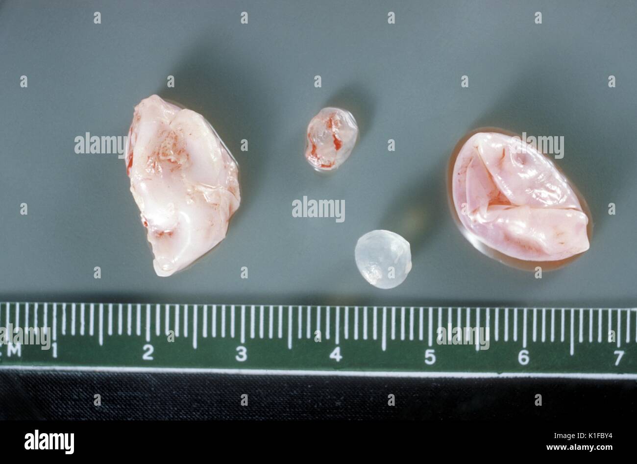 Gross pathology of membrane and hydatid daughter cysts from human lung. Parasite. Image courtesy CDC/Dr. I. Kagan, 1961. Stock Photohttps://www.alamy.com/image-license-details/?v=1https://www.alamy.com/gross-pathology-of-membrane-and-hydatid-daughter-cysts-from-human-image155846632.html
RMK1FBY4–Gross pathology of membrane and hydatid daughter cysts from human lung. Parasite. Image courtesy CDC/Dr. I. Kagan, 1961.
 Close-up of a gross specimen showing the surface of a human kidney in a case of autosomal dominant polycystic kidney disease (ADPKD). The surface is f Stock Photohttps://www.alamy.com/image-license-details/?v=1https://www.alamy.com/close-up-of-a-gross-specimen-showing-the-surface-of-a-human-kidney-in-a-case-of-autosomal-dominant-polycystic-kidney-disease-adpkd-the-surface-is-f-image501929168.html
RF2M4GRNM–Close-up of a gross specimen showing the surface of a human kidney in a case of autosomal dominant polycystic kidney disease (ADPKD). The surface is f
 Alzheimer's Disease Progression Stock Photohttps://www.alamy.com/image-license-details/?v=1https://www.alamy.com/alzheimers-disease-progression-image353184388.html
RM2BEGX84–Alzheimer's Disease Progression
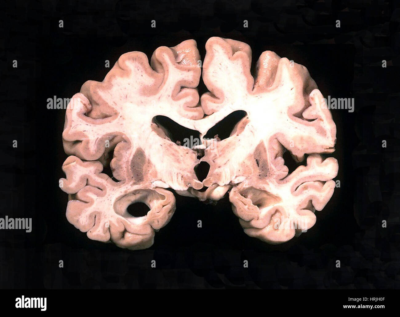 Alzheimer Brain, Gross Specimen Stock Photohttps://www.alamy.com/image-license-details/?v=1https://www.alamy.com/stock-photo-alzheimer-brain-gross-specimen-135018143.html
RMHRJH0F–Alzheimer Brain, Gross Specimen
 Scurvy past and present . ave beendescribed. Central Nervous System.—The most frequent abnormal-ity of the central nervous system is, as would be expected,hemorrhage; this has been discussed in the section dealingwith gross pathology. No specific changes have beenfound in nerve-cells or fibres of the brain. In a case of fatal scurvy in an infant a focal de-generation of the lumbar cord has been described,extending for a distance of about a quarter of an inch(Hess). The lesion differed from that of poliomyelitisin the absence of round-celled infiltration and of the char-acteristic changes in th Stock Photohttps://www.alamy.com/image-license-details/?v=1https://www.alamy.com/scurvy-past-and-present-ave-beendescribed-central-nervous-systemthe-most-frequent-abnormal-ity-of-the-central-nervous-system-is-as-would-be-expectedhemorrhage-this-has-been-discussed-in-the-section-dealingwith-gross-pathology-no-specific-changes-have-beenfound-in-nerve-cells-or-fibres-of-the-brain-in-a-case-of-fatal-scurvy-in-an-infant-a-focal-de-generation-of-the-lumbar-cord-has-been-describedextending-for-a-distance-of-about-a-quarter-of-an-inchhess-the-lesion-differed-from-that-of-poliomyelitisin-the-absence-of-round-celled-infiltration-and-of-the-char-acteristic-changes-in-th-image339120593.html
RM2AKM7NN–Scurvy past and present . ave beendescribed. Central Nervous System.—The most frequent abnormal-ity of the central nervous system is, as would be expected,hemorrhage; this has been discussed in the section dealingwith gross pathology. No specific changes have beenfound in nerve-cells or fibres of the brain. In a case of fatal scurvy in an infant a focal de-generation of the lumbar cord has been described,extending for a distance of about a quarter of an inch(Hess). The lesion differed from that of poliomyelitisin the absence of round-celled infiltration and of the char-acteristic changes in th
 Selecting tissue sample, Anatomic Pathology, Hospital Donostia, San Sebastian, Gipuzkoa, Basque Country, Spain. Stock Photohttps://www.alamy.com/image-license-details/?v=1https://www.alamy.com/selecting-tissue-sample-anatomic-pathology-hospital-donostia-san-sebastian-gipuzkoa-basque-country-spain-image624981546.html
RM2Y8PA9E–Selecting tissue sample, Anatomic Pathology, Hospital Donostia, San Sebastian, Gipuzkoa, Basque Country, Spain.
 Man with smoker's lungs, illustration Stock Photohttps://www.alamy.com/image-license-details/?v=1https://www.alamy.com/man-with-smokers-lungs-illustration-image590681933.html
RF2W8YTR9–Man with smoker's lungs, illustration
 Hand tendon disease. Cropped woman hand with dense node under finger. Dupuytren muscular contracture Stock Photohttps://www.alamy.com/image-license-details/?v=1https://www.alamy.com/hand-tendon-disease-cropped-woman-hand-with-dense-node-under-finger-dupuytren-muscular-contracture-image620759042.html
RF2Y1X0DP–Hand tendon disease. Cropped woman hand with dense node under finger. Dupuytren muscular contracture
 A 3D medical illustration displaying a patient's hand with Dupuytren's contracture, emphasizing the affected tendons and palmar fascia to illustrate t Stock Photohttps://www.alamy.com/image-license-details/?v=1https://www.alamy.com/a-3d-medical-illustration-displaying-a-patients-hand-with-dupuytrens-contracture-emphasizing-the-affected-tendons-and-palmar-fascia-to-illustrate-t-image571986313.html
RF2T6G6AH–A 3D medical illustration displaying a patient's hand with Dupuytren's contracture, emphasizing the affected tendons and palmar fascia to illustrate t
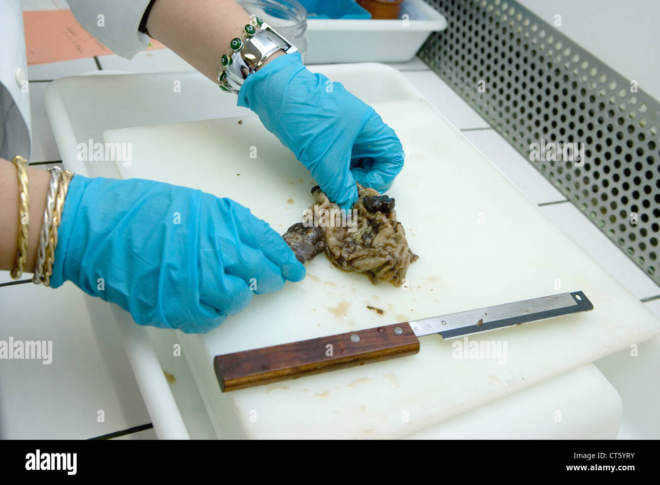 ANATOMIC PATHOLOGY Stock Photohttps://www.alamy.com/image-license-details/?v=1https://www.alamy.com/stock-photo-anatomic-pathology-49304079.html
RMCT5YRY–ANATOMIC PATHOLOGY
 Gross pathology of membrane and hydatid daughter cysts from human lung. Parasite. Image courtesy CDC/Dr. I. Kagan, 1961. Stock Photohttps://www.alamy.com/image-license-details/?v=1https://www.alamy.com/gross-pathology-of-membrane-and-hydatid-daughter-cysts-from-human-image155846620.html
RMK1FBXM–Gross pathology of membrane and hydatid daughter cysts from human lung. Parasite. Image courtesy CDC/Dr. I. Kagan, 1961.
 Gross pathology of liver containing amebic abscess. Image courtesy CDC/Dr. Mae Melvin, Dr. E. West of Mobile, AL, 1962. Stock Photohttps://www.alamy.com/image-license-details/?v=1https://www.alamy.com/gross-pathology-of-liver-containing-amebic-abscess-image-courtesy-image155846473.html
RMK1FBND–Gross pathology of liver containing amebic abscess. Image courtesy CDC/Dr. Mae Melvin, Dr. E. West of Mobile, AL, 1962.
 Close-up of a gross specimen of a human kidney in a case of autosomal dominant polycystic kidney disease (ADPKD) showing an almost complete replacemen Stock Photohttps://www.alamy.com/image-license-details/?v=1https://www.alamy.com/close-up-of-a-gross-specimen-of-a-human-kidney-in-a-case-of-autosomal-dominant-polycystic-kidney-disease-adpkd-showing-an-almost-complete-replacemen-image501929157.html
RF2M4GRN9–Close-up of a gross specimen of a human kidney in a case of autosomal dominant polycystic kidney disease (ADPKD) showing an almost complete replacemen
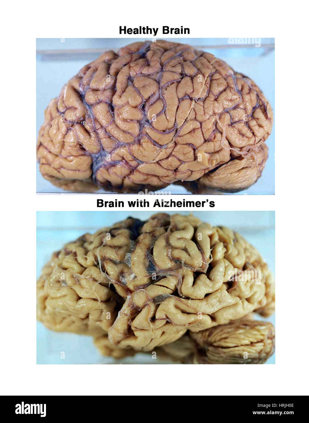 Healthy and Alzheimer Brains, Gross Specimens Stock Photohttps://www.alamy.com/image-license-details/?v=1https://www.alamy.com/stock-photo-healthy-and-alzheimer-brains-gross-specimens-135018142.html
RMHRJH0E–Healthy and Alzheimer Brains, Gross Specimens
 . Journal of radiology . Case I.—Fig. IV.—Another view ofrejected jejunum. Arrow points tostricture. the stricture. The small jejunum be-low the stricture was inverted as theappendix is done, the larger closed,as colon. A lateral anastomosis,Bloodgoods method, was done, theends pointing in opposite directions,interrupted silk sutures being used.The abdominal wound was closed withburied catgut. Silver wire through and Watch The Journal—It Leads! iNTESTlNAL OBSTRUCTION—KAHN 47 through. No shock. Gross Pathology.At the position of the stricture therewas a small area like cancer. Thefrozen section Stock Photohttps://www.alamy.com/image-license-details/?v=1https://www.alamy.com/journal-of-radiology-case-ifig-ivanother-view-ofrejected-jejunum-arrow-points-tostricture-the-stricture-the-small-jejunum-be-low-the-stricture-was-inverted-as-theappendix-is-done-the-larger-closedas-colon-a-lateral-anastomosisbloodgoods-method-was-done-theends-pointing-in-opposite-directionsinterrupted-silk-sutures-being-usedthe-abdominal-wound-was-closed-withburied-catgut-silver-wire-through-and-watch-the-journalit-leads!-intestlnal-obstructionkahn-47-through-no-shock-gross-pathologyat-the-position-of-the-stricture-therewas-a-small-area-like-cancer-thefrozen-section-image375995028.html
RM2CRM1DT–. Journal of radiology . Case I.—Fig. IV.—Another view ofrejected jejunum. Arrow points tostricture. the stricture. The small jejunum be-low the stricture was inverted as theappendix is done, the larger closed,as colon. A lateral anastomosis,Bloodgoods method, was done, theends pointing in opposite directions,interrupted silk sutures being used.The abdominal wound was closed withburied catgut. Silver wire through and Watch The Journal—It Leads! iNTESTlNAL OBSTRUCTION—KAHN 47 through. No shock. Gross Pathology.At the position of the stricture therewas a small area like cancer. Thefrozen section
 Selecting tissue sample, Anatomic Pathology, Hospital Donostia, San Sebastian, Gipuzkoa, Basque Country, Spain. Stock Photohttps://www.alamy.com/image-license-details/?v=1https://www.alamy.com/selecting-tissue-sample-anatomic-pathology-hospital-donostia-san-sebastian-gipuzkoa-basque-country-spain-image602130625.html
RM2WYHBN5–Selecting tissue sample, Anatomic Pathology, Hospital Donostia, San Sebastian, Gipuzkoa, Basque Country, Spain.
 Man with smoker's lungs, illustration Stock Photohttps://www.alamy.com/image-license-details/?v=1https://www.alamy.com/man-with-smokers-lungs-illustration-image590681964.html
RF2W8YTTC–Man with smoker's lungs, illustration
 Hand tendon disease. Cropped woman hand with dense node under finger. Dupuytren muscular contracture Stock Photohttps://www.alamy.com/image-license-details/?v=1https://www.alamy.com/hand-tendon-disease-cropped-woman-hand-with-dense-node-under-finger-dupuytren-muscular-contracture-image620241181.html
RF2Y12BXN–Hand tendon disease. Cropped woman hand with dense node under finger. Dupuytren muscular contracture
 A 3D medical illustration displaying a patient's hand with Dupuytren's contracture, emphasizing the affected tendons and palmar fascia to illustrate t Stock Photohttps://www.alamy.com/image-license-details/?v=1https://www.alamy.com/a-3d-medical-illustration-displaying-a-patients-hand-with-dupuytrens-contracture-emphasizing-the-affected-tendons-and-palmar-fascia-to-illustrate-t-image571986317.html
RF2T6G6AN–A 3D medical illustration displaying a patient's hand with Dupuytren's contracture, emphasizing the affected tendons and palmar fascia to illustrate t
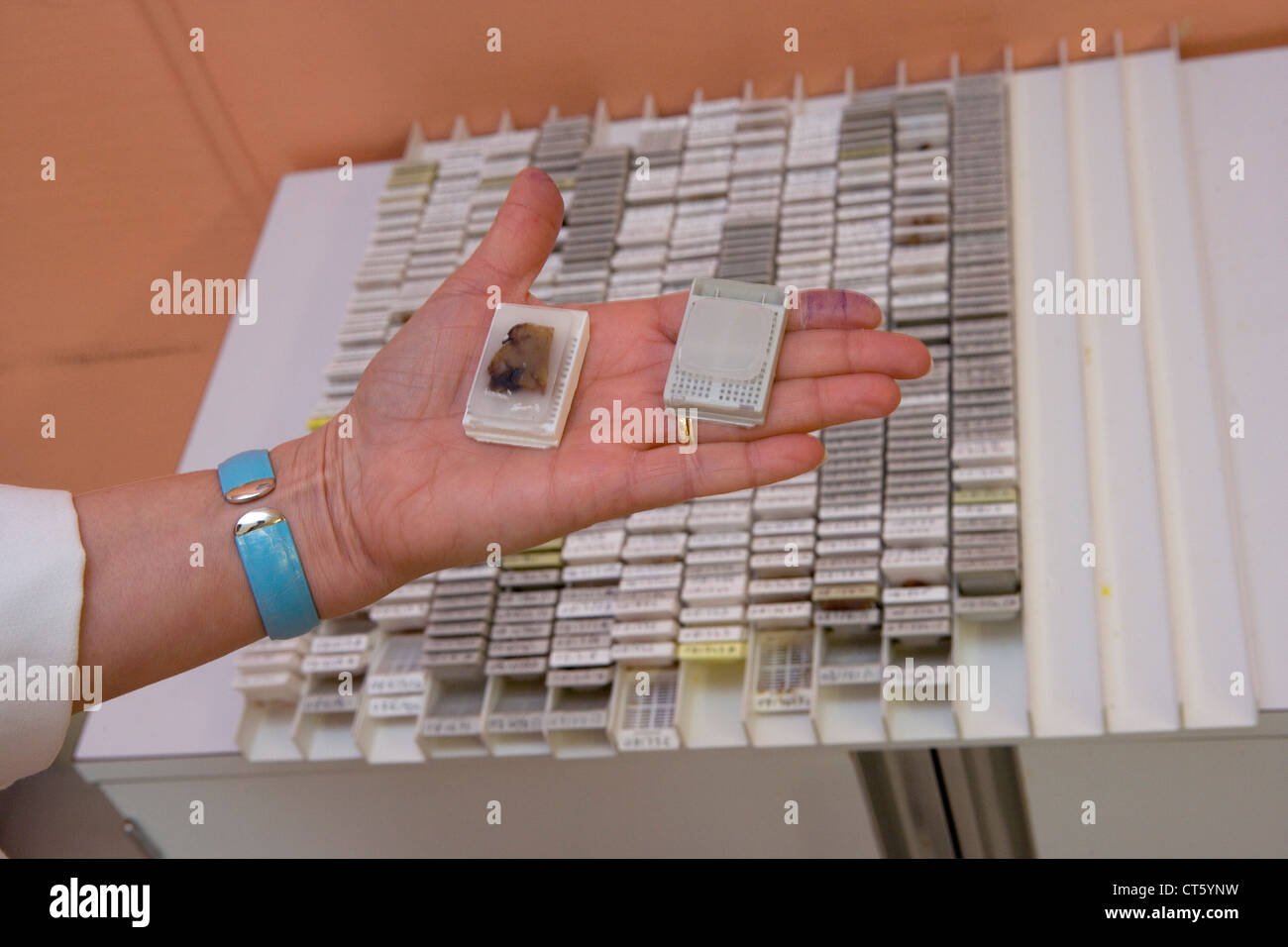 ANATOMIC PATHOLOGY Stock Photohttps://www.alamy.com/image-license-details/?v=1https://www.alamy.com/stock-photo-anatomic-pathology-49304021.html
RMCT5YNW–ANATOMIC PATHOLOGY
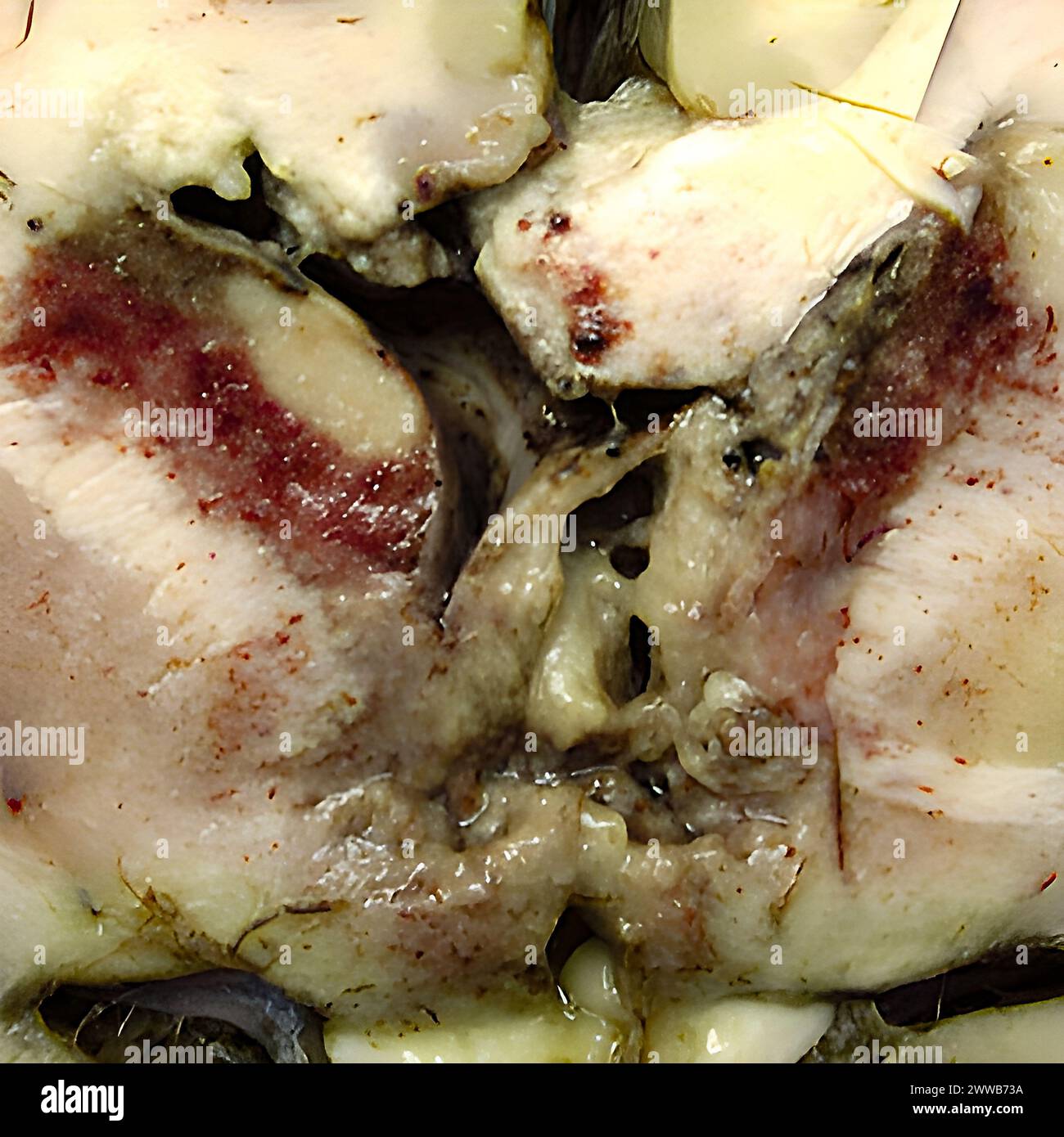 Gross specimen of brain tissue from a patient who died of granulomatous amebic encephalitis caused by Balamuthia mandrillaris. Stock Photohttps://www.alamy.com/image-license-details/?v=1https://www.alamy.com/gross-specimen-of-brain-tissue-from-a-patient-who-died-of-granulomatous-amebic-encephalitis-caused-by-balamuthia-mandrillaris-image600765966.html
RM2WWB73A–Gross specimen of brain tissue from a patient who died of granulomatous amebic encephalitis caused by Balamuthia mandrillaris.
 Gross pathology of liver containing amebic abscess. Image courtesy CDC/Dr. Mae Melvin, Dr. E. West of Mobile, AL, 1962. Stock Photohttps://www.alamy.com/image-license-details/?v=1https://www.alamy.com/gross-pathology-of-liver-containing-amebic-abscess-image-courtesy-image155846490.html
RMK1FBP2–Gross pathology of liver containing amebic abscess. Image courtesy CDC/Dr. Mae Melvin, Dr. E. West of Mobile, AL, 1962.
 Gross specimen of a human kidney in a case of autosomal dominant polycystic kidney disease (ADPKD). This is the left kidney split in half showing an a Stock Photohttps://www.alamy.com/image-license-details/?v=1https://www.alamy.com/gross-specimen-of-a-human-kidney-in-a-case-of-autosomal-dominant-polycystic-kidney-disease-adpkd-this-is-the-left-kidney-split-in-half-showing-an-a-image501929167.html
RF2M4GRNK–Gross specimen of a human kidney in a case of autosomal dominant polycystic kidney disease (ADPKD). This is the left kidney split in half showing an a
 Impetigo Stock Photohttps://www.alamy.com/image-license-details/?v=1https://www.alamy.com/stock-photo-impetigo-134944736.html
RMHRF7AT–Impetigo
 . American practice of surgery ; a complete system of the science and art of surgery . us. Symptoms.—The varied gross pathology of actmomycosis gives rise toequally varied symptoms, which are more marked in mixed than in simple infec- * North Am. Pract., Chicago, 1891, iii., 593. ACTINOMYCOSIS. 59 tion, and more serious the deeper and more numerous and extensive the foci ofthe disease. Ruhriih has collected over a thousand reported cases in man, andfinds that the head and neck are affected in fifty-six per cent, the digestive tractin twenty per cent, the lungs in fifteen per cent, and the skin Stock Photohttps://www.alamy.com/image-license-details/?v=1https://www.alamy.com/american-practice-of-surgery-a-complete-system-of-the-science-and-art-of-surgery-us-symptomsthe-varied-gross-pathology-of-actmomycosis-gives-rise-toequally-varied-symptoms-which-are-more-marked-in-mixed-than-in-simple-infec-north-am-pract-chicago-1891-iii-593-actinomycosis-59-tion-and-more-serious-the-deeper-and-more-numerous-and-extensive-the-foci-ofthe-disease-ruhriih-has-collected-over-a-thousand-reported-cases-in-man-andfinds-that-the-head-and-neck-are-affected-in-fifty-six-per-cent-the-digestive-tractin-twenty-per-cent-the-lungs-in-fifteen-per-cent-and-the-skin-image370152813.html
RM2CE5WK9–. American practice of surgery ; a complete system of the science and art of surgery . us. Symptoms.—The varied gross pathology of actmomycosis gives rise toequally varied symptoms, which are more marked in mixed than in simple infec- * North Am. Pract., Chicago, 1891, iii., 593. ACTINOMYCOSIS. 59 tion, and more serious the deeper and more numerous and extensive the foci ofthe disease. Ruhriih has collected over a thousand reported cases in man, andfinds that the head and neck are affected in fifty-six per cent, the digestive tractin twenty per cent, the lungs in fifteen per cent, and the skin
 Cryostat for Standard Applications in the Clinical Histopathology Laboratory, Anatomic Pathology, Hospital Donostia, San Sebastian, Gipuzkoa, Basque Country, Spain. Stock Photohttps://www.alamy.com/image-license-details/?v=1https://www.alamy.com/cryostat-for-standard-applications-in-the-clinical-histopathology-laboratory-anatomic-pathology-hospital-donostia-san-sebastian-gipuzkoa-basque-country-spain-image624976406.html
RM2Y8P3NX–Cryostat for Standard Applications in the Clinical Histopathology Laboratory, Anatomic Pathology, Hospital Donostia, San Sebastian, Gipuzkoa, Basque Country, Spain.
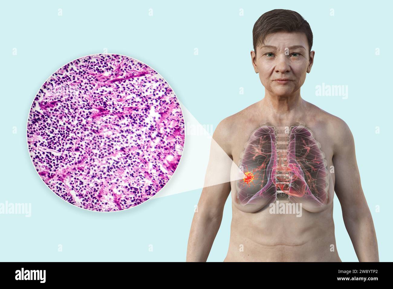 Woman with lung cancer, illustration Stock Photohttps://www.alamy.com/image-license-details/?v=1https://www.alamy.com/woman-with-lung-cancer-illustration-image590681898.html
RF2W8YTP2–Woman with lung cancer, illustration
 Unseen old woman rubs, feels tender hand with stiff knot under ring finger. Dupuytren contracture Stock Photohttps://www.alamy.com/image-license-details/?v=1https://www.alamy.com/unseen-old-woman-rubs-feels-tender-hand-with-stiff-knot-under-ring-finger-dupuytren-contracture-image620416299.html
RF2Y1AB8Y–Unseen old woman rubs, feels tender hand with stiff knot under ring finger. Dupuytren contracture
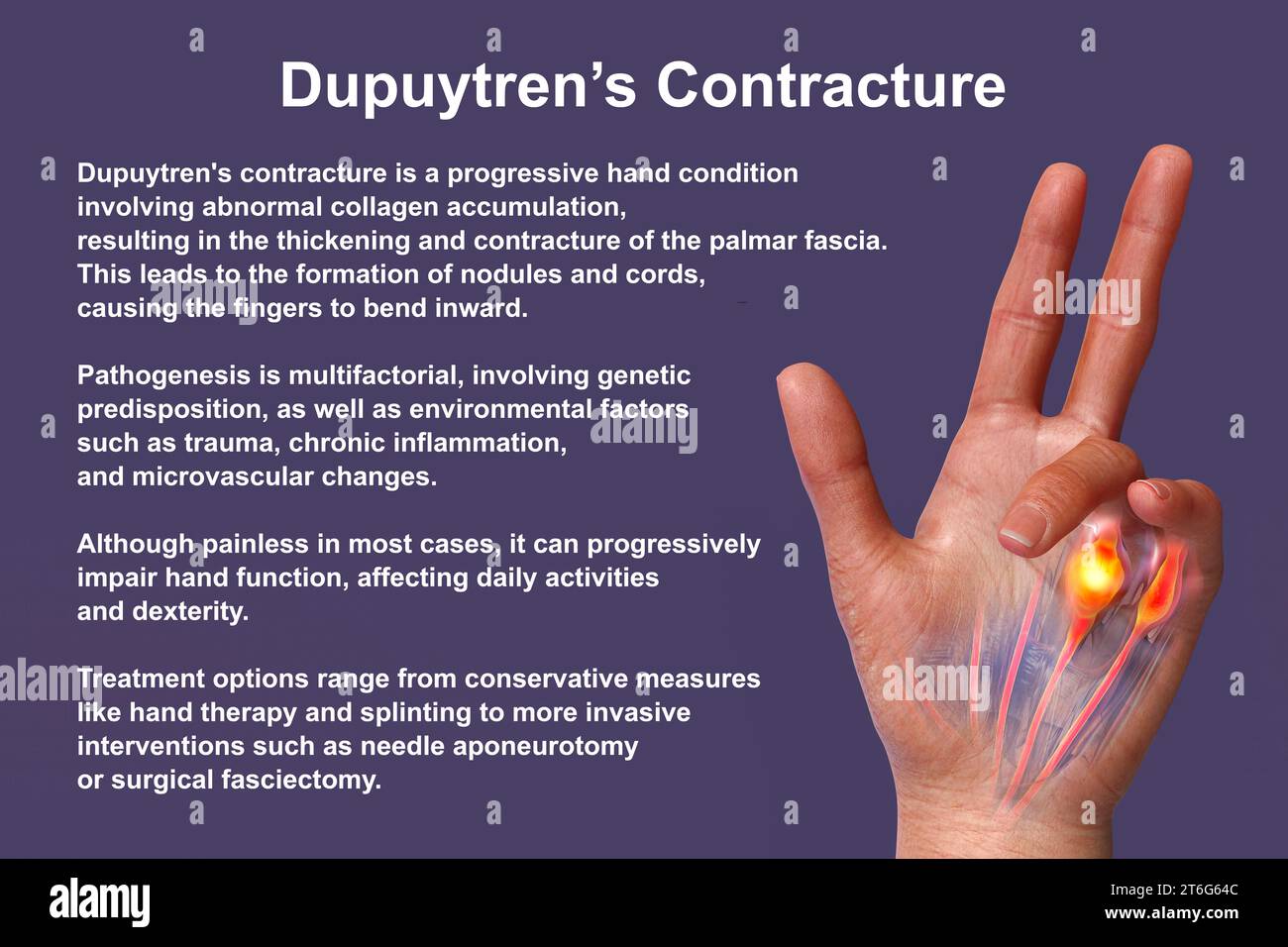 A 3D medical illustration displaying a patient's hand with Dupuytren's contracture, emphasizing the affected tendons and palmar fascia to illustrate t Stock Photohttps://www.alamy.com/image-license-details/?v=1https://www.alamy.com/a-3d-medical-illustration-displaying-a-patients-hand-with-dupuytrens-contracture-emphasizing-the-affected-tendons-and-palmar-fascia-to-illustrate-t-image571986140.html
RF2T6G64C–A 3D medical illustration displaying a patient's hand with Dupuytren's contracture, emphasizing the affected tendons and palmar fascia to illustrate t
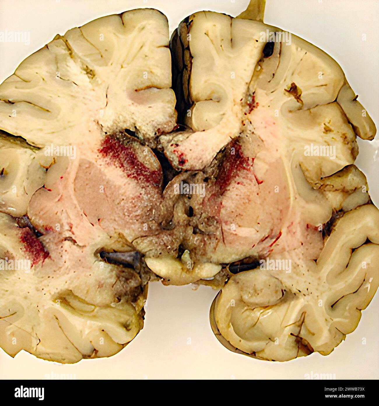 Gross specimen of brain tissue from a patient who died of granulomatous amebic encephalitis caused by Balamuthia mandrillaris. Stock Photohttps://www.alamy.com/image-license-details/?v=1https://www.alamy.com/gross-specimen-of-brain-tissue-from-a-patient-who-died-of-granulomatous-amebic-encephalitis-caused-by-balamuthia-mandrillaris-image600765982.html
RM2WWB73X–Gross specimen of brain tissue from a patient who died of granulomatous amebic encephalitis caused by Balamuthia mandrillaris.
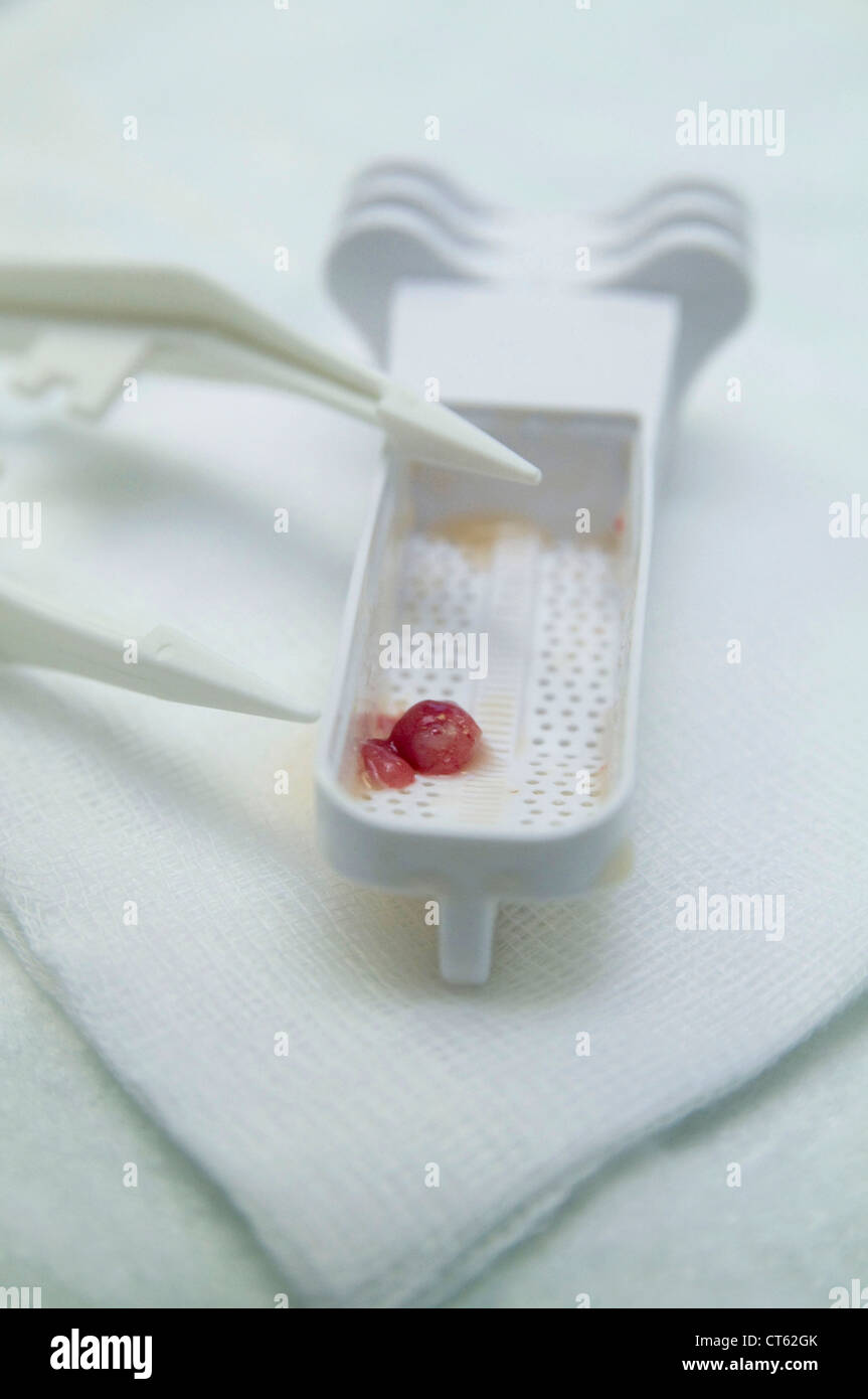 POLYP IN THE COLON, ANATOMY Stock Photohttps://www.alamy.com/image-license-details/?v=1https://www.alamy.com/stock-photo-polyp-in-the-colon-anatomy-49306227.html
RMCT62GK–POLYP IN THE COLON, ANATOMY
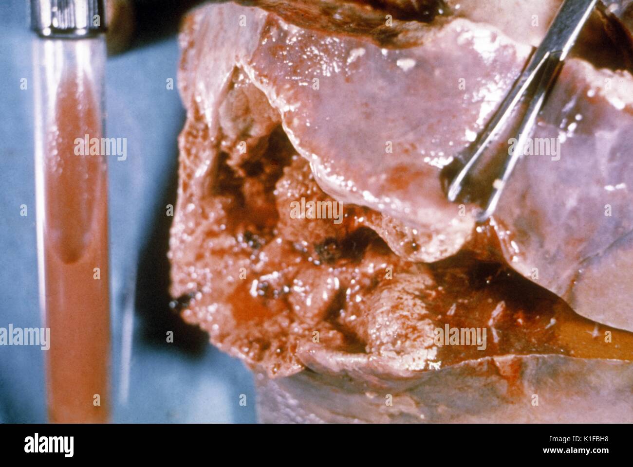 Gross pathology of amebic abscess of liver. Tube of 'chocolate' pus from abscess. Image courtesy CDC/Dr. Mae Melvin, Dr. E. West of Mobile, AL, 1962. Stock Photohttps://www.alamy.com/image-license-details/?v=1https://www.alamy.com/gross-pathology-of-amebic-abscess-of-liver-tube-of-chocolate-pus-from-image155846356.html
RMK1FBH8–Gross pathology of amebic abscess of liver. Tube of 'chocolate' pus from abscess. Image courtesy CDC/Dr. Mae Melvin, Dr. E. West of Mobile, AL, 1962.
 ENDOMETRIOSIS SURGERY Stock Photohttps://www.alamy.com/image-license-details/?v=1https://www.alamy.com/endometriosis-surgery-image184709486.html
RMMME6RA–ENDOMETRIOSIS SURGERY
 Gross pathology of polyp, colon, Incidental autopsy finding of a pedunculated polyp during a gross pathologic exam of a colon specimen. Image courtesy CDC/Dr. Edwin P. Ewing, Jr, 1972. Stock Photohttps://www.alamy.com/image-license-details/?v=1https://www.alamy.com/gross-pathology-of-polyp-colon-incidental-autopsy-finding-of-a-pedunculated-image155848534.html
RMK1FEB2–Gross pathology of polyp, colon, Incidental autopsy finding of a pedunculated polyp during a gross pathologic exam of a colon specimen. Image courtesy CDC/Dr. Edwin P. Ewing, Jr, 1972.
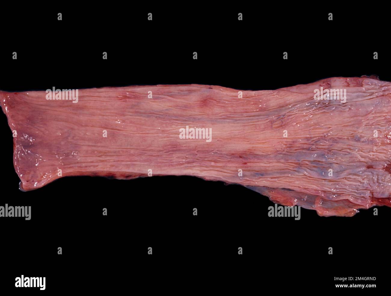 Opened human oesophagus showing oesophageal varices. They are dilated submucosal veins located in the lower third of the oesophagus. They are a conseq Stock Photohttps://www.alamy.com/image-license-details/?v=1https://www.alamy.com/opened-human-oesophagus-showing-oesophageal-varices-they-are-dilated-submucosal-veins-located-in-the-lower-third-of-the-oesophagus-they-are-a-conseq-image501929161.html
RF2M4GRND–Opened human oesophagus showing oesophageal varices. They are dilated submucosal veins located in the lower third of the oesophagus. They are a conseq
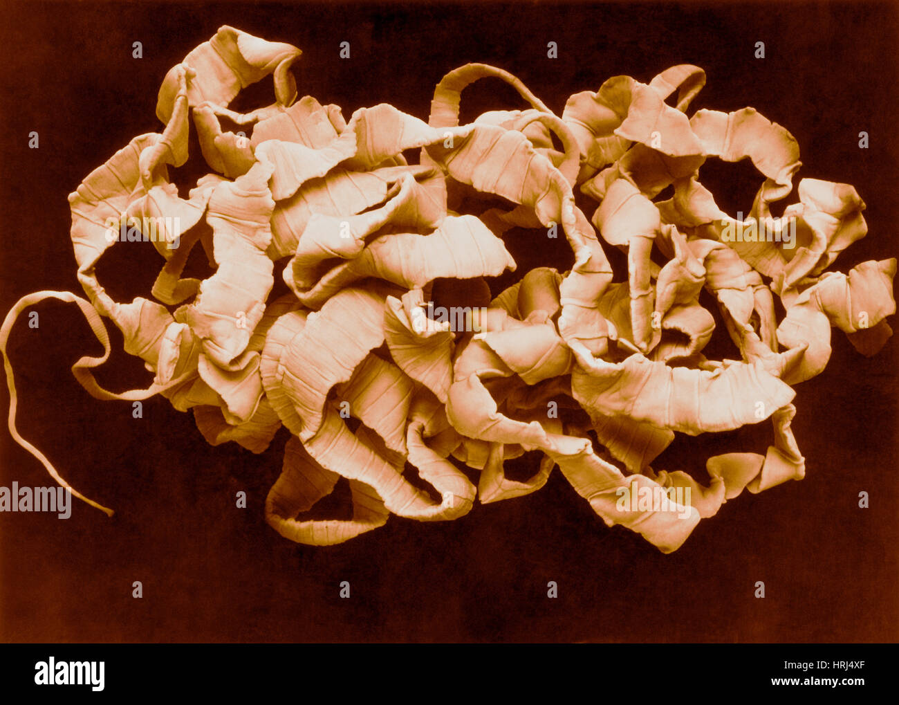 Human Tapeworm Stock Photohttps://www.alamy.com/image-license-details/?v=1https://www.alamy.com/stock-photo-human-tapeworm-135008679.html
RMHRJ4XF–Human Tapeworm
 Coronal section through a human brain at the thalamus level showing a large abscess, probably of bacterial origin, located in the temporal lobe. The g Stock Photohttps://www.alamy.com/image-license-details/?v=1https://www.alamy.com/coronal-section-through-a-human-brain-at-the-thalamus-level-showing-a-large-abscess-probably-of-bacterial-origin-located-in-the-temporal-lobe-the-g-image501929164.html
RF2M4GRNG–Coronal section through a human brain at the thalamus level showing a large abscess, probably of bacterial origin, located in the temporal lobe. The g
 Fibroids and allied tumours (myoma and adenomyoma) : their pathology, clinical features and surgical treatment . ii6 GROSS LESIONS OF APPENDAGES chap.. h^ V.;;.^;^ VI MALIGNANT OVARIAN GROWTHS 117 Adenocarcinoma of the Ovary.—Cullen found malig-nant disease of the ovaries in 8 out of 934 cases of myoma Stock Photohttps://www.alamy.com/image-license-details/?v=1https://www.alamy.com/fibroids-and-allied-tumours-myoma-and-adenomyoma-their-pathology-clinical-features-and-surgical-treatment-ii6-gross-lesions-of-appendages-chap-h-v-vi-malignant-ovarian-growths-117-adenocarcinoma-of-the-ovarycullen-found-malig-nant-disease-of-the-ovaries-in-8-out-of-934-cases-of-myoma-image340202610.html
RM2ANDFW6–Fibroids and allied tumours (myoma and adenomyoma) : their pathology, clinical features and surgical treatment . ii6 GROSS LESIONS OF APPENDAGES chap.. h^ V.;;.^;^ VI MALIGNANT OVARIAN GROWTHS 117 Adenocarcinoma of the Ovary.—Cullen found malig-nant disease of the ovaries in 8 out of 934 cases of myoma
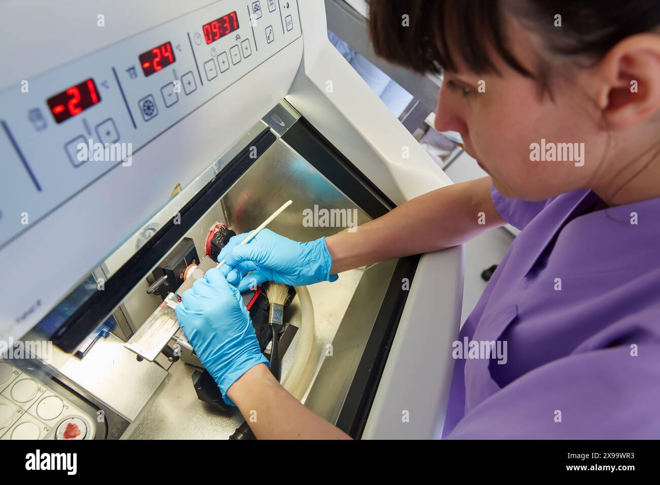 Cryostat for Standard Applications in the Clinical Histopathology Laboratory, Anatomic Pathology, Hospital Donostia, San Sebastian, Gipuzkoa, Basque Country, Spain. Stock Photohttps://www.alamy.com/image-license-details/?v=1https://www.alamy.com/cryostat-for-standard-applications-in-the-clinical-histopathology-laboratory-anatomic-pathology-hospital-donostia-san-sebastian-gipuzkoa-basque-country-spain-image608112599.html
RM2X99WR3–Cryostat for Standard Applications in the Clinical Histopathology Laboratory, Anatomic Pathology, Hospital Donostia, San Sebastian, Gipuzkoa, Basque Country, Spain.
 Man with smoker's lungs, illustration Stock Photohttps://www.alamy.com/image-license-details/?v=1https://www.alamy.com/man-with-smokers-lungs-illustration-image590681971.html
RF2W8YTTK–Man with smoker's lungs, illustration
 A 3D medical illustration displaying a patient's hand with Dupuytren's contracture, emphasizing the affected tendons and palmar fascia to illustrate t Stock Photohttps://www.alamy.com/image-license-details/?v=1https://www.alamy.com/a-3d-medical-illustration-displaying-a-patients-hand-with-dupuytrens-contracture-emphasizing-the-affected-tendons-and-palmar-fascia-to-illustrate-t-image571986146.html
RF2T6G64J–A 3D medical illustration displaying a patient's hand with Dupuytren's contracture, emphasizing the affected tendons and palmar fascia to illustrate t
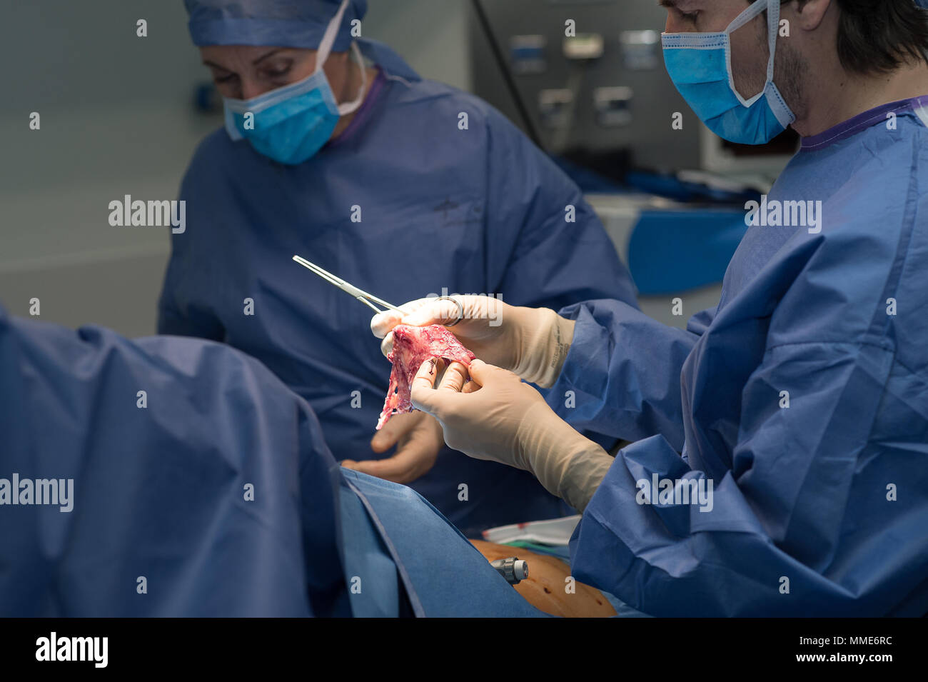 ENDOMETRIOSIS SURGERY Stock Photohttps://www.alamy.com/image-license-details/?v=1https://www.alamy.com/endometriosis-surgery-image184709488.html
RMMME6RC–ENDOMETRIOSIS SURGERY
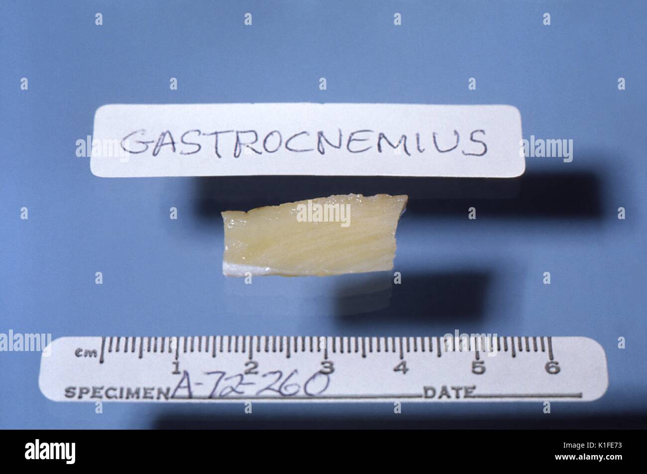 Gross pathology of skeletal muscle in fatal Duchenne muscular dystrophy, This gross fixed autopsy specimen of the gastrocnemius muscle was harvested from a patient who died of pseudohypertrophic muscular dystrophy, Duchenne type. In this form of the disease, yellowish-white fat replaces normally reddish-brown skeletal muscle. Image courtesy CDC/Dr. Edwin P. Ewing, Jr, 1972. Stock Photohttps://www.alamy.com/image-license-details/?v=1https://www.alamy.com/gross-pathology-of-skeletal-muscle-in-fatal-duchenne-muscular-dystrophy-image155848423.html
RMK1FE73–Gross pathology of skeletal muscle in fatal Duchenne muscular dystrophy, This gross fixed autopsy specimen of the gastrocnemius muscle was harvested from a patient who died of pseudohypertrophic muscular dystrophy, Duchenne type. In this form of the disease, yellowish-white fat replaces normally reddish-brown skeletal muscle. Image courtesy CDC/Dr. Edwin P. Ewing, Jr, 1972.
 Gross pathology of fixed, cut brain showing hemorrhagic meningitis due to inhalation anthrax Image courtesy CDC, 1966. Stock Photohttps://www.alamy.com/image-license-details/?v=1https://www.alamy.com/gross-pathology-of-fixed-cut-brain-showing-hemorrhagic-meningitis-image155845038.html
RMK1F9X6–Gross pathology of fixed, cut brain showing hemorrhagic meningitis due to inhalation anthrax Image courtesy CDC, 1966.
 Coronal section through a human brain at the thalamus level showing a large abscess, probably of bacterial origin, located in the temporal lobe. The g Stock Photohttps://www.alamy.com/image-license-details/?v=1https://www.alamy.com/coronal-section-through-a-human-brain-at-the-thalamus-level-showing-a-large-abscess-probably-of-bacterial-origin-located-in-the-temporal-lobe-the-g-image501929162.html
RF2M4GRNE–Coronal section through a human brain at the thalamus level showing a large abscess, probably of bacterial origin, located in the temporal lobe. The g
 Gross pathology of intestinal ulcers due to amebiasis. Image courtesy CDC/Dr. Mae Melvin, Dr. E. West of Mobile, AL, 1962. Stock Photohttps://www.alamy.com/image-license-details/?v=1https://www.alamy.com/gross-pathology-of-intestinal-ulcers-due-to-amebiasis-image-courtesy-image155846394.html
RMK1FBJJ–Gross pathology of intestinal ulcers due to amebiasis. Image courtesy CDC/Dr. Mae Melvin, Dr. E. West of Mobile, AL, 1962.
 Close-up of a brain slice showing two large intraparenchymal haemorrhages (one of the partially organized). Hypertension is the most frequent cause of Stock Photohttps://www.alamy.com/image-license-details/?v=1https://www.alamy.com/close-up-of-a-brain-slice-showing-two-large-intraparenchymal-haemorrhages-one-of-the-partially-organized-hypertension-is-the-most-frequent-cause-of-image501929160.html
RF2M4GRNC–Close-up of a brain slice showing two large intraparenchymal haemorrhages (one of the partially organized). Hypertension is the most frequent cause of
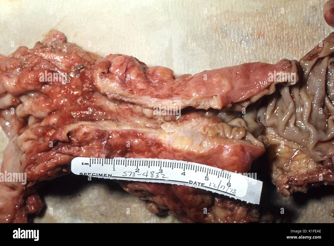 Gross pathology of adenocarcinoma, colon, 'Napkin-ring' configuration of colonic adenocarcinoma, which was revealed in this surgical specimen. Image courtesy CDC/Dr. Edwin P. Ewing, Jr., 1974. Stock Photohttps://www.alamy.com/image-license-details/?v=1https://www.alamy.com/gross-pathology-of-adenocarcinoma-colon-napkin-ring-configuration-image155848518.html
RMK1FEAE–Gross pathology of adenocarcinoma, colon, 'Napkin-ring' configuration of colonic adenocarcinoma, which was revealed in this surgical specimen. Image courtesy CDC/Dr. Edwin P. Ewing, Jr., 1974.
 Histology, Sample preparation, Anatomic Pathology, Hospital Donostia, San Sebastian, Gipuzkoa, Basque Country, Spain. Stock Photohttps://www.alamy.com/image-license-details/?v=1https://www.alamy.com/histology-sample-preparation-anatomic-pathology-hospital-donostia-san-sebastian-gipuzkoa-basque-country-spain-image608117022.html
RM2X9A3D2–Histology, Sample preparation, Anatomic Pathology, Hospital Donostia, San Sebastian, Gipuzkoa, Basque Country, Spain.
 Man with smoker's lungs, illustration Stock Photohttps://www.alamy.com/image-license-details/?v=1https://www.alamy.com/man-with-smokers-lungs-illustration-image590681966.html
RF2W8YTTE–Man with smoker's lungs, illustration
 Gross pathology of adenocarcinoma, stomach, Gastric adenocarcinoma with ulcerated surface and rolled border in a surgical specimen of the stomach. Image courtesy CDC/Dr. Edwin P. Ewing, Jr., 1973. Stock Photohttps://www.alamy.com/image-license-details/?v=1https://www.alamy.com/gross-pathology-of-adenocarcinoma-stomach-gastric-adenocarcinoma-with-image155848532.html
RMK1FEB0–Gross pathology of adenocarcinoma, stomach, Gastric adenocarcinoma with ulcerated surface and rolled border in a surgical specimen of the stomach. Image courtesy CDC/Dr. Edwin P. Ewing, Jr., 1973.
 A 3D medical illustration displaying a patient's hand with Dupuytren's contracture, emphasizing the affected tendons and palmar fascia to illustrate t Stock Photohttps://www.alamy.com/image-license-details/?v=1https://www.alamy.com/a-3d-medical-illustration-displaying-a-patients-hand-with-dupuytrens-contracture-emphasizing-the-affected-tendons-and-palmar-fascia-to-illustrate-t-image571986155.html
RF2T6G64Y–A 3D medical illustration displaying a patient's hand with Dupuytren's contracture, emphasizing the affected tendons and palmar fascia to illustrate t
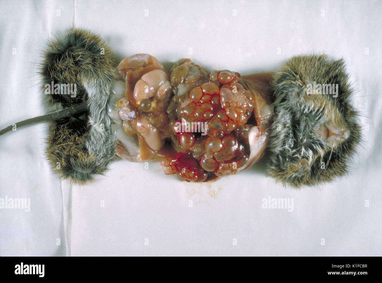 Gross pathology of cotton rat infected with Echinococcus multilocularis, Gross pathology of cotton rat infected with Echinococcus multilocularis . First E. locularis isolated in the United States proper. Necropsy. Image courtesy CDC/Dr. I. Kagan, 1964. Stock Photohttps://www.alamy.com/image-license-details/?v=1https://www.alamy.com/gross-pathology-of-cotton-rat-infected-with-echinococcus-multilocularis-image155846987.html
RMK1FCBR–Gross pathology of cotton rat infected with Echinococcus multilocularis, Gross pathology of cotton rat infected with Echinococcus multilocularis . First E. locularis isolated in the United States proper. Necropsy. Image courtesy CDC/Dr. I. Kagan, 1964.
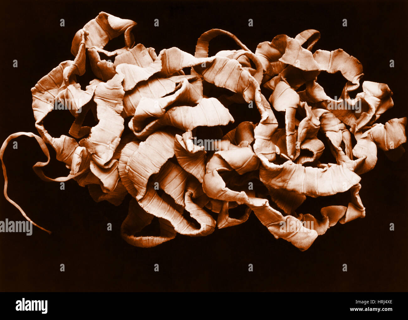 Human Tapeworm Stock Photohttps://www.alamy.com/image-license-details/?v=1https://www.alamy.com/stock-photo-human-tapeworm-135008678.html
RMHRJ4XE–Human Tapeworm
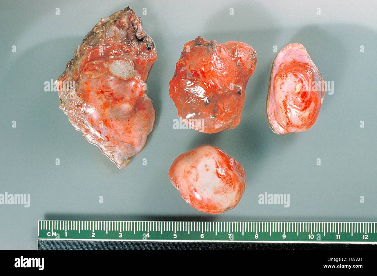 Gross pathology photograph of the membrane and hydatid daughter cysts excised from a human lung, 1961. Hydatid disease is a parasitic infestation by a tapeworm of the genus Echinococcus. Endemic areas usually involve third world countries. The liver is the most common organ involved, followed by the lungs. Image courtesy CDC/Dr. Kagan. Stock Photohttps://www.alamy.com/image-license-details/?v=1https://www.alamy.com/gross-pathology-photograph-of-the-membrane-and-hydatid-daughter-cysts-excised-from-a-human-lung-1961-hydatid-disease-is-a-parasitic-infestation-by-a-tapeworm-of-the-genus-echinococcus-endemic-areas-usually-involve-third-world-countries-the-liver-is-the-most-common-organ-involved-followed-by-the-lungs-image-courtesy-cdcdr-kagan-image244837036.html
RMT6983T–Gross pathology photograph of the membrane and hydatid daughter cysts excised from a human lung, 1961. Hydatid disease is a parasitic infestation by a tapeworm of the genus Echinococcus. Endemic areas usually involve third world countries. The liver is the most common organ involved, followed by the lungs. Image courtesy CDC/Dr. Kagan.
 Fibroids and allied tumours (myoma and adenomyoma) : their pathology, clinical features and surgical treatment . It > papuliferous ovarian cyst. The accompanying myoma ofthe uterus was not diagnosed before operation. OVARIAN CYSTS 113. 114 GROSS LESIONS OF APPENDAGES chap. Broad-Ligament Cysts.—Figure 125 shows an intra-ligamentary cystoma which arose on the right side, and by Stock Photohttps://www.alamy.com/image-license-details/?v=1https://www.alamy.com/fibroids-and-allied-tumours-myoma-and-adenomyoma-their-pathology-clinical-features-and-surgical-treatment-it-gt-papuliferous-ovarian-cyst-the-accompanying-myoma-ofthe-uterus-was-not-diagnosed-before-operation-ovarian-cysts-113-114-gross-lesions-of-appendages-chap-broad-ligament-cystsfigure-125-shows-an-intra-ligamentary-cystoma-which-arose-on-the-right-side-and-by-image340203705.html
RM2ANDH89–Fibroids and allied tumours (myoma and adenomyoma) : their pathology, clinical features and surgical treatment . It > papuliferous ovarian cyst. The accompanying myoma ofthe uterus was not diagnosed before operation. OVARIAN CYSTS 113. 114 GROSS LESIONS OF APPENDAGES chap. Broad-Ligament Cysts.—Figure 125 shows an intra-ligamentary cystoma which arose on the right side, and by
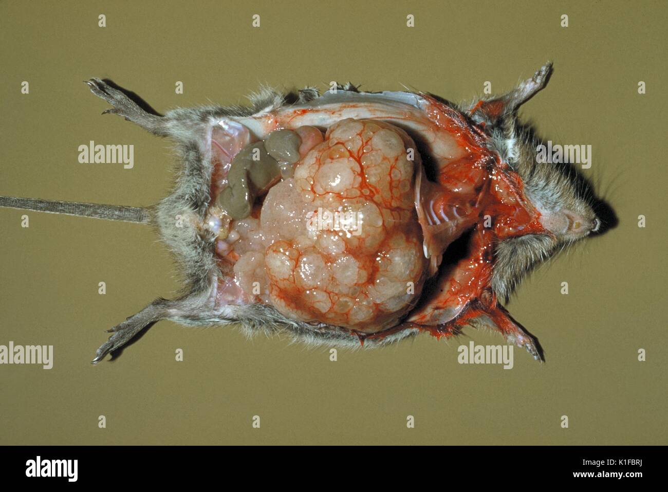 Dissected rat showing evidence of echinococcosis due to Echinococcus multilocularis in organs at 45 days. Dissected rat showing evidence of echinococcosis due to Echinococcus multilocularis in organs at 45 days. Gross pathology. Image courtesy CDC/Dr. Irving Kagan, 1960. Stock Photohttps://www.alamy.com/image-license-details/?v=1https://www.alamy.com/dissected-rat-showing-evidence-of-echinococcosis-due-to-echinococcus-image155846534.html
RMK1FBRJ–Dissected rat showing evidence of echinococcosis due to Echinococcus multilocularis in organs at 45 days. Dissected rat showing evidence of echinococcosis due to Echinococcus multilocularis in organs at 45 days. Gross pathology. Image courtesy CDC/Dr. Irving Kagan, 1960.
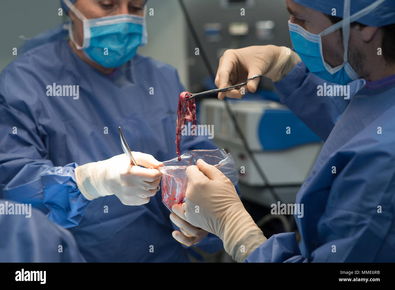 ENDOMETRIOSIS SURGERY Stock Photohttps://www.alamy.com/image-license-details/?v=1https://www.alamy.com/endometriosis-surgery-image184709487.html
RMMME6RB–ENDOMETRIOSIS SURGERY
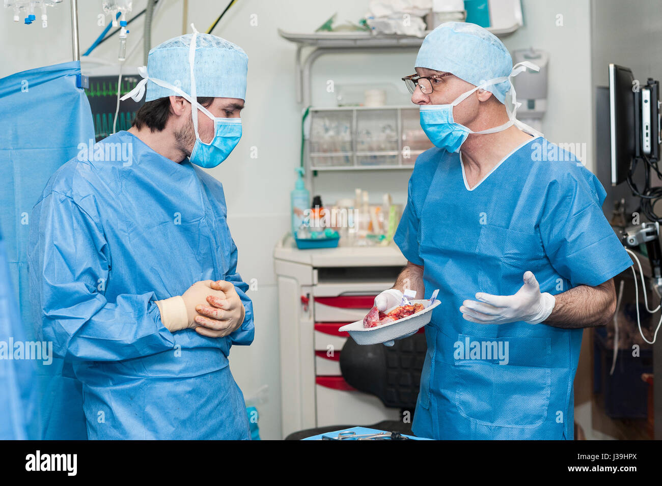 GYNECOLOGICAL SURGERY Stock Photohttps://www.alamy.com/image-license-details/?v=1https://www.alamy.com/stock-photo-gynecological-surgery-139738450.html
RMJ39HPX–GYNECOLOGICAL SURGERY
 Histology, Sample preparation, Anatomic Pathology, Hospital Donostia, San Sebastian, Gipuzkoa, Basque Country, Spain. Stock Photohttps://www.alamy.com/image-license-details/?v=1https://www.alamy.com/histology-sample-preparation-anatomic-pathology-hospital-donostia-san-sebastian-gipuzkoa-basque-country-spain-image624977417.html
RM2Y8P521–Histology, Sample preparation, Anatomic Pathology, Hospital Donostia, San Sebastian, Gipuzkoa, Basque Country, Spain.
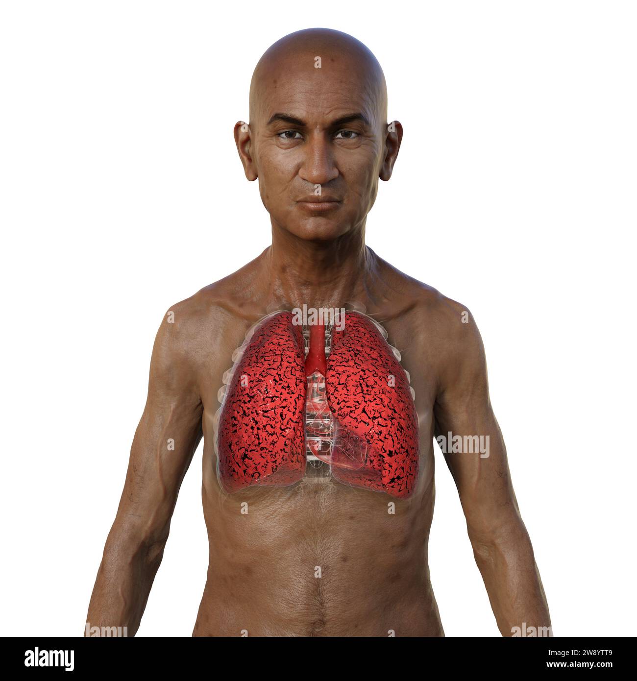 Man with smoker's lungs, illustration Stock Photohttps://www.alamy.com/image-license-details/?v=1https://www.alamy.com/man-with-smokers-lungs-illustration-image590681961.html
RF2W8YTT9–Man with smoker's lungs, illustration
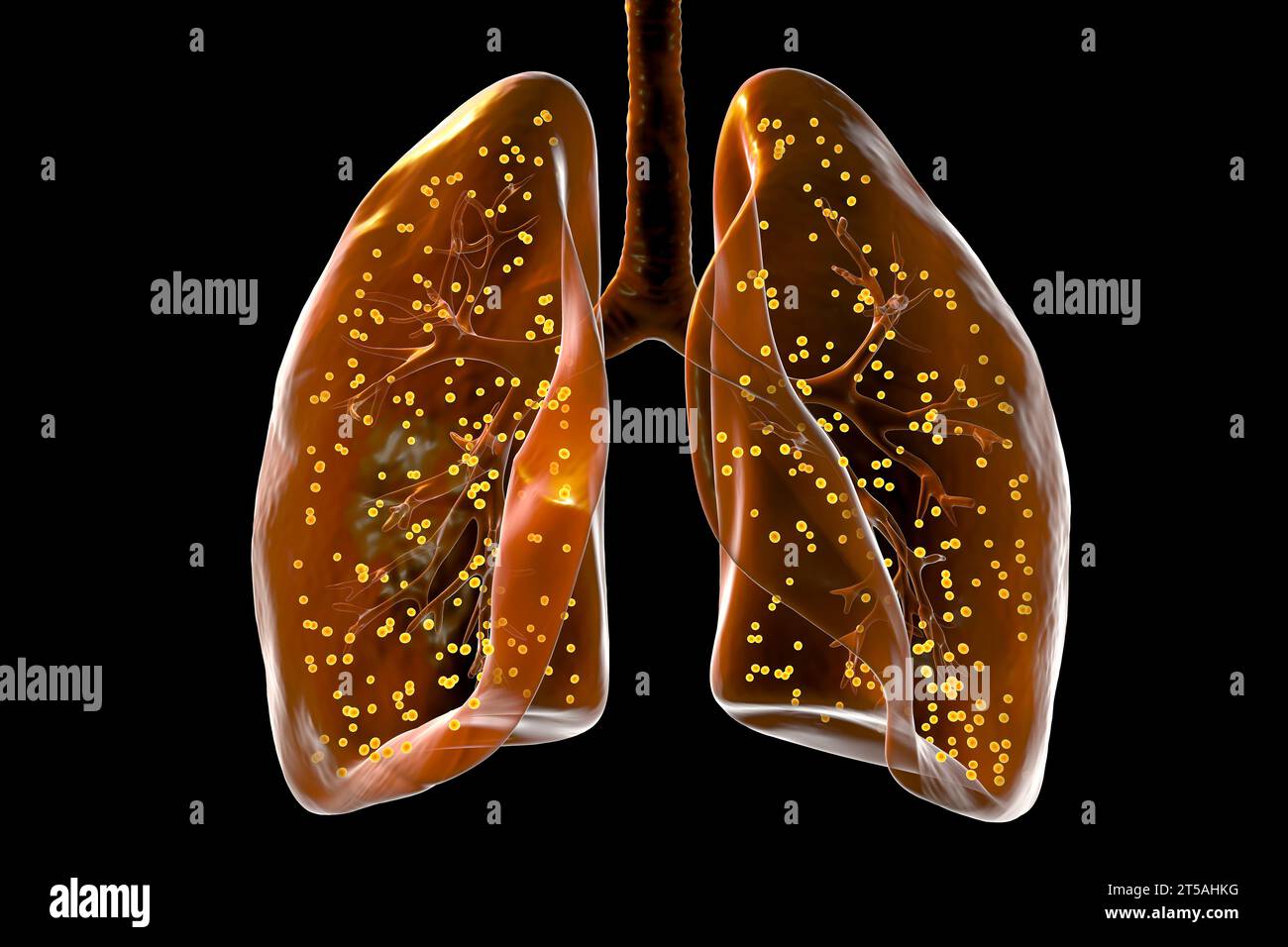 Lungs affected by miliary tuberculosis, illustration Stock Photohttps://www.alamy.com/image-license-details/?v=1https://www.alamy.com/lungs-affected-by-miliary-tuberculosis-illustration-image571248820.html
RF2T5AHKG–Lungs affected by miliary tuberculosis, illustration
 A 3D medical illustration displaying a patient's hand with Dupuytren's contracture, emphasizing the affected tendons and palmar fascia to illustrate t Stock Photohttps://www.alamy.com/image-license-details/?v=1https://www.alamy.com/a-3d-medical-illustration-displaying-a-patients-hand-with-dupuytrens-contracture-emphasizing-the-affected-tendons-and-palmar-fascia-to-illustrate-t-image571986160.html
RF2T6G654–A 3D medical illustration displaying a patient's hand with Dupuytren's contracture, emphasizing the affected tendons and palmar fascia to illustrate t
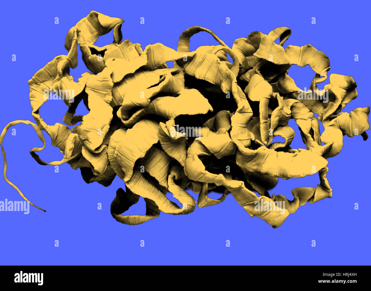 Human Tapeworm Stock Photohttps://www.alamy.com/image-license-details/?v=1https://www.alamy.com/stock-photo-human-tapeworm-135008681.html
RMHRJ4XH–Human Tapeworm
 Fibroids and allied tumours (myoma and adenomyoma) : their pathology, clinical features and surgical treatment . r-formed for the same tumour at a time when it was so bulkyas to cause considerable abdominal enlargement.^ If furthersupport were needed to prove the effects of the ovaries onthe growth of fibroids, the frequency of gross anatomicalchanges in the ovaries themselves in cases of uterine myomamight be mentioned. Histology. — Myomas are formed of two mesoblasticstructures—muscle-bundles and fibrous tissue ; the formerare arranged transversely and longitudinally and are boundtogether by Stock Photohttps://www.alamy.com/image-license-details/?v=1https://www.alamy.com/fibroids-and-allied-tumours-myoma-and-adenomyoma-their-pathology-clinical-features-and-surgical-treatment-r-formed-for-the-same-tumour-at-a-time-when-it-was-so-bulkyas-to-cause-considerable-abdominal-enlargement-if-furthersupport-were-needed-to-prove-the-effects-of-the-ovaries-onthe-growth-of-fibroids-the-frequency-of-gross-anatomicalchanges-in-the-ovaries-themselves-in-cases-of-uterine-myomamight-be-mentioned-histology-myomas-are-formed-of-two-mesoblasticstructuresmuscle-bundles-and-fibrous-tissue-the-formerare-arranged-transversely-and-longitudinally-and-are-boundtogether-by-image340238570.html
RM2ANF5NE–Fibroids and allied tumours (myoma and adenomyoma) : their pathology, clinical features and surgical treatment . r-formed for the same tumour at a time when it was so bulkyas to cause considerable abdominal enlargement.^ If furthersupport were needed to prove the effects of the ovaries onthe growth of fibroids, the frequency of gross anatomicalchanges in the ovaries themselves in cases of uterine myomamight be mentioned. Histology. — Myomas are formed of two mesoblasticstructures—muscle-bundles and fibrous tissue ; the formerare arranged transversely and longitudinally and are boundtogether by
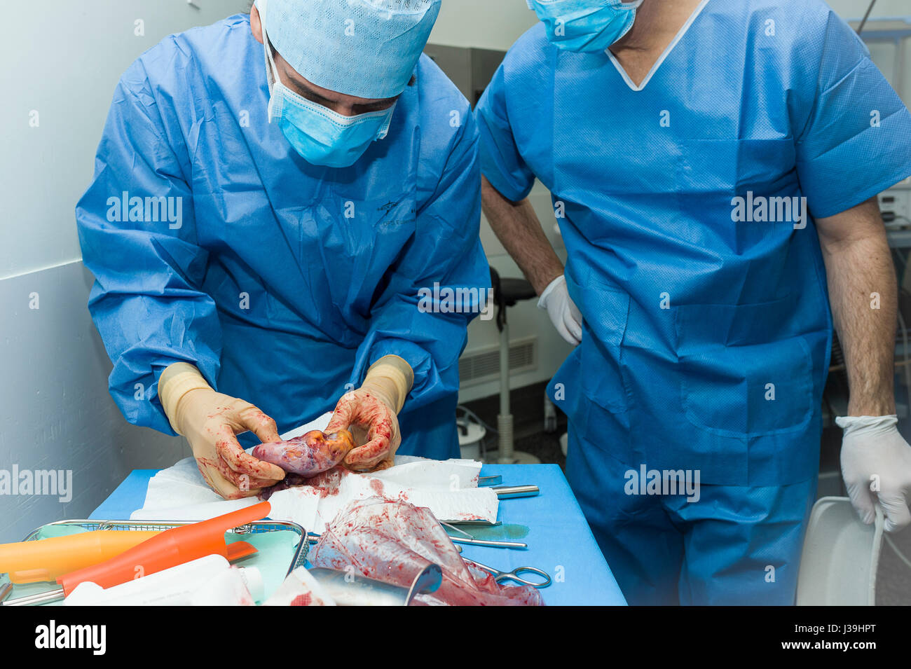 GYNECOLOGICAL SURGERY Stock Photohttps://www.alamy.com/image-license-details/?v=1https://www.alamy.com/stock-photo-gynecological-surgery-139738448.html
RMJ39HPT–GYNECOLOGICAL SURGERY
 ANATOMIC PATHOLOGY Stock Photohttps://www.alamy.com/image-license-details/?v=1https://www.alamy.com/stock-photo-anatomic-pathology-49185959.html
RMCT0H5B–ANATOMIC PATHOLOGY
 Selecting brain tissue sample, Anatomic Pathology, Hospital Donostia, San Sebastian, Gipuzkoa, Basque Country, Spain. Stock Photohttps://www.alamy.com/image-license-details/?v=1https://www.alamy.com/selecting-brain-tissue-sample-anatomic-pathology-hospital-donostia-san-sebastian-gipuzkoa-basque-country-spain-image608110081.html
RM2X99PH5–Selecting brain tissue sample, Anatomic Pathology, Hospital Donostia, San Sebastian, Gipuzkoa, Basque Country, Spain.
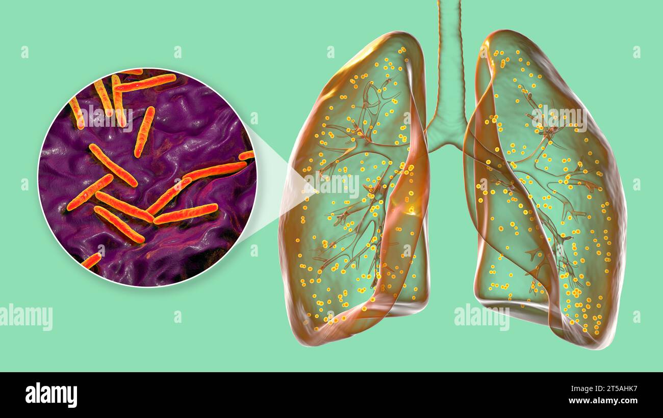 Lungs affected by miliary tuberculosis, illustration Stock Photohttps://www.alamy.com/image-license-details/?v=1https://www.alamy.com/lungs-affected-by-miliary-tuberculosis-illustration-image571248811.html
RF2T5AHK7–Lungs affected by miliary tuberculosis, illustration
 A 3D medical illustration displaying a patient's hand with Dupuytren's contracture, emphasizing the affected tendons and palmar fascia to illustrate t Stock Photohttps://www.alamy.com/image-license-details/?v=1https://www.alamy.com/a-3d-medical-illustration-displaying-a-patients-hand-with-dupuytrens-contracture-emphasizing-the-affected-tendons-and-palmar-fascia-to-illustrate-t-image571986156.html
RF2T6G650–A 3D medical illustration displaying a patient's hand with Dupuytren's contracture, emphasizing the affected tendons and palmar fascia to illustrate t