Quick filters:
Head of femur Stock Photos and Images
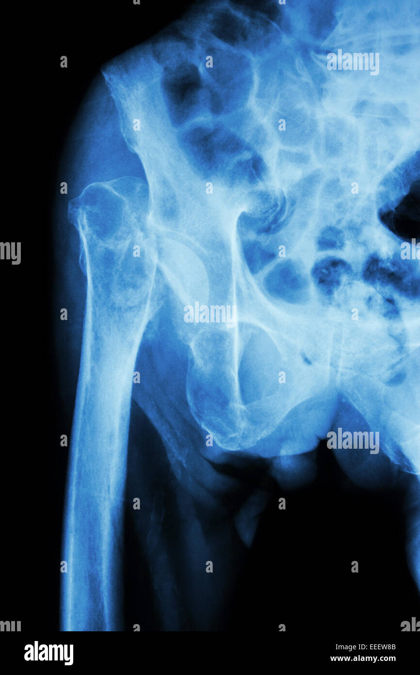 X-ray pelvis & hip joint : Fracture head of femur (thigh bone) Stock Photohttps://www.alamy.com/image-license-details/?v=1https://www.alamy.com/stock-photo-x-ray-pelvis-hip-joint-fracture-head-of-femur-thigh-bone-77773819.html
RFEEEW8B–X-ray pelvis & hip joint : Fracture head of femur (thigh bone)
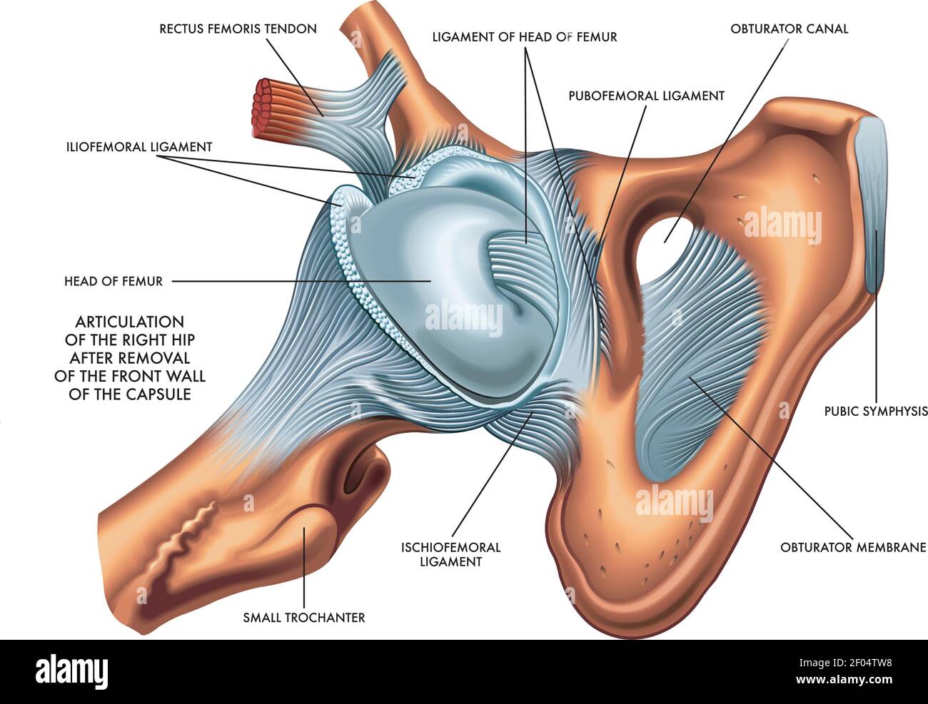 Medical illustration of articulation of the Right Hip whit annotations. Stock Vectorhttps://www.alamy.com/image-license-details/?v=1https://www.alamy.com/medical-illustration-of-articulation-of-the-right-hip-whit-annotations-image413156164.html
RF2F04TW8–Medical illustration of articulation of the Right Hip whit annotations.
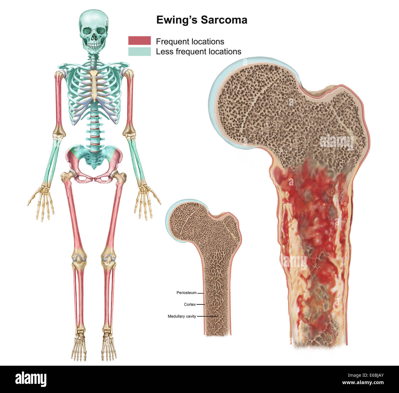 Ewings sarcoma locations on the skeleton and detail of tumor on head of femur. Stock Photohttps://www.alamy.com/image-license-details/?v=1https://www.alamy.com/stock-photo-ewings-sarcoma-locations-on-the-skeleton-and-detail-of-tumor-on-head-72785299.html
RME6BJAY–Ewings sarcoma locations on the skeleton and detail of tumor on head of femur.
 FRACTURED FEMUR Stock Photohttps://www.alamy.com/image-license-details/?v=1https://www.alamy.com/stock-photo-fractured-femur-93467973.html
RMFC1R9W–FRACTURED FEMUR
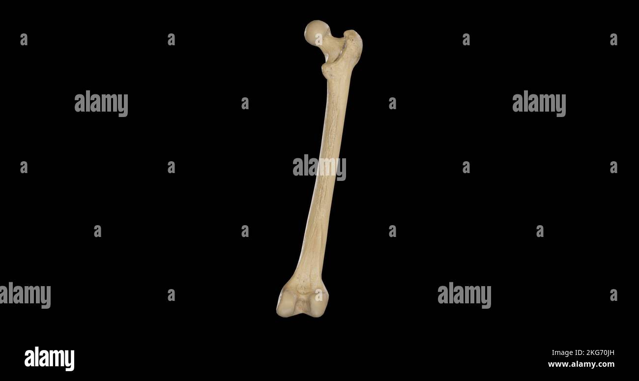 Posterior view of Right Femur Stock Photohttps://www.alamy.com/image-license-details/?v=1https://www.alamy.com/posterior-view-of-right-femur-image491878985.html
RF2KG70JH–Posterior view of Right Femur
 This is a joint head of a femur of a juvenile woolly mammoth (Mammuthus primigenius). The whole is very weathered and in many places the spongy bone is visible., Bone, joint head, bone, L: 15.0 cm, W: 13.5 cm, D: 10.0 cm, Prehistory, Netherlands, North Holland , Texel, Texel Stock Photohttps://www.alamy.com/image-license-details/?v=1https://www.alamy.com/this-is-a-joint-head-of-a-femur-of-a-juvenile-woolly-mammoth-mammuthus-primigenius-the-whole-is-very-weathered-and-in-many-places-the-spongy-bone-is-visible-bone-joint-head-bone-l-150-cm-w-135-cm-d-100-cm-prehistory-netherlands-north-holland-texel-texel-image344496678.html
RM2B0D50P–This is a joint head of a femur of a juvenile woolly mammoth (Mammuthus primigenius). The whole is very weathered and in many places the spongy bone is visible., Bone, joint head, bone, L: 15.0 cm, W: 13.5 cm, D: 10.0 cm, Prehistory, Netherlands, North Holland , Texel, Texel
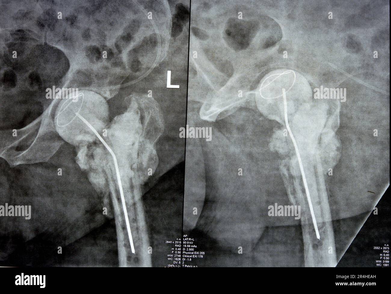 Plain X ray hip joint show left trans cervical fracture of the head of femur with temporary antibiotic loader spacer antibiotic-loaded bone cement aft Stock Photohttps://www.alamy.com/image-license-details/?v=1https://www.alamy.com/plain-x-ray-hip-joint-show-left-trans-cervical-fracture-of-the-head-of-femur-with-temporary-antibiotic-loader-spacer-antibiotic-loaded-bone-cement-aft-image553574857.html
RF2R4HEAH–Plain X ray hip joint show left trans cervical fracture of the head of femur with temporary antibiotic loader spacer antibiotic-loaded bone cement aft
 X-ray of a cat with a hip luxation, with displacement of head of femur from the acetabular socket caused by trauma. R indicates the right side Stock Photohttps://www.alamy.com/image-license-details/?v=1https://www.alamy.com/x-ray-of-a-cat-with-a-hip-luxation-with-displacement-of-head-of-femur-from-the-acetabular-socket-caused-by-trauma-r-indicates-the-right-side-image362127868.html
RF2C149P4–X-ray of a cat with a hip luxation, with displacement of head of femur from the acetabular socket caused by trauma. R indicates the right side
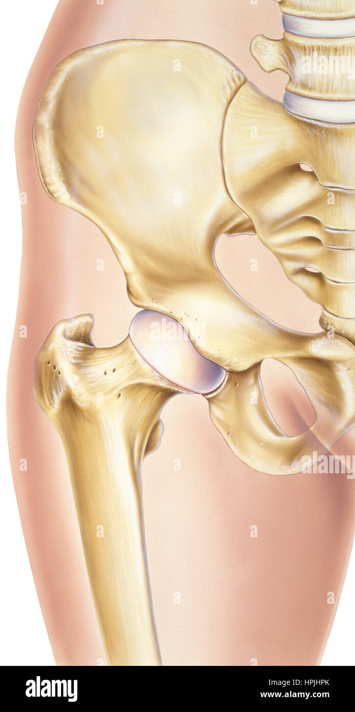 Shown is a normal human hip with the bones and joints visible, specifically the pelvis, acetabular (socket), femoral head (ball), femur, cartilag eand Stock Photohttps://www.alamy.com/image-license-details/?v=1https://www.alamy.com/stock-photo-shown-is-a-normal-human-hip-with-the-bones-and-joints-visible-specifically-134404107.html
RFHPJHPK–Shown is a normal human hip with the bones and joints visible, specifically the pelvis, acetabular (socket), femoral head (ball), femur, cartilag eand
 cave of the discovery of the head of a femur of a young Neanderthal man. Abruzzo, Italy, Europe Stock Photohttps://www.alamy.com/image-license-details/?v=1https://www.alamy.com/cave-of-the-discovery-of-the-head-of-a-femur-of-a-young-neanderthal-man-abruzzo-italy-europe-image380549041.html
RM2D33E55–cave of the discovery of the head of a femur of a young Neanderthal man. Abruzzo, Italy, Europe
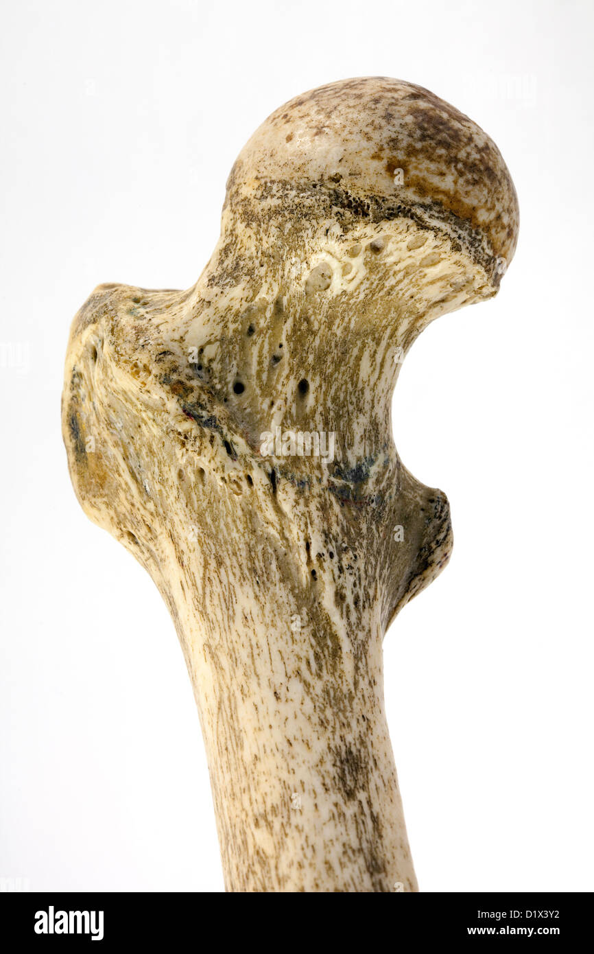 Human femur bone, close up to show the neck of femur, the commonest area to fracture in a fall, Right femur, anterior view Stock Photohttps://www.alamy.com/image-license-details/?v=1https://www.alamy.com/stock-photo-human-femur-bone-close-up-to-show-the-neck-of-femur-the-commonest-52819622.html
RMD1X3Y2–Human femur bone, close up to show the neck of femur, the commonest area to fracture in a fall, Right femur, anterior view
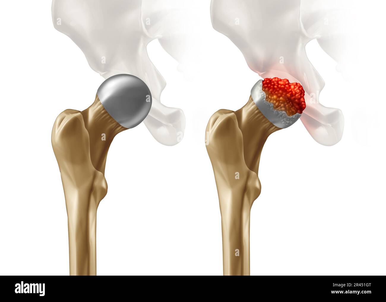 Femoral Head Disease and osteonecrosis or avascular necrosis and aseptic necrosis with a healthy hip compared to an osteoarthritis damaged pelvic join Stock Photohttps://www.alamy.com/image-license-details/?v=1https://www.alamy.com/femoral-head-disease-and-osteonecrosis-or-avascular-necrosis-and-aseptic-necrosis-with-a-healthy-hip-compared-to-an-osteoarthritis-damaged-pelvic-join-image553301416.html
RF2R451GT–Femoral Head Disease and osteonecrosis or avascular necrosis and aseptic necrosis with a healthy hip compared to an osteoarthritis damaged pelvic join
 Human femur head computer artwork. Stock Photohttps://www.alamy.com/image-license-details/?v=1https://www.alamy.com/human-femur-head-computer-artwork-image69878638.html
RFE1K6WJ–Human femur head computer artwork.
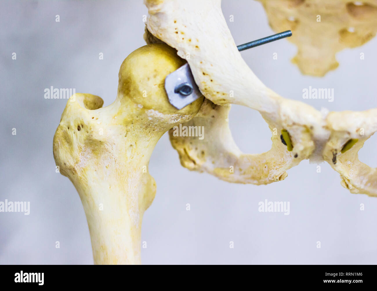 articulated hip bone and head of femur showing human hip joint anatomy in white background Stock Photohttps://www.alamy.com/image-license-details/?v=1https://www.alamy.com/articulated-hip-bone-and-head-of-femur-showing-human-hip-joint-anatomy-in-white-background-image238334214.html
RFRRN1M6–articulated hip bone and head of femur showing human hip joint anatomy in white background
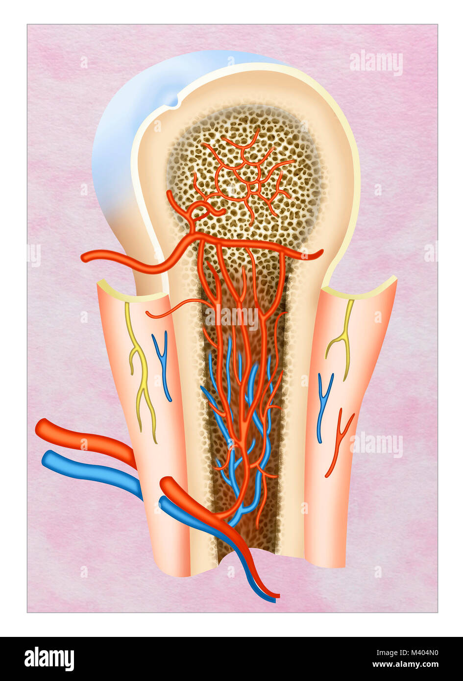 The process of blood formation is called hematopoiesis. This is found in the spongy tissue of the bones. The red bone marrow becomes the primary hemop Stock Photohttps://www.alamy.com/image-license-details/?v=1https://www.alamy.com/stock-photo-the-process-of-blood-formation-is-called-hematopoiesis-this-is-found-174566028.html
RFM404N0–The process of blood formation is called hematopoiesis. This is found in the spongy tissue of the bones. The red bone marrow becomes the primary hemop
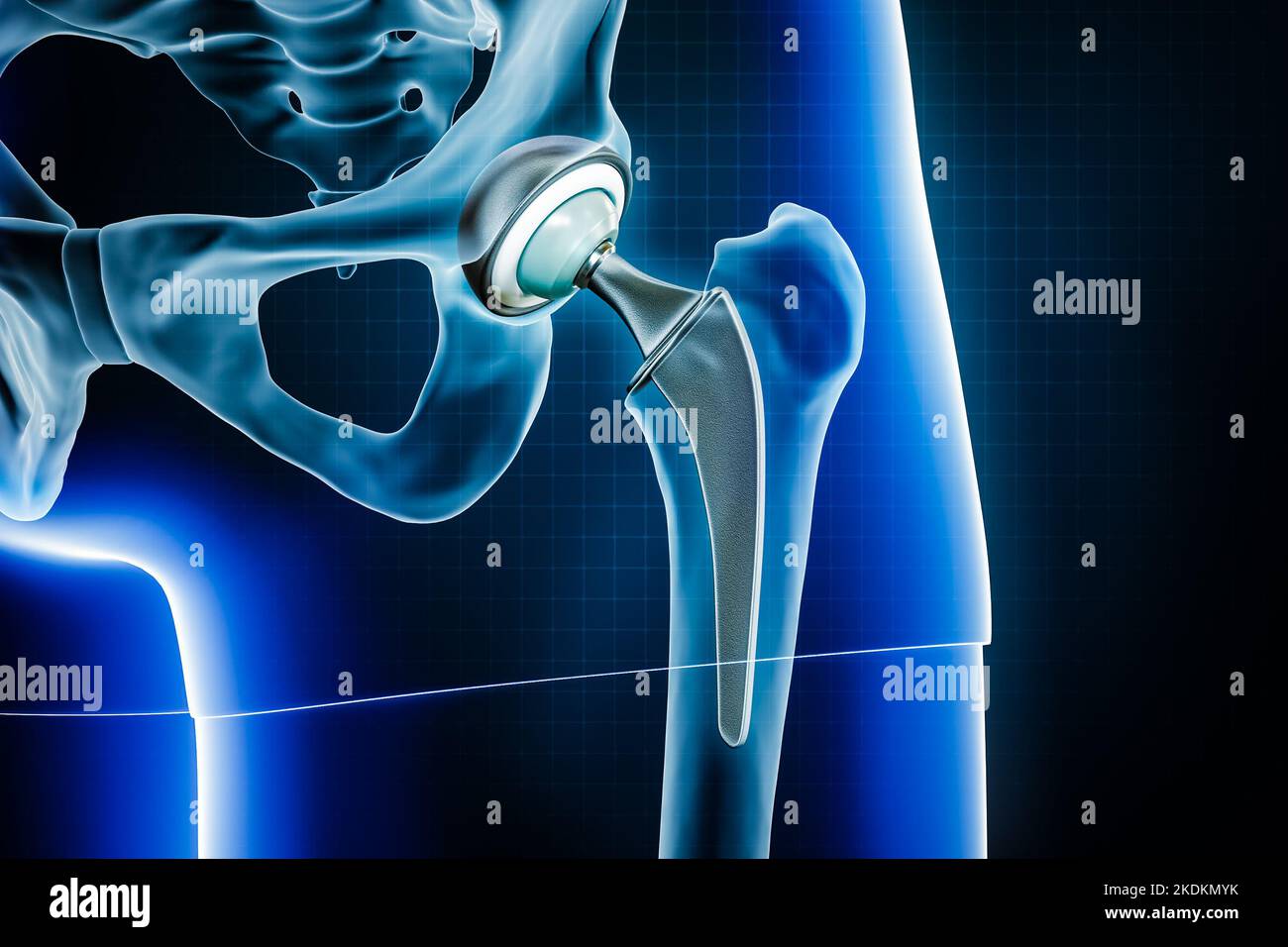 Femoral head hip prosthesis or implant. Total hip joint replacement surgery or arthroplasty 3D rendering illustration. Medical and healthcare, arthrit Stock Photohttps://www.alamy.com/image-license-details/?v=1https://www.alamy.com/femoral-head-hip-prosthesis-or-implant-total-hip-joint-replacement-surgery-or-arthroplasty-3d-rendering-illustration-medical-and-healthcare-arthrit-image490314375.html
RF2KDKMYK–Femoral head hip prosthesis or implant. Total hip joint replacement surgery or arthroplasty 3D rendering illustration. Medical and healthcare, arthrit
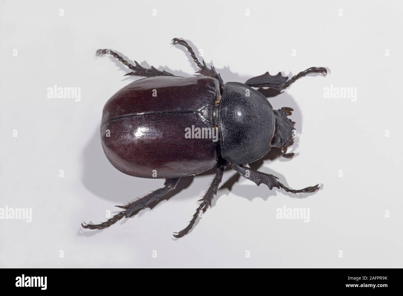 BEETLE (Xylotrupes sp.). Large 40mm long, head to tip of abdomen. Cuc Phuong National Park, Vietnam. Stock Photohttps://www.alamy.com/image-license-details/?v=1https://www.alamy.com/beetle-xylotrupes-sp-large-40mm-long-head-to-tip-of-abdomen-cuc-phuong-national-park-vietnam-image336718079.html
RM2AFPR9K–BEETLE (Xylotrupes sp.). Large 40mm long, head to tip of abdomen. Cuc Phuong National Park, Vietnam.
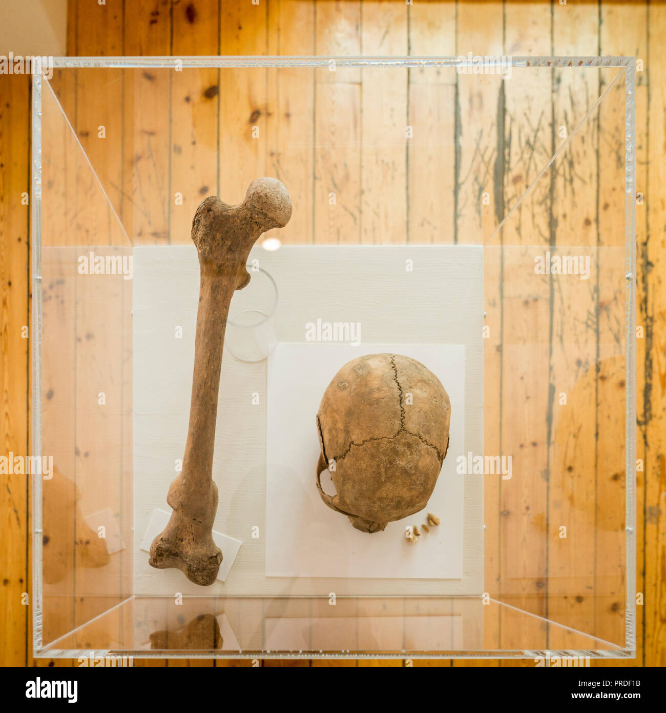 Human remains on display at Skriduklaustur, Eastern Iceland. Stock Photohttps://www.alamy.com/image-license-details/?v=1https://www.alamy.com/human-remains-on-display-at-skriduklaustur-eastern-iceland-image220958679.html
RMPRDF1B–Human remains on display at Skriduklaustur, Eastern Iceland.
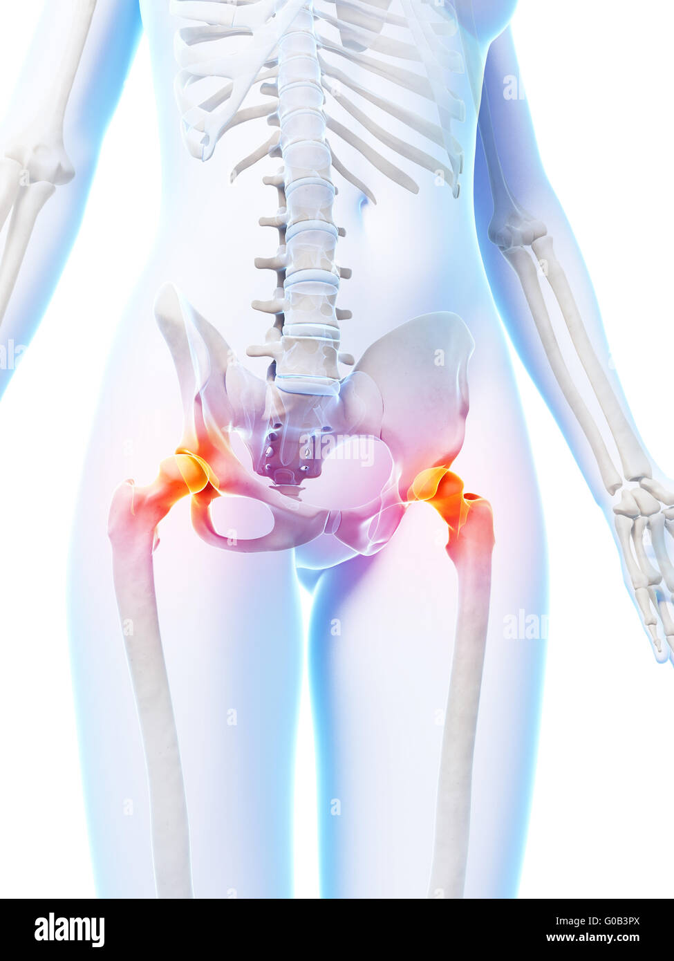 3d rendered illustration of painful hip joints Stock Photohttps://www.alamy.com/image-license-details/?v=1https://www.alamy.com/stock-photo-3d-rendered-illustration-of-painful-hip-joints-103506674.html
RMG0B3PX–3d rendered illustration of painful hip joints
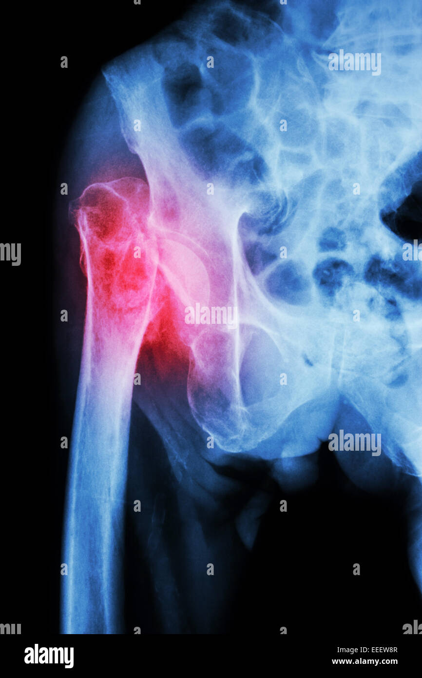 X-ray pelvis & hip joint : Fracture head of femur (thigh bone) Stock Photohttps://www.alamy.com/image-license-details/?v=1https://www.alamy.com/stock-photo-x-ray-pelvis-hip-joint-fracture-head-of-femur-thigh-bone-77773831.html
RFEEEW8R–X-ray pelvis & hip joint : Fracture head of femur (thigh bone)
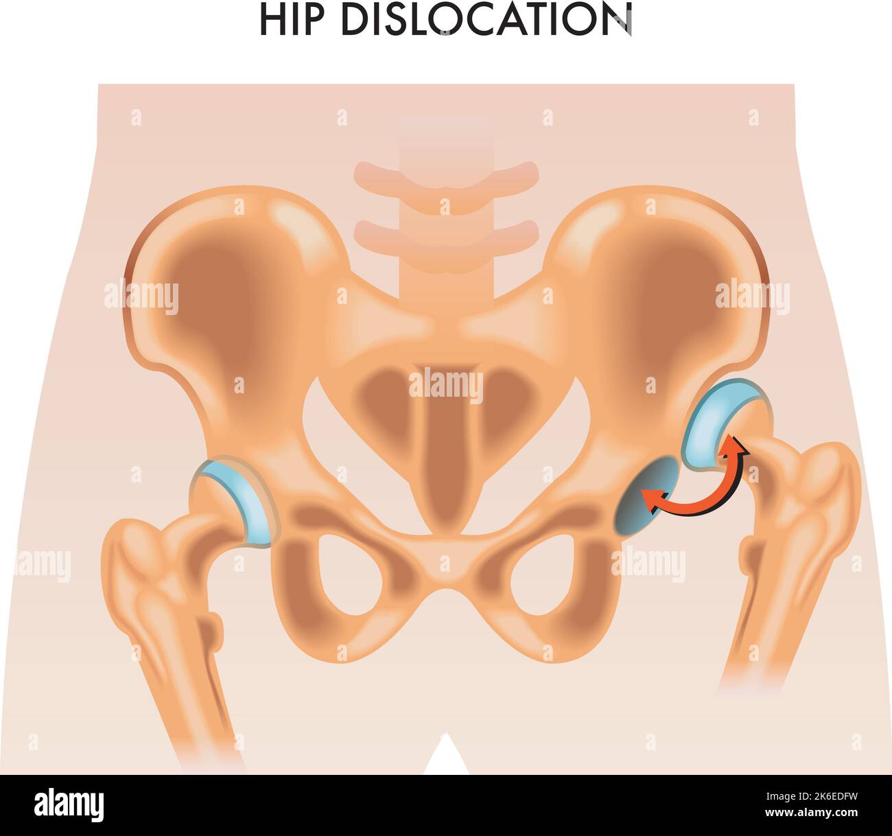 Medical illustration of the hip dislocation. Stock Vectorhttps://www.alamy.com/image-license-details/?v=1https://www.alamy.com/medical-illustration-of-the-hip-dislocation-image485896205.html
RF2K6EDFW–Medical illustration of the hip dislocation.
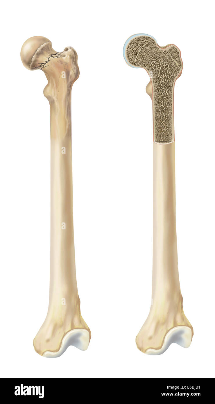 Head of femur fracture. Stock Photohttps://www.alamy.com/image-license-details/?v=1https://www.alamy.com/stock-photo-head-of-femur-fracture-72785301.html
RME6BJB1–Head of femur fracture.
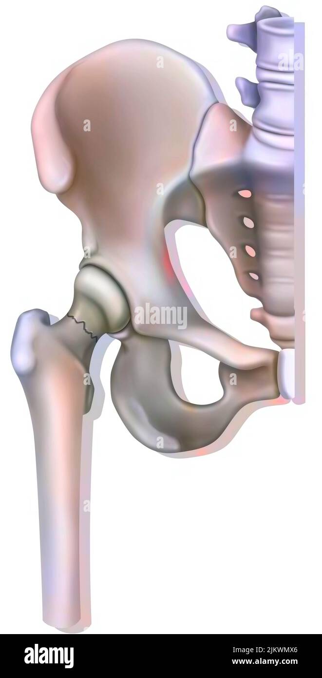 Bone system: fracture of the neck of the femur, linked to osteoporosis. Stock Photohttps://www.alamy.com/image-license-details/?v=1https://www.alamy.com/bone-system-fracture-of-the-neck-of-the-femur-linked-to-osteoporosis-image476923614.html
RF2JKWMX6–Bone system: fracture of the neck of the femur, linked to osteoporosis.
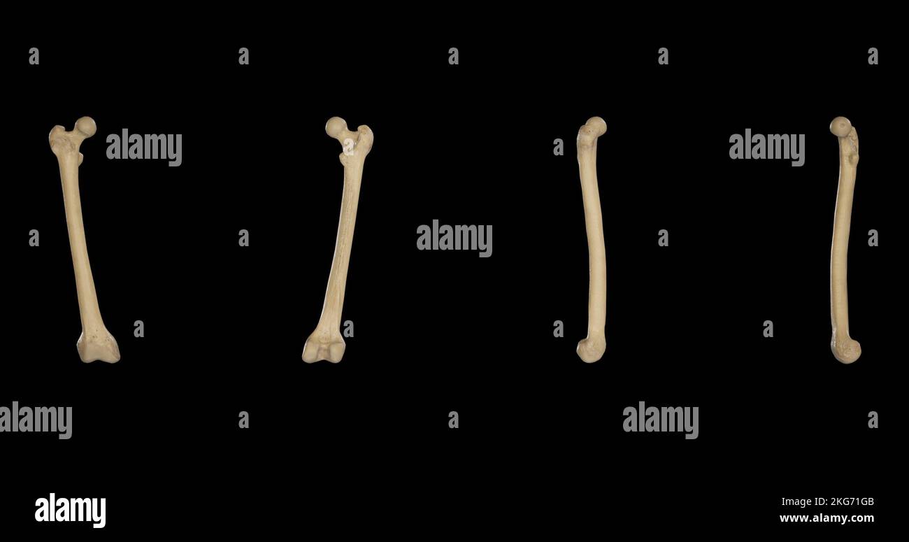 Right Femur from multiple sides Stock Photohttps://www.alamy.com/image-license-details/?v=1https://www.alamy.com/right-femur-from-multiple-sides-image491879707.html
RF2KG71GB–Right Femur from multiple sides
 Standing warrior with raised right arm and bent left arm. Shield and lance have been lost. On the head a point helmet. Furthermore cuirass with belt, short armor (subligaculum) and shin guards. Probably the war god Laran. On the right femur Etruscan inscription., Figurine, male statue, bronze, 36,8 x 11,7 x 11 cm., Fixed base (HxWxD): 10 x 9,3 x 9,3 cm., Transport size object (lxwxd): 48 , 5 x 12 x 11 cm., Mid-archaic 540-520 BC, Italy, Italy Stock Photohttps://www.alamy.com/image-license-details/?v=1https://www.alamy.com/standing-warrior-with-raised-right-arm-and-bent-left-arm-shield-and-lance-have-been-lost-on-the-head-a-point-helmet-furthermore-cuirass-with-belt-short-armor-subligaculum-and-shin-guards-probably-the-war-god-laran-on-the-right-femur-etruscan-inscription-figurine-male-statue-bronze-368-x-117-x-11-cm-fixed-base-hxwxd-10-x-93-x-93-cm-transport-size-object-lxwxd-48-5-x-12-x-11-cm-mid-archaic-540-520-bc-italy-italy-image344529349.html
RM2B0EJKH–Standing warrior with raised right arm and bent left arm. Shield and lance have been lost. On the head a point helmet. Furthermore cuirass with belt, short armor (subligaculum) and shin guards. Probably the war god Laran. On the right femur Etruscan inscription., Figurine, male statue, bronze, 36,8 x 11,7 x 11 cm., Fixed base (HxWxD): 10 x 9,3 x 9,3 cm., Transport size object (lxwxd): 48 , 5 x 12 x 11 cm., Mid-archaic 540-520 BC, Italy, Italy
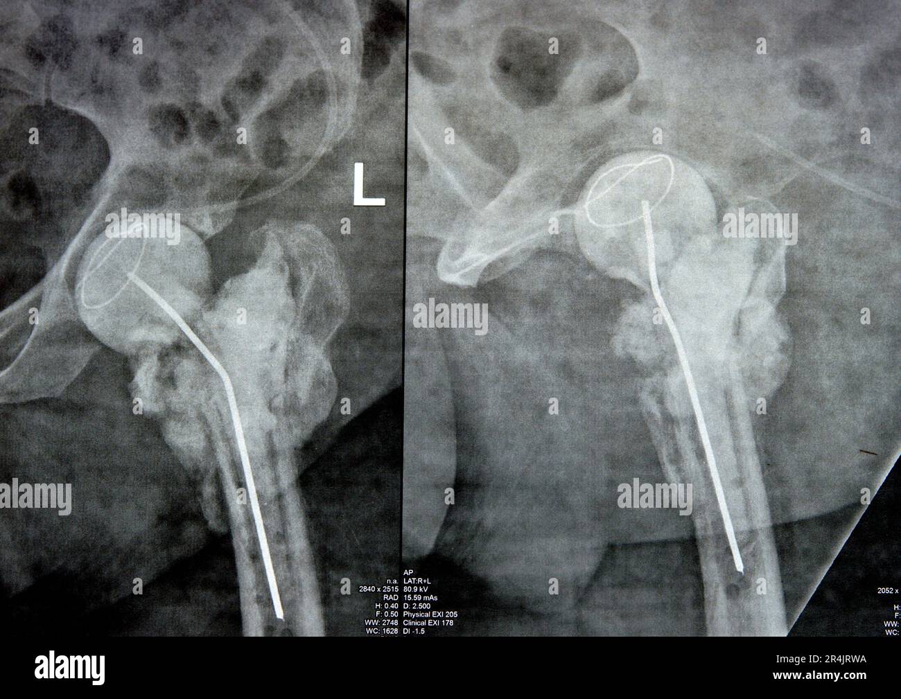 Plain X ray hip joint show left trans cervical fracture of the head of femur with temporary antibiotic loader spacer antibiotic-loaded bone cement aft Stock Photohttps://www.alamy.com/image-license-details/?v=1https://www.alamy.com/plain-x-ray-hip-joint-show-left-trans-cervical-fracture-of-the-head-of-femur-with-temporary-antibiotic-loader-spacer-antibiotic-loaded-bone-cement-aft-image553604278.html
RF2R4JRWA–Plain X ray hip joint show left trans cervical fracture of the head of femur with temporary antibiotic loader spacer antibiotic-loaded bone cement aft
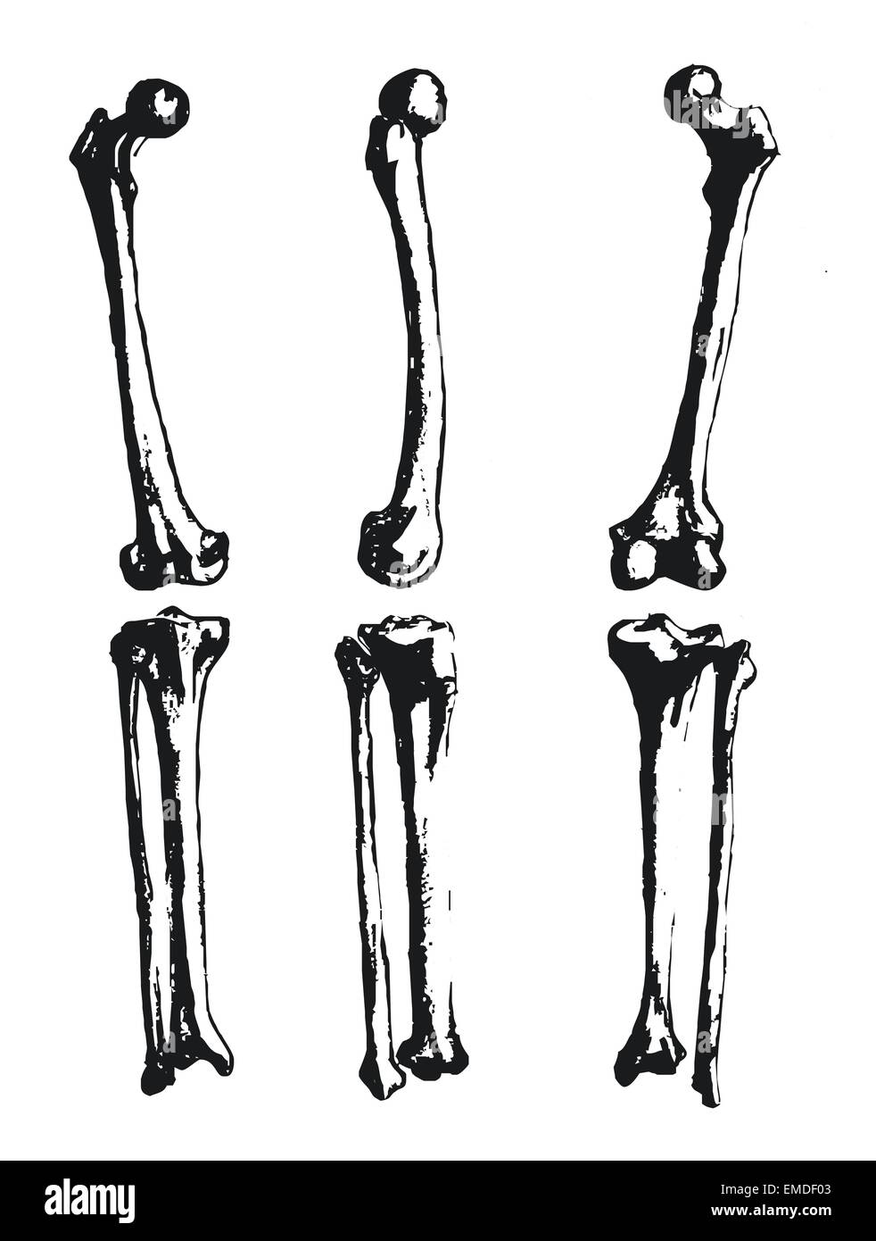 Hand drawn fibula and femur Stock Vectorhttps://www.alamy.com/image-license-details/?v=1https://www.alamy.com/stock-photo-hand-drawn-fibula-and-femur-81431731.html
RFEMDF03–Hand drawn fibula and femur
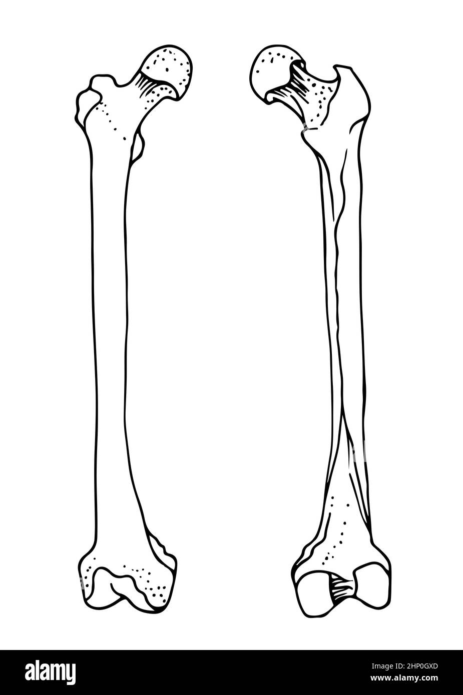 Human femur bones, vector hand drawn illustration isolated on a white background, orthopedics medicine anatomy sketch Stock Vectorhttps://www.alamy.com/image-license-details/?v=1https://www.alamy.com/human-femur-bones-vector-hand-drawn-illustration-isolated-on-a-white-background-orthopedics-medicine-anatomy-sketch-image461005285.html
RF2HP0GXD–Human femur bones, vector hand drawn illustration isolated on a white background, orthopedics medicine anatomy sketch
 cave of the discovery of the head of a femur of a young Neanderthal man. Abruzzo, Italy, Europe Stock Photohttps://www.alamy.com/image-license-details/?v=1https://www.alamy.com/cave-of-the-discovery-of-the-head-of-a-femur-of-a-young-neanderthal-man-abruzzo-italy-europe-image380549281.html
RM2D33EDN–cave of the discovery of the head of a femur of a young Neanderthal man. Abruzzo, Italy, Europe
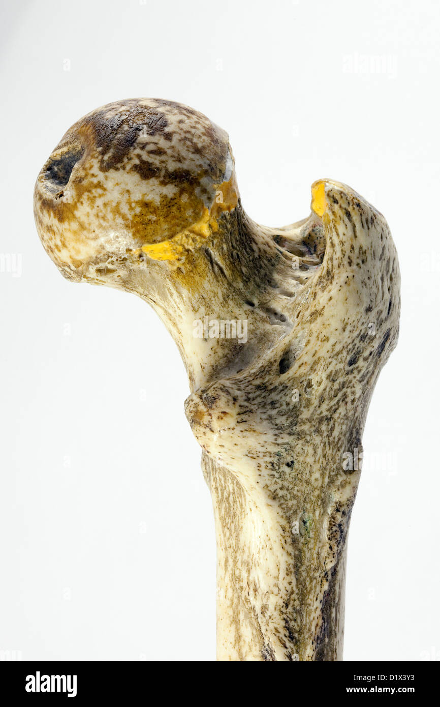 Femur bone human, close up of upper part to show the neck of femur, the commonest part to fracture - Right femur, posterior view Stock Photohttps://www.alamy.com/image-license-details/?v=1https://www.alamy.com/stock-photo-femur-bone-human-close-up-of-upper-part-to-show-the-neck-of-femur-52819623.html
RMD1X3Y3–Femur bone human, close up of upper part to show the neck of femur, the commonest part to fracture - Right femur, posterior view
 Intertrochanteric fracture on the femur head skeleton 3D medical illustration Stock Photohttps://www.alamy.com/image-license-details/?v=1https://www.alamy.com/intertrochanteric-fracture-on-the-femur-head-skeleton-3d-medical-illustration-image596268173.html
RF2WJ2A3W–Intertrochanteric fracture on the femur head skeleton 3D medical illustration
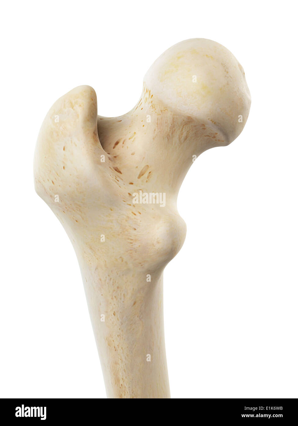 Human femur head computer artwork. Stock Photohttps://www.alamy.com/image-license-details/?v=1https://www.alamy.com/human-femur-head-computer-artwork-image69878631.html
RFE1K6WB–Human femur head computer artwork.
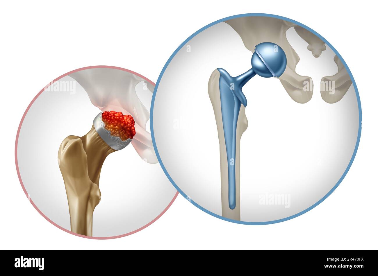 Hip Surgery and pelvic joint Replacement concept as an artificial joint or prosthesis with orthopedic surgery inserting a metal ball and socket Stock Photohttps://www.alamy.com/image-license-details/?v=1https://www.alamy.com/hip-surgery-and-pelvic-joint-replacement-concept-as-an-artificial-joint-or-prosthesis-with-orthopedic-surgery-inserting-a-metal-ball-and-socket-image553344510.html
RF2R470FX–Hip Surgery and pelvic joint Replacement concept as an artificial joint or prosthesis with orthopedic surgery inserting a metal ball and socket
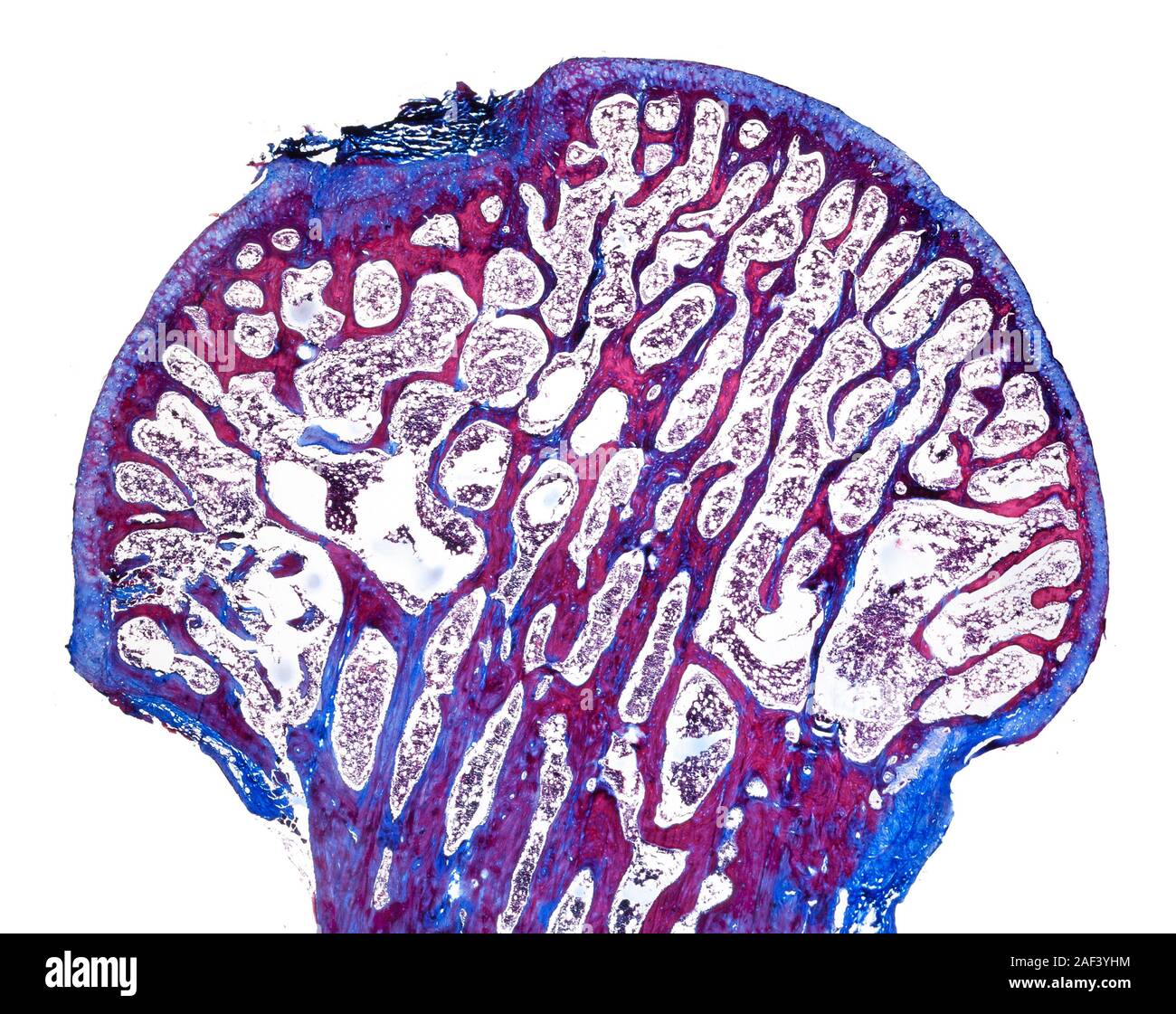 Typical small mammal bone structure, femur bone head, stained LS Stock Photohttps://www.alamy.com/image-license-details/?v=1https://www.alamy.com/typical-small-mammal-bone-structure-femur-bone-head-stained-ls-image336304352.html
RM2AF3YHM–Typical small mammal bone structure, femur bone head, stained LS
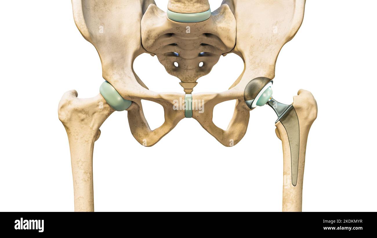 Hip prosthesis or implant isolated on white background. Hip joint or femoral head replacement 3D rendering illustration. Medicine, medical and healthc Stock Photohttps://www.alamy.com/image-license-details/?v=1https://www.alamy.com/hip-prosthesis-or-implant-isolated-on-white-background-hip-joint-or-femoral-head-replacement-3d-rendering-illustration-medicine-medical-and-healthc-image490314379.html
RF2KDKMYR–Hip prosthesis or implant isolated on white background. Hip joint or femoral head replacement 3D rendering illustration. Medicine, medical and healthc
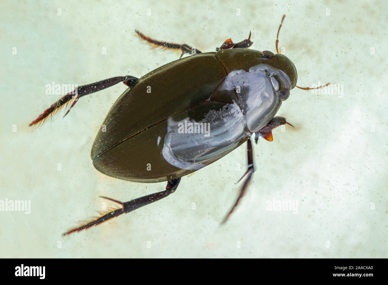 Silver Water Beetle (Hydrophilus piceus). Dorsal view. In a pond dipping identification tray. Showing body sections, head, thorax, abdomen protected Stock Photohttps://www.alamy.com/image-license-details/?v=1https://www.alamy.com/silver-water-beetle-hydrophilus-piceus-dorsal-view-in-a-pond-dipping-identification-tray-showing-body-sections-head-thorax-abdomen-protected-image329739704.html
RM2A4CXA0–Silver Water Beetle (Hydrophilus piceus). Dorsal view. In a pond dipping identification tray. Showing body sections, head, thorax, abdomen protected
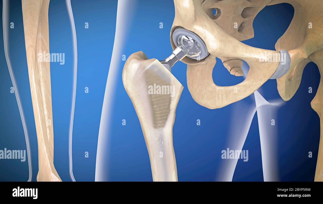 Function of a hip joint implant or hip prosthesis in frontal view - 3d illustration Stock Photohttps://www.alamy.com/image-license-details/?v=1https://www.alamy.com/function-of-a-hip-joint-implant-or-hip-prosthesis-in-frontal-view-3d-illustration-image361290600.html
RF2BYP5RM–Function of a hip joint implant or hip prosthesis in frontal view - 3d illustration
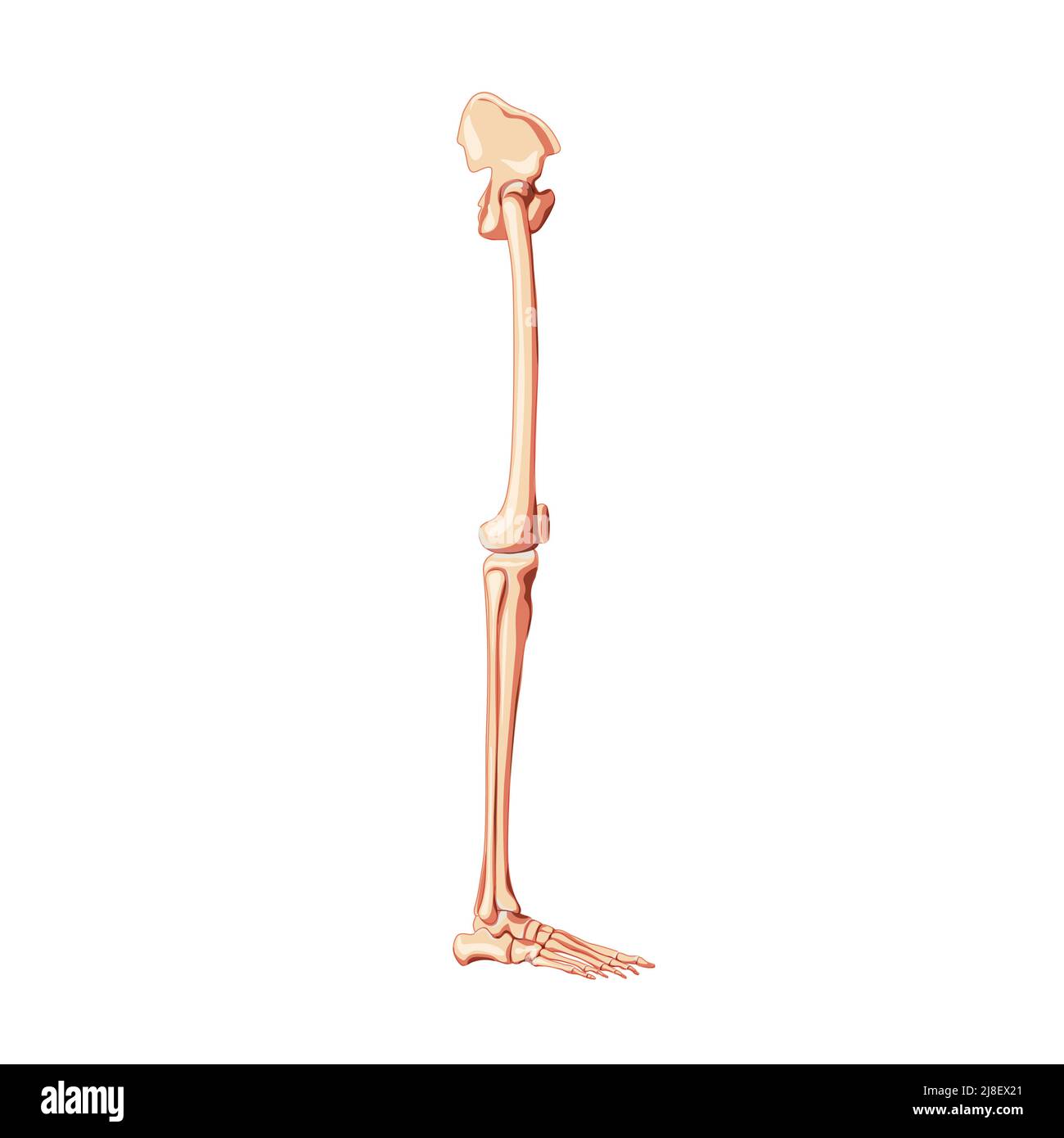 Human Pelvis with legs Skeleton side view with hip bone, thighs, foot, femur, patella, knee, fibula. Anatomically correct 3D realistic flat concept Vector illustration isolated on white background Stock Vectorhttps://www.alamy.com/image-license-details/?v=1https://www.alamy.com/human-pelvis-with-legs-skeleton-side-view-with-hip-bone-thighs-foot-femur-patella-knee-fibula-anatomically-correct-3d-realistic-flat-concept-vector-illustration-isolated-on-white-background-image469924953.html
RF2J8EX21–Human Pelvis with legs Skeleton side view with hip bone, thighs, foot, femur, patella, knee, fibula. Anatomically correct 3D realistic flat concept Vector illustration isolated on white background
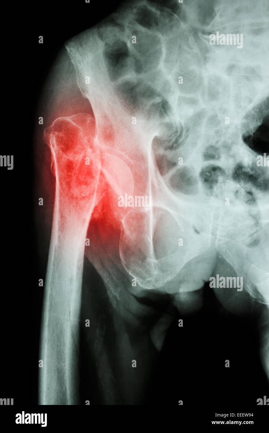 X-ray pelvis & hip joint : Fracture head of femur (thigh bone) Stock Photohttps://www.alamy.com/image-license-details/?v=1https://www.alamy.com/stock-photo-x-ray-pelvis-hip-joint-fracture-head-of-femur-thigh-bone-77773840.html
RFEEEW94–X-ray pelvis & hip joint : Fracture head of femur (thigh bone)
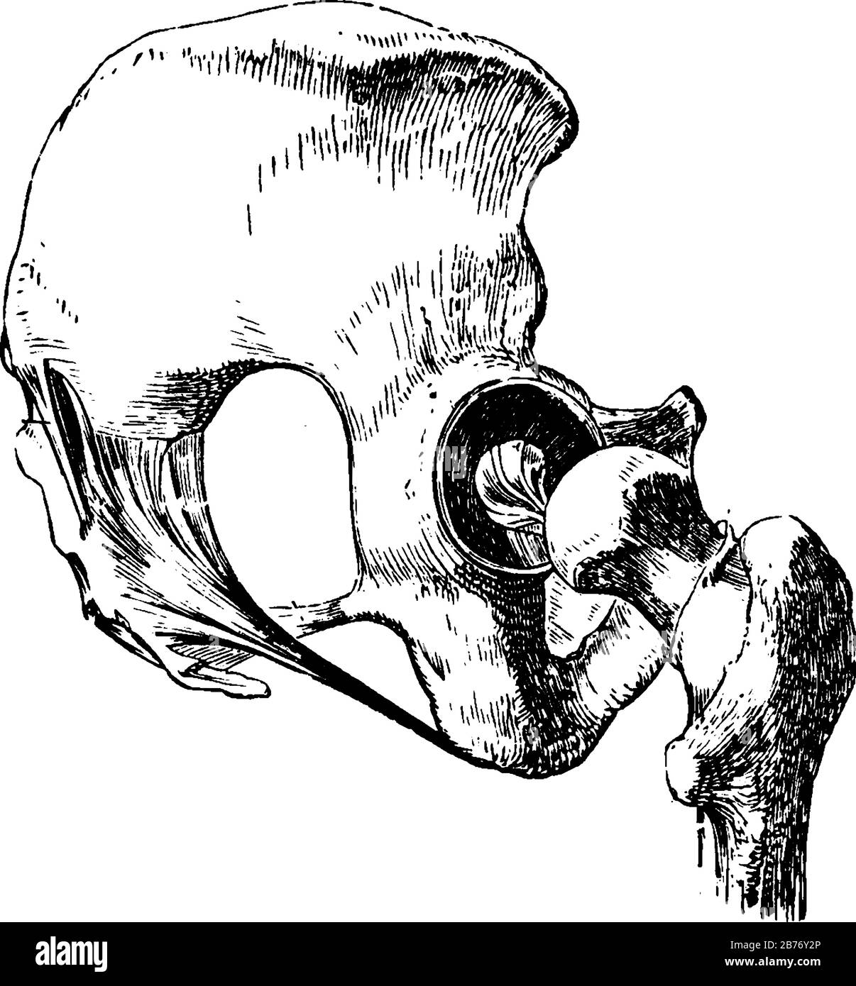 Hip Joint, the ball-and-socket joint connecting a leg to the trunk of the body, with ligaments removed, except the one on the head of the femur, vinta Stock Vectorhttps://www.alamy.com/image-license-details/?v=1https://www.alamy.com/hip-joint-the-ball-and-socket-joint-connecting-a-leg-to-the-trunk-of-the-body-with-ligaments-removed-except-the-one-on-the-head-of-the-femur-vinta-image348662910.html
RF2B76Y2P–Hip Joint, the ball-and-socket joint connecting a leg to the trunk of the body, with ligaments removed, except the one on the head of the femur, vinta
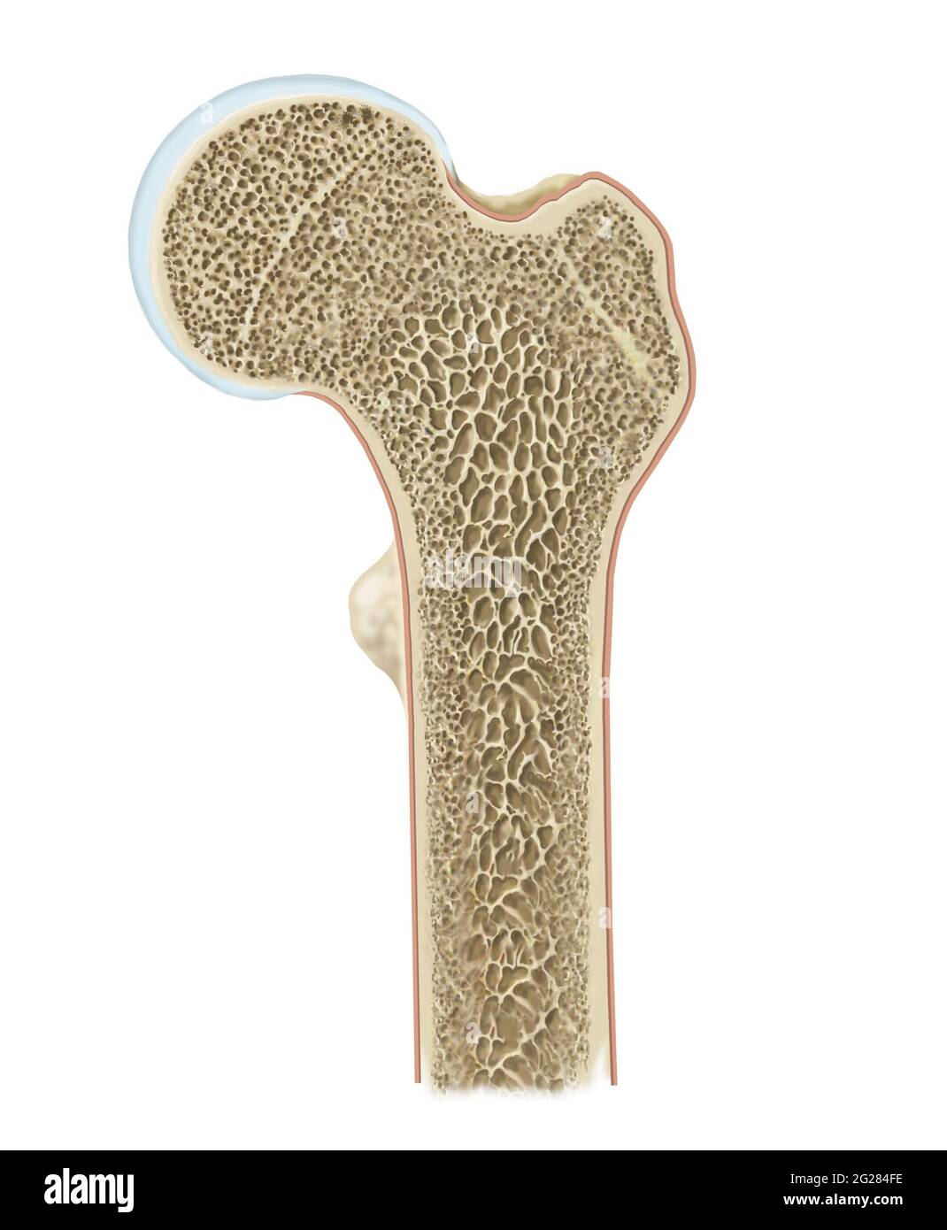 Detailed cross section of head of femur. Stock Photohttps://www.alamy.com/image-license-details/?v=1https://www.alamy.com/detailed-cross-section-of-head-of-femur-image431667698.html
RF2G284FE–Detailed cross section of head of femur.
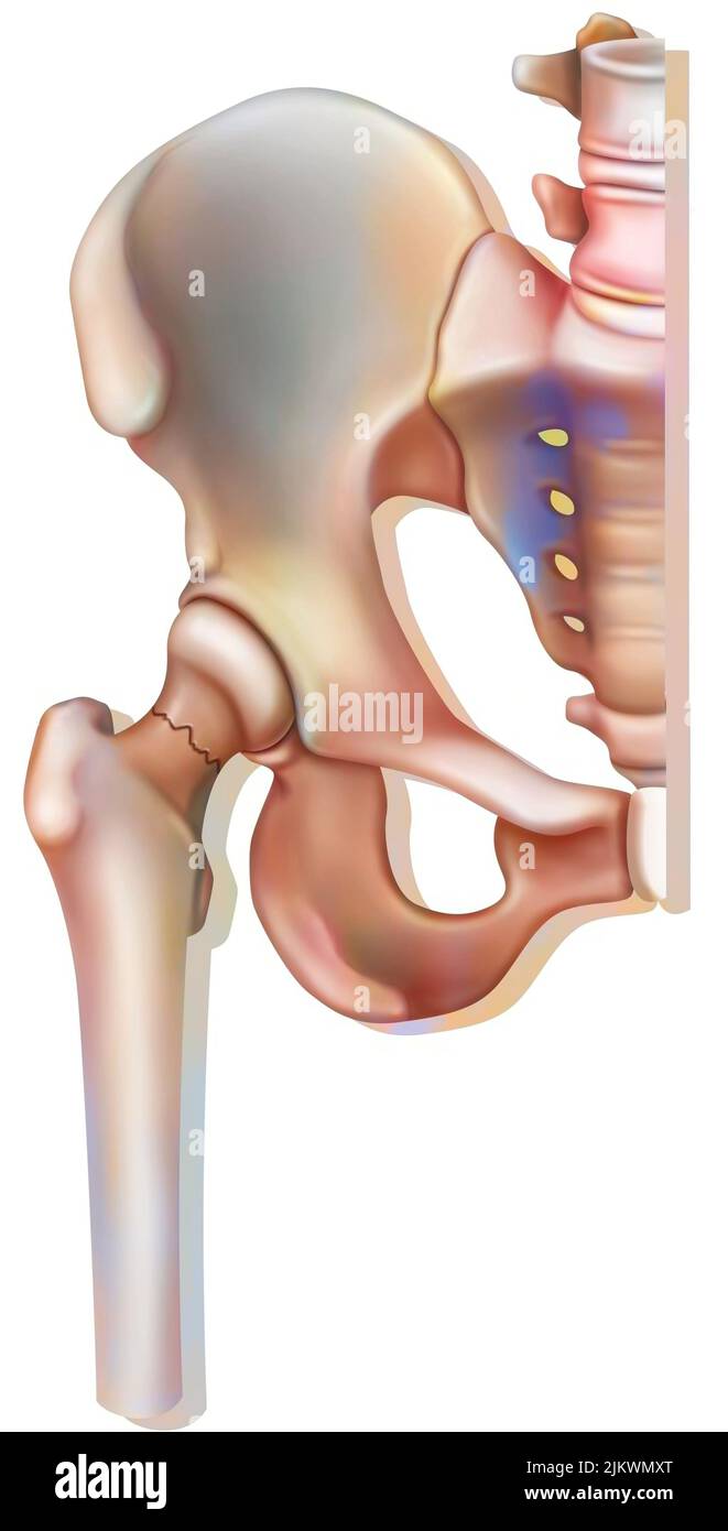 Bone system: fracture of the neck of the femur, linked to osteoporosis. Stock Photohttps://www.alamy.com/image-license-details/?v=1https://www.alamy.com/bone-system-fracture-of-the-neck-of-the-femur-linked-to-osteoporosis-image476923632.html
RF2JKWMXT–Bone system: fracture of the neck of the femur, linked to osteoporosis.
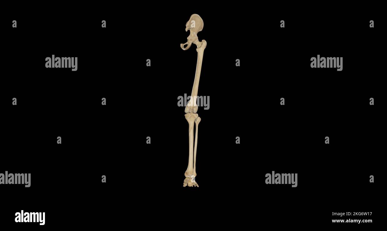 Bones of Right Lower Limb - Posterior View Stock Photohttps://www.alamy.com/image-license-details/?v=1https://www.alamy.com/bones-of-right-lower-limb-posterior-view-image491876147.html
RF2KG6W17–Bones of Right Lower Limb - Posterior View
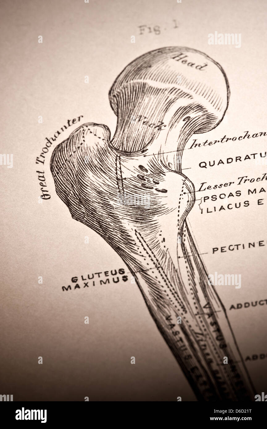 A vintage illustration of a hip joint. Stock Photohttps://www.alamy.com/image-license-details/?v=1https://www.alamy.com/stock-photo-a-vintage-illustration-of-a-hip-joint-55606036.html
RFD6D21T–A vintage illustration of a hip joint.
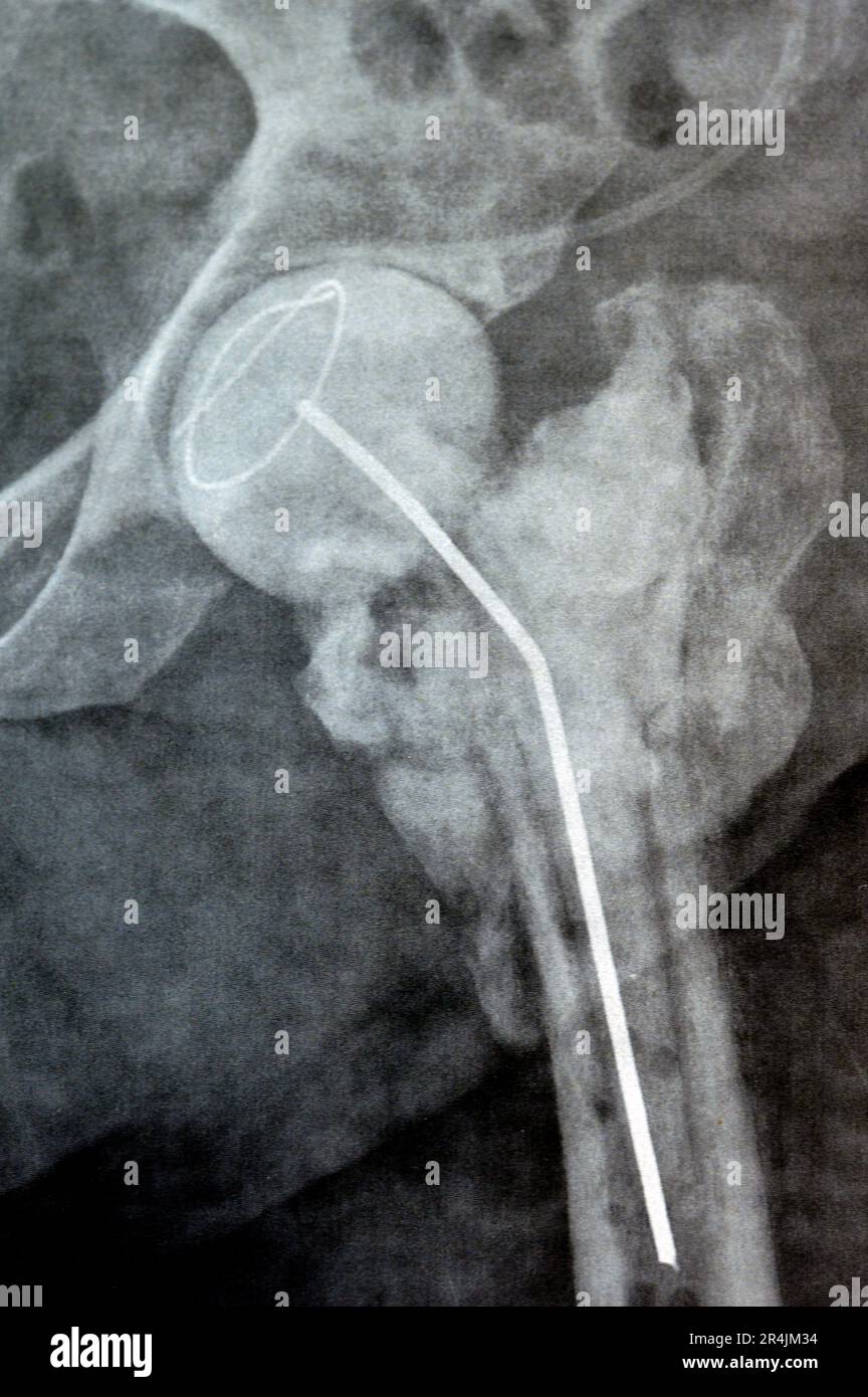 Plain X ray hip joint show left trans cervical fracture of the head of femur with temporary antibiotic loader spacer antibiotic-loaded bone cement aft Stock Photohttps://www.alamy.com/image-license-details/?v=1https://www.alamy.com/plain-x-ray-hip-joint-show-left-trans-cervical-fracture-of-the-head-of-femur-with-temporary-antibiotic-loader-spacer-antibiotic-loaded-bone-cement-aft-image553601304.html
RF2R4JM34–Plain X ray hip joint show left trans cervical fracture of the head of femur with temporary antibiotic loader spacer antibiotic-loaded bone cement aft
 Painted Backbone Pieces From Roadkill Stock Photohttps://www.alamy.com/image-license-details/?v=1https://www.alamy.com/stock-photo-painted-backbone-pieces-from-roadkill-102844242.html
RFFY8XTJ–Painted Backbone Pieces From Roadkill
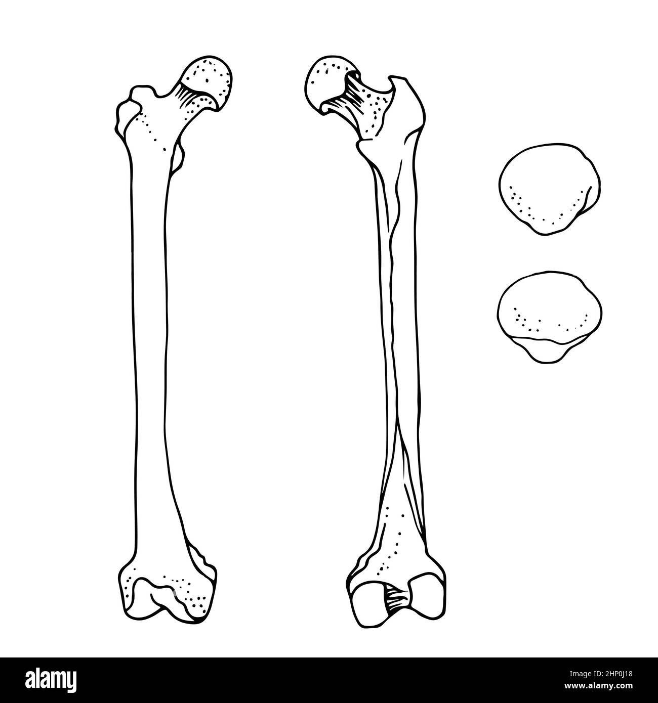 Human femur and patella, vector hand drawn illustration isolated on a white background, orthopedics medicine anatomy sketch Stock Vectorhttps://www.alamy.com/image-license-details/?v=1https://www.alamy.com/human-femur-and-patella-vector-hand-drawn-illustration-isolated-on-a-white-background-orthopedics-medicine-anatomy-sketch-image461006148.html
RF2HP0J18–Human femur and patella, vector hand drawn illustration isolated on a white background, orthopedics medicine anatomy sketch
 cave of the discovery of the head of a femur of a young Neanderthal man. Abruzzo, Italy, Europe Stock Photohttps://www.alamy.com/image-license-details/?v=1https://www.alamy.com/cave-of-the-discovery-of-the-head-of-a-femur-of-a-young-neanderthal-man-abruzzo-italy-europe-image380549174.html
RM2D33E9X–cave of the discovery of the head of a femur of a young Neanderthal man. Abruzzo, Italy, Europe
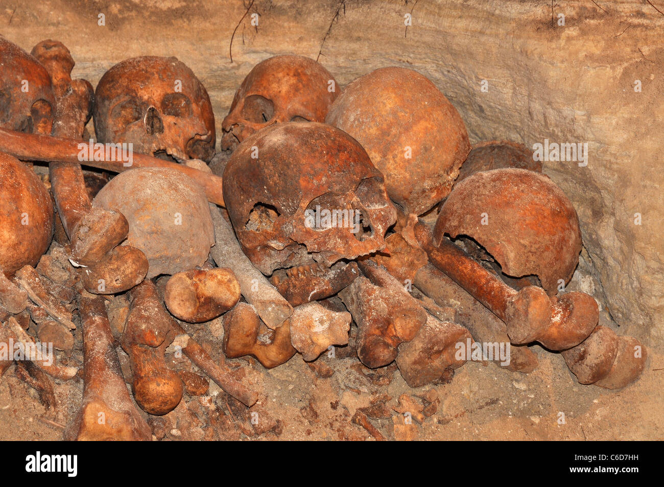 Group of skulls and bones from an unknown skeleton Stock Photohttps://www.alamy.com/image-license-details/?v=1https://www.alamy.com/stock-photo-group-of-skulls-and-bones-from-an-unknown-skeleton-38400029.html
RFC6D7HH–Group of skulls and bones from an unknown skeleton
 Intertrochanteric fracture on the femur head skeleton 3D medical illustration Stock Photohttps://www.alamy.com/image-license-details/?v=1https://www.alamy.com/intertrochanteric-fracture-on-the-femur-head-skeleton-3d-medical-illustration-image596268214.html
RF2WJ2A5A–Intertrochanteric fracture on the femur head skeleton 3D medical illustration
 Human femur head computer artwork. Stock Photohttps://www.alamy.com/image-license-details/?v=1https://www.alamy.com/human-femur-head-computer-artwork-image69878417.html
RFE1K6HN–Human femur head computer artwork.
 Set of human skull splash art Stock Vectorhttps://www.alamy.com/image-license-details/?v=1https://www.alamy.com/set-of-human-skull-splash-art-image614190689.html
RF2XK6PDN–Set of human skull splash art
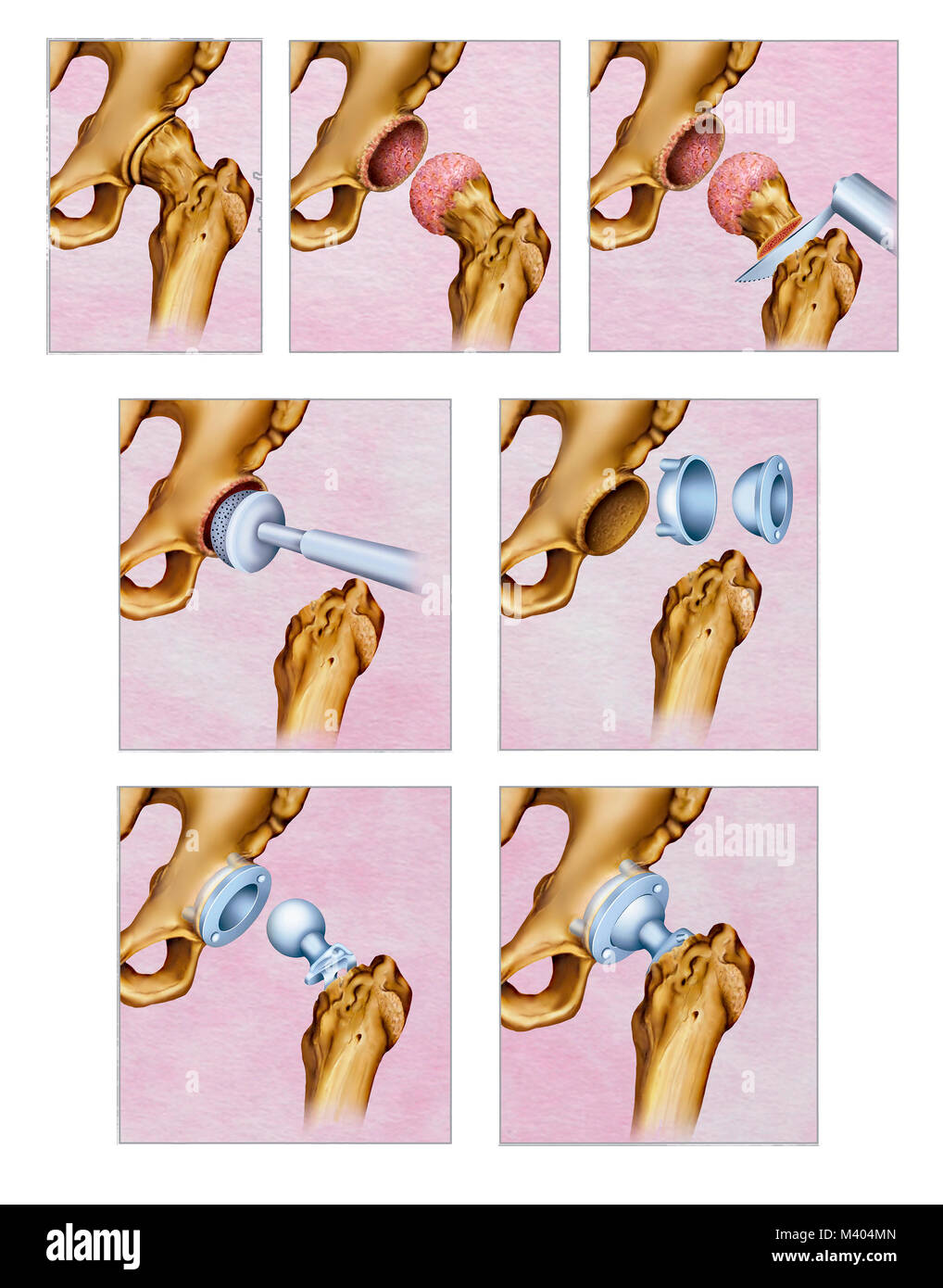 Ilustración sobre el tratamiento quirúrgico por artrosis en la articulación de la cadera. La artrosis de cadera es el desgaste del cartílago de la art Stock Photohttps://www.alamy.com/image-license-details/?v=1https://www.alamy.com/stock-photo-ilustracin-sobre-el-tratamiento-quirrgico-por-artrosis-en-la-articulacin-174566021.html
RFM404MN–Ilustración sobre el tratamiento quirúrgico por artrosis en la articulación de la cadera. La artrosis de cadera es el desgaste del cartílago de la art
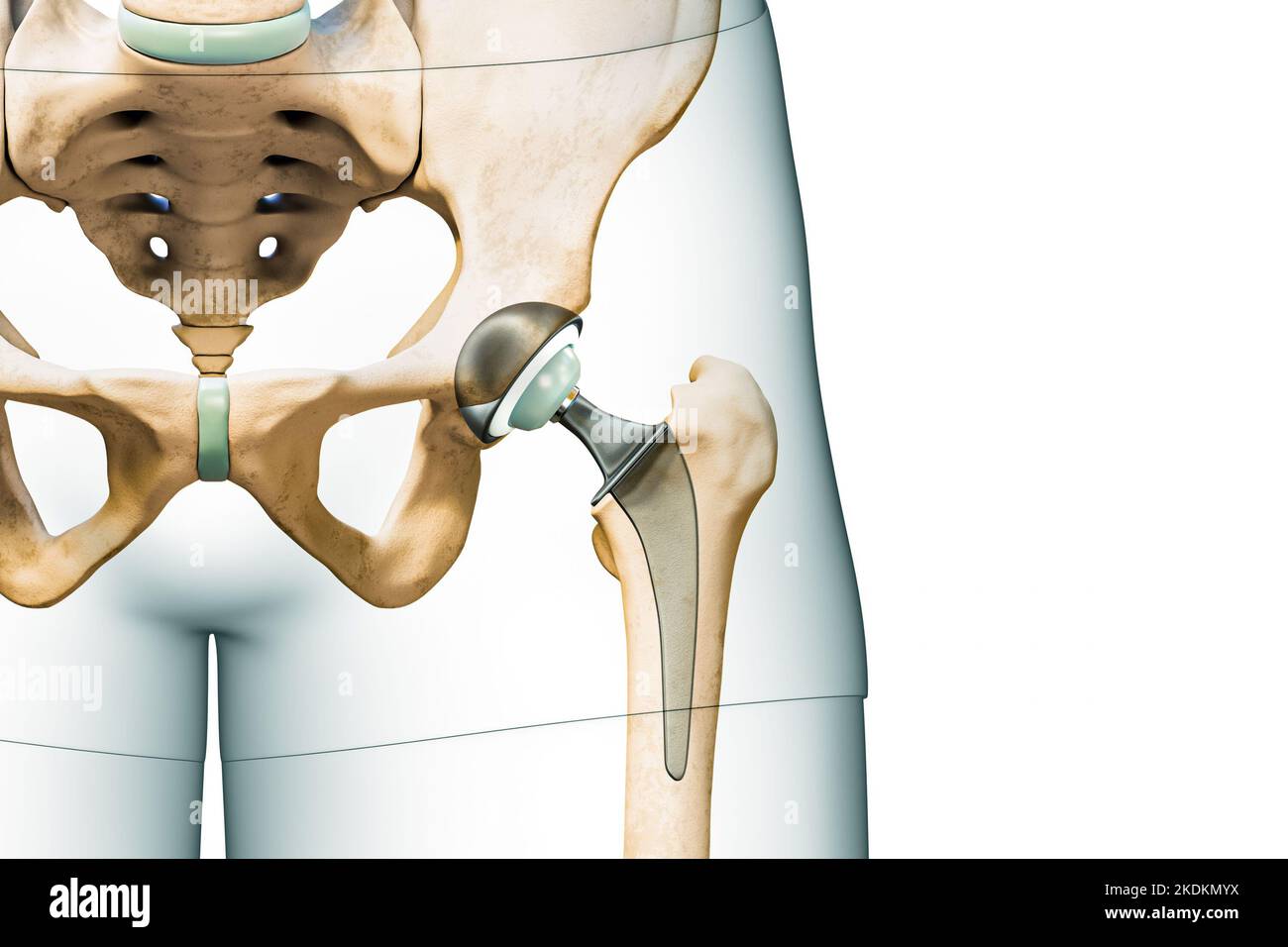 Hip prosthesis or implant isolated on white background with copy space and body contours. Hip joint or femoral head replacement 3D rendering illustrat Stock Photohttps://www.alamy.com/image-license-details/?v=1https://www.alamy.com/hip-prosthesis-or-implant-isolated-on-white-background-with-copy-space-and-body-contours-hip-joint-or-femoral-head-replacement-3d-rendering-illustrat-image490314382.html
RF2KDKMYX–Hip prosthesis or implant isolated on white background with copy space and body contours. Hip joint or femoral head replacement 3D rendering illustrat
 Silver Water Beetle (Hydrophilus piceus). Ventral, or underside view. In a pond dipping net. Showing body sections, head, thorax, abdomen, legs. Stock Photohttps://www.alamy.com/image-license-details/?v=1https://www.alamy.com/silver-water-beetle-hydrophilus-piceus-ventral-or-underside-view-in-a-pond-dipping-net-showing-body-sections-head-thorax-abdomen-legs-image329739768.html
RM2A4CXC8–Silver Water Beetle (Hydrophilus piceus). Ventral, or underside view. In a pond dipping net. Showing body sections, head, thorax, abdomen, legs.
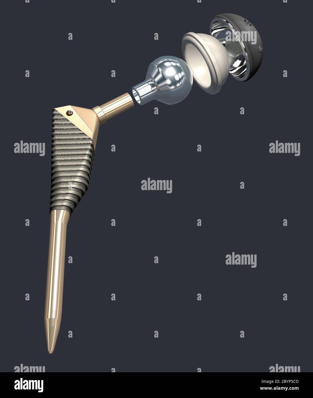 Function of a hip joint implant or hip prosthesis in frontal view - 3d illustration Stock Photohttps://www.alamy.com/image-license-details/?v=1https://www.alamy.com/function-of-a-hip-joint-implant-or-hip-prosthesis-in-frontal-view-3d-illustration-image361290285.html
RF2BYP5CD–Function of a hip joint implant or hip prosthesis in frontal view - 3d illustration
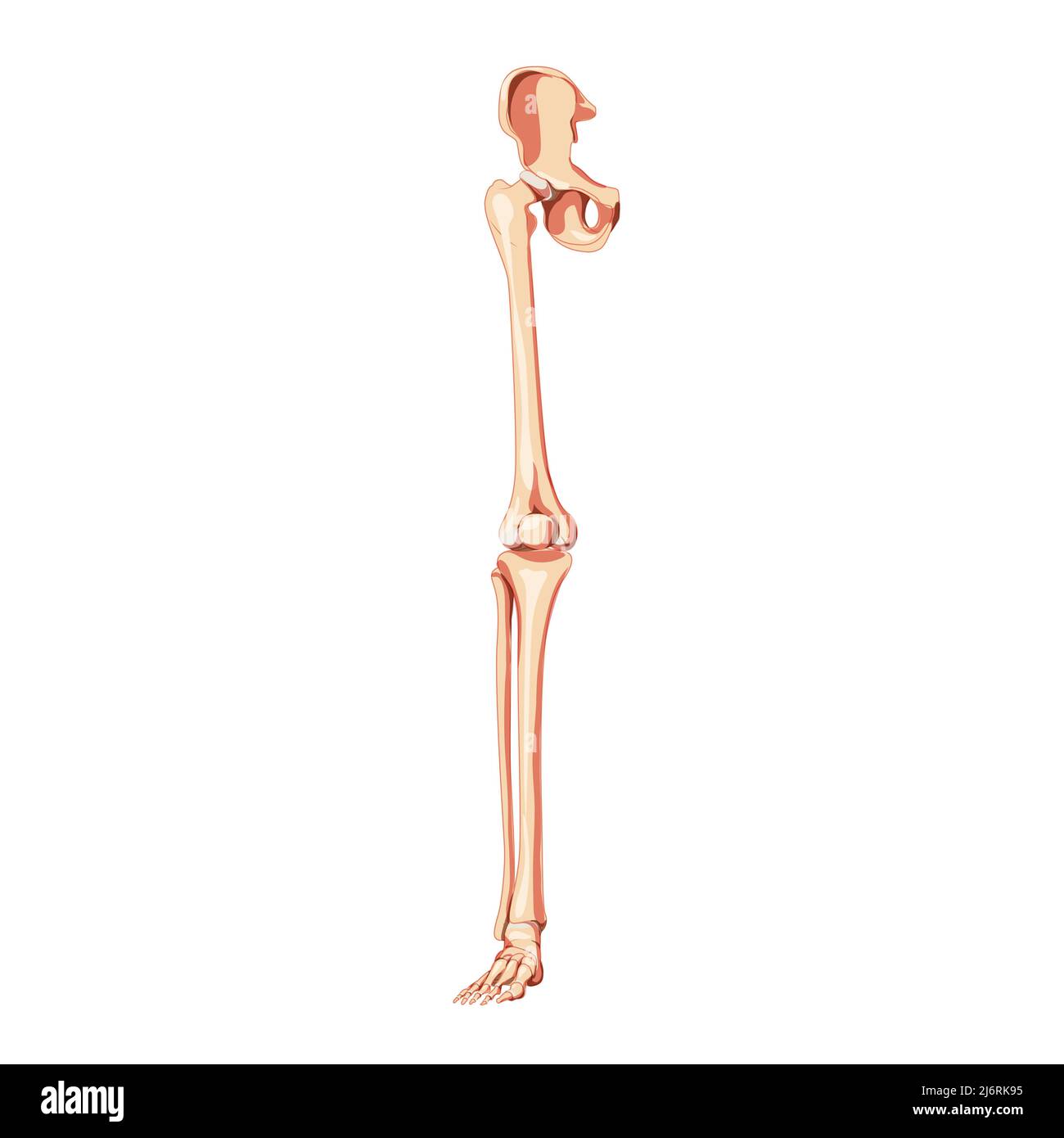 Human Pelvis with leg Skeleton front view with hip bone, thighs, foot, femur. Anatomically correct 3D realistic flat natural color concept Vector illustration of anatomy isolated on white background Stock Vectorhttps://www.alamy.com/image-license-details/?v=1https://www.alamy.com/human-pelvis-with-leg-skeleton-front-view-with-hip-bone-thighs-foot-femur-anatomically-correct-3d-realistic-flat-natural-color-concept-vector-illustration-of-anatomy-isolated-on-white-background-image468887921.html
RF2J6RK95–Human Pelvis with leg Skeleton front view with hip bone, thighs, foot, femur. Anatomically correct 3D realistic flat natural color concept Vector illustration of anatomy isolated on white background
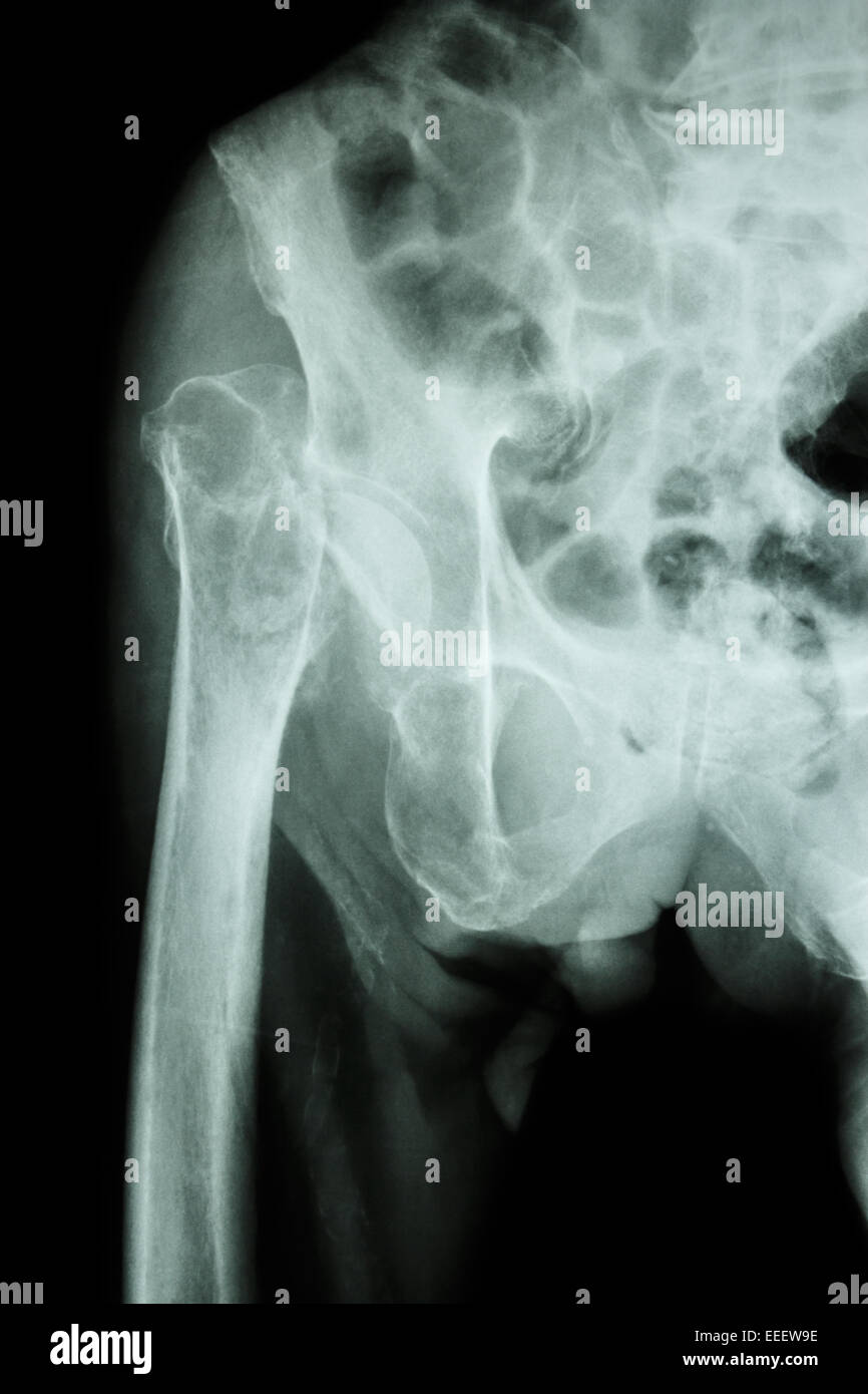 X-ray pelvis & hip joint : Fracture head of femur (thigh bone) Stock Photohttps://www.alamy.com/image-license-details/?v=1https://www.alamy.com/stock-photo-x-ray-pelvis-hip-joint-fracture-head-of-femur-thigh-bone-77773850.html
RFEEEW9E–X-ray pelvis & hip joint : Fracture head of femur (thigh bone)
 The head of the femur articulates with the acetabulum in the pelvic bone forms the hip joint. Shown here is a femur sawed lengthwise, vintage line dra Stock Vectorhttps://www.alamy.com/image-license-details/?v=1https://www.alamy.com/the-head-of-the-femur-articulates-with-the-acetabulum-in-the-pelvic-bone-forms-the-hip-joint-shown-here-is-a-femur-sawed-lengthwise-vintage-line-dra-image348662876.html
RF2B76Y1G–The head of the femur articulates with the acetabulum in the pelvic bone forms the hip joint. Shown here is a femur sawed lengthwise, vintage line dra
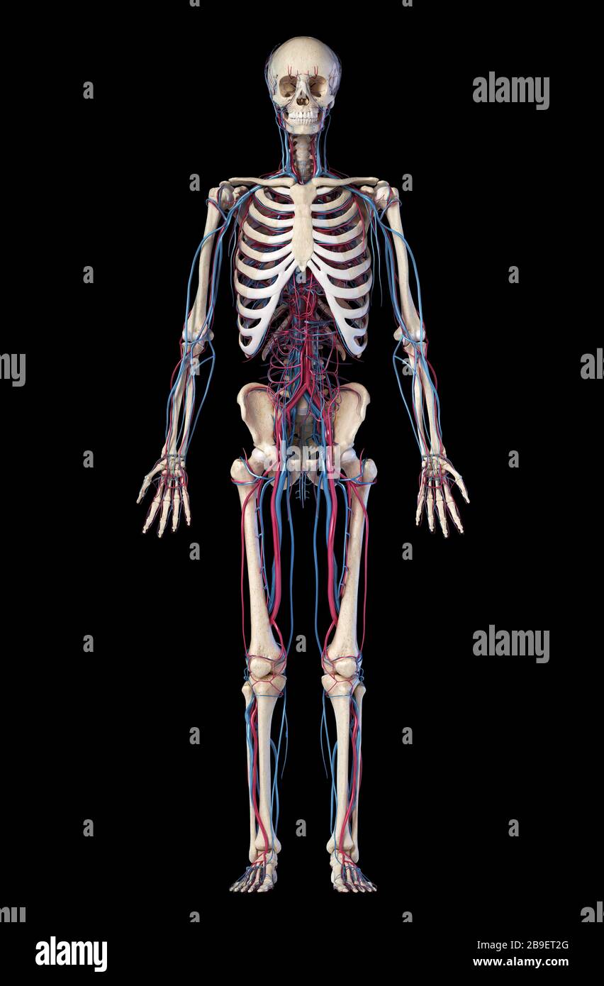 Anatomy of human skeleton with veins and arteries. Front view on black background. Stock Photohttps://www.alamy.com/image-license-details/?v=1https://www.alamy.com/anatomy-of-human-skeleton-with-veins-and-arteries-front-view-on-black-background-image350065480.html
RF2B9ET2G–Anatomy of human skeleton with veins and arteries. Front view on black background.
 Bone system: the bony joint of the hip. Stock Photohttps://www.alamy.com/image-license-details/?v=1https://www.alamy.com/bone-system-the-bony-joint-of-the-hip-image476923639.html
RF2JKWMY3–Bone system: the bony joint of the hip.
 Bones of Right Lower Limb - Anterior View Stock Photohttps://www.alamy.com/image-license-details/?v=1https://www.alamy.com/bones-of-right-lower-limb-anterior-view-image491876141.html
RF2KG6W11–Bones of Right Lower Limb - Anterior View
 3d rendered illustration of a painful hip joint Stock Photohttps://www.alamy.com/image-license-details/?v=1https://www.alamy.com/stock-photo-3d-rendered-illustration-of-a-painful-hip-joint-103506642.html
RMG0B3NP–3d rendered illustration of a painful hip joint
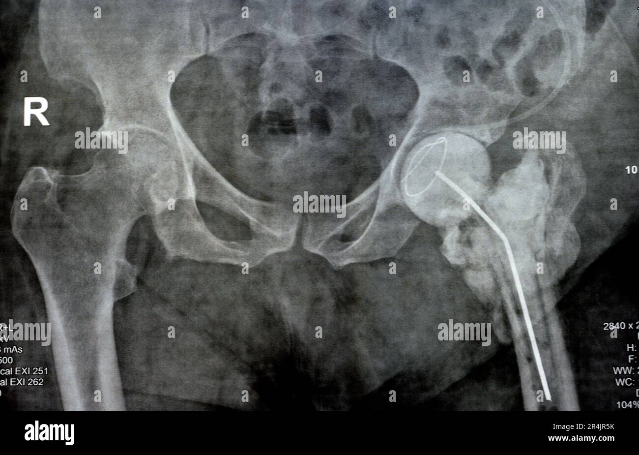 Plain X ray hip joint show left trans cervical fracture of the head of femur with temporary antibiotic loader spacer antibiotic-loaded bone cement aft Stock Photohttps://www.alamy.com/image-license-details/?v=1https://www.alamy.com/plain-x-ray-hip-joint-show-left-trans-cervical-fracture-of-the-head-of-femur-with-temporary-antibiotic-loader-spacer-antibiotic-loaded-bone-cement-aft-image553603727.html
RF2R4JR5K–Plain X ray hip joint show left trans cervical fracture of the head of femur with temporary antibiotic loader spacer antibiotic-loaded bone cement aft
 ***MARCH 28, 2012, FILE PHOTO*** Czech singer Hana Hegerova died at the age of 89 in the Na Homolce hospital in Prague today, on Tuesday, March 23, 2021, her friend, publisher Tomas Padevet has confirmed the news. Hegerova, widely considered the 'First Lady of the Czechoslovak Chanson,' recently suffered from a number of health problems after the head of her femur was broken. Hegerova, born as Carmen Farkasova-Celkova, hails from Slovakia. She started her career as an actress in the theatre in Zilina, north Slovakia. At the end of the 1950s and beginning of the 1960s, she worked in the Pragu Stock Photohttps://www.alamy.com/image-license-details/?v=1https://www.alamy.com/march-28-2012-file-photo-czech-singer-hana-hegerova-died-at-the-age-of-89-in-the-na-homolce-hospital-in-prague-today-on-tuesday-march-23-2021-her-friend-publisher-tomas-padevet-has-confirmed-the-news-hegerova-widely-considered-the-first-lady-of-the-czechoslovak-chanson-recently-suffered-from-a-number-of-health-problems-after-the-head-of-her-femur-was-broken-hegerova-born-as-carmen-farkasova-celkova-hails-from-slovakia-she-started-her-career-as-an-actress-in-the-theatre-in-zilina-north-slovakia-at-the-end-of-the-1950s-and-beginning-of-the-1960s-she-worked-in-the-pragu-image416055465.html
RM2F4TXYN–***MARCH 28, 2012, FILE PHOTO*** Czech singer Hana Hegerova died at the age of 89 in the Na Homolce hospital in Prague today, on Tuesday, March 23, 2021, her friend, publisher Tomas Padevet has confirmed the news. Hegerova, widely considered the 'First Lady of the Czechoslovak Chanson,' recently suffered from a number of health problems after the head of her femur was broken. Hegerova, born as Carmen Farkasova-Celkova, hails from Slovakia. She started her career as an actress in the theatre in Zilina, north Slovakia. At the end of the 1950s and beginning of the 1960s, she worked in the Pragu
 Cat Skeleton Anatomy. Side view Stock Photohttps://www.alamy.com/image-license-details/?v=1https://www.alamy.com/cat-skeleton-anatomy-side-view-image450097981.html
RF2H47MFW–Cat Skeleton Anatomy. Side view
 cave of the discovery of the head of a femur of a young Neanderthal man. Abruzzo, Italy, Europe Stock Photohttps://www.alamy.com/image-license-details/?v=1https://www.alamy.com/cave-of-the-discovery-of-the-head-of-a-femur-of-a-young-neanderthal-man-abruzzo-italy-europe-image462506637.html
RM2HTCYX5–cave of the discovery of the head of a femur of a young Neanderthal man. Abruzzo, Italy, Europe
 Human skulls in the catacombs of Paris, France Stock Photohttps://www.alamy.com/image-license-details/?v=1https://www.alamy.com/human-skulls-in-the-catacombs-of-paris-france-image243069227.html
RMT3CN7R–Human skulls in the catacombs of Paris, France
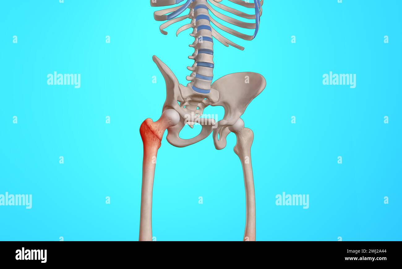 Intertrochanteric fracture on the femur head skeleton 3D medical illustration Stock Photohttps://www.alamy.com/image-license-details/?v=1https://www.alamy.com/intertrochanteric-fracture-on-the-femur-head-skeleton-3d-medical-illustration-image596268180.html
RF2WJ2A44–Intertrochanteric fracture on the femur head skeleton 3D medical illustration
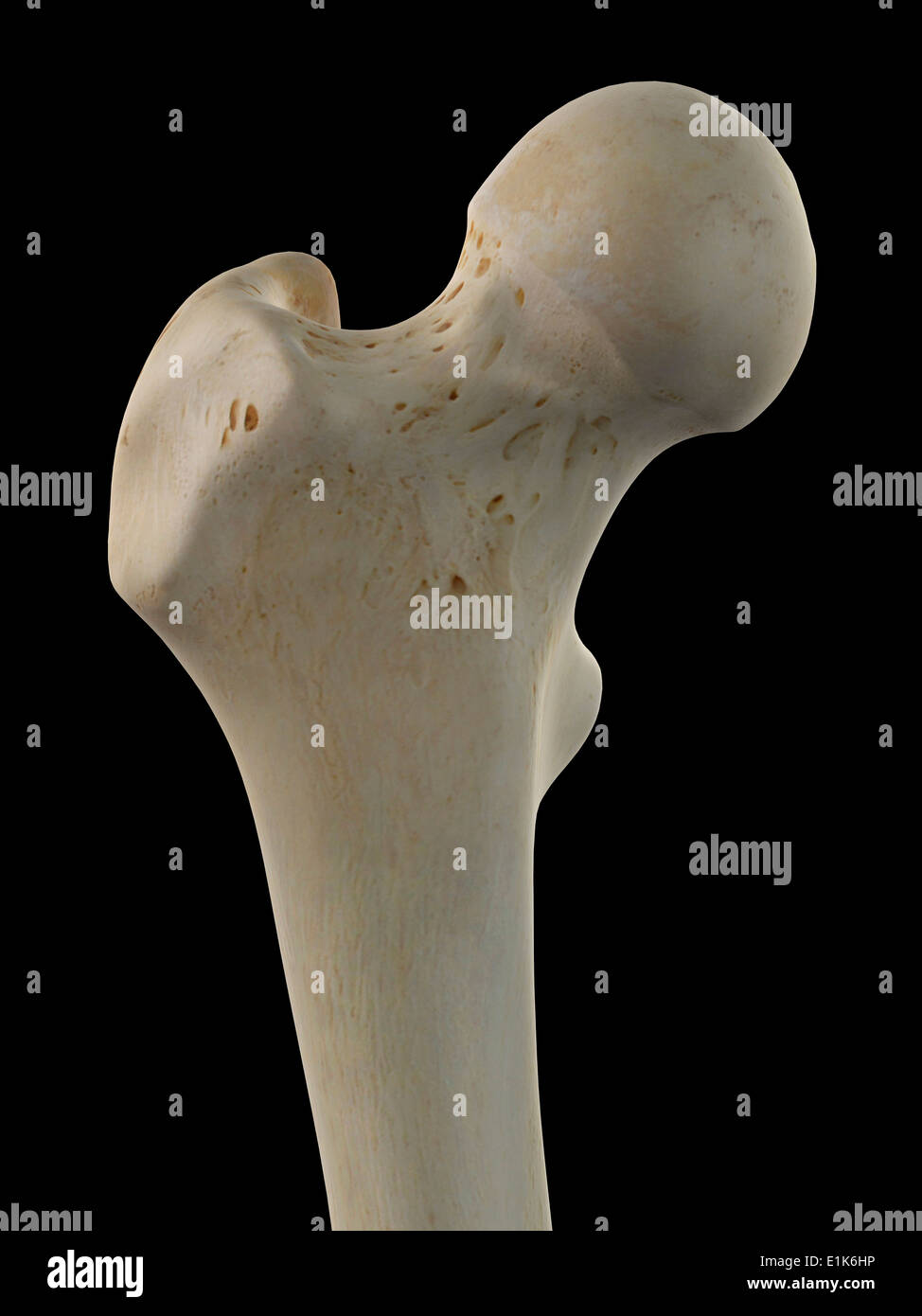 Human femur head computer artwork. Stock Photohttps://www.alamy.com/image-license-details/?v=1https://www.alamy.com/human-femur-head-computer-artwork-image69878418.html
RFE1K6HP–Human femur head computer artwork.
 Human skull isolated on white background Stock Vectorhttps://www.alamy.com/image-license-details/?v=1https://www.alamy.com/human-skull-isolated-on-white-background-image543507698.html
RF2PG6WH6–Human skull isolated on white background
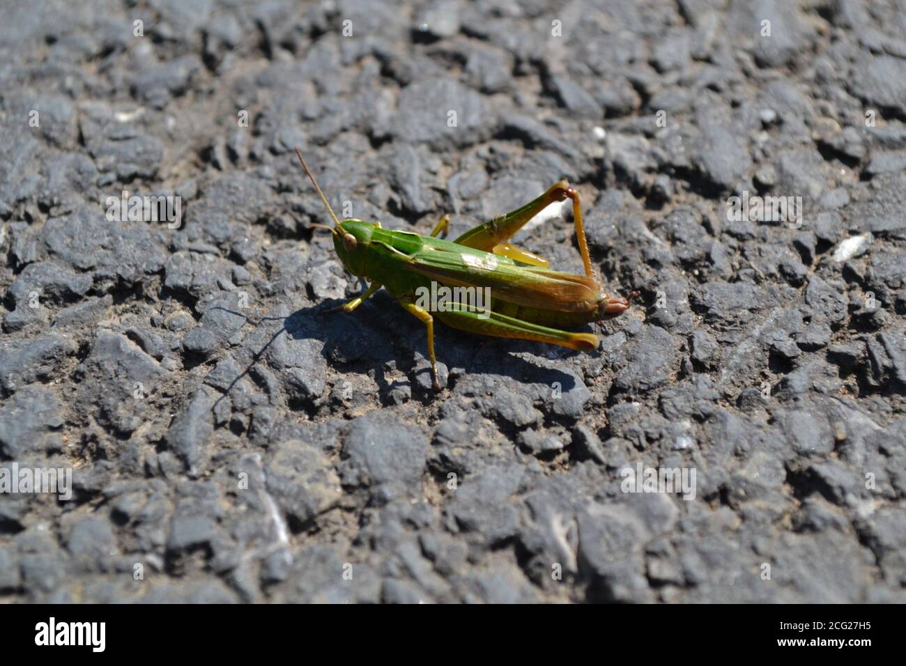 Green Grasshopper pausing on tarmac between leaps. Stock Photohttps://www.alamy.com/image-license-details/?v=1https://www.alamy.com/green-grasshopper-pausing-on-tarmac-between-leaps-image371302097.html
RF2CG27H5–Green Grasshopper pausing on tarmac between leaps.
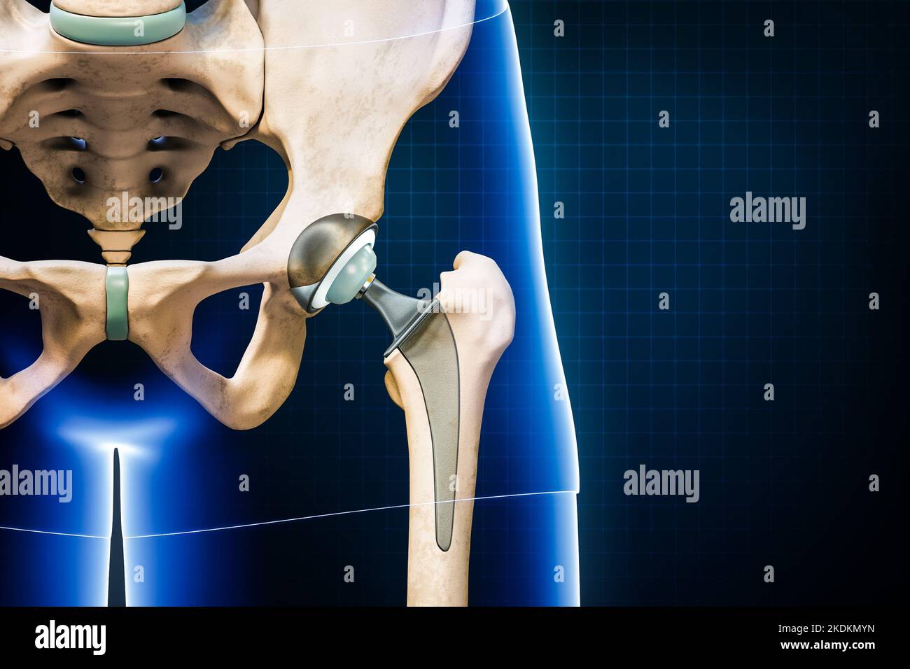 Hip prosthesis or implant isolated on blue background with copy space. Hip joint or femoral head replacement 3D rendering illustration. Medicine, medi Stock Photohttps://www.alamy.com/image-license-details/?v=1https://www.alamy.com/hip-prosthesis-or-implant-isolated-on-blue-background-with-copy-space-hip-joint-or-femoral-head-replacement-3d-rendering-illustration-medicine-medi-image490314377.html
RF2KDKMYN–Hip prosthesis or implant isolated on blue background with copy space. Hip joint or femoral head replacement 3D rendering illustration. Medicine, medi
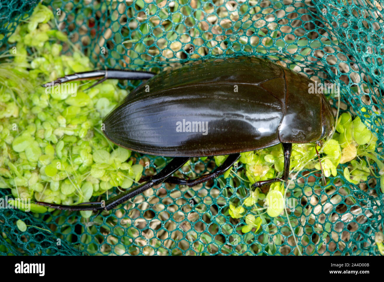 Great Silver Water Beetle (Hydrophilus piceus). The bulkiest British and European beetle reaching a length of 5 centimetres. A male. Stock Photohttps://www.alamy.com/image-license-details/?v=1https://www.alamy.com/great-silver-water-beetle-hydrophilus-piceus-the-bulkiest-british-and-european-beetle-reaching-a-length-of-5-centimetres-a-male-image329741003.html
RM2A4D00B–Great Silver Water Beetle (Hydrophilus piceus). The bulkiest British and European beetle reaching a length of 5 centimetres. A male.
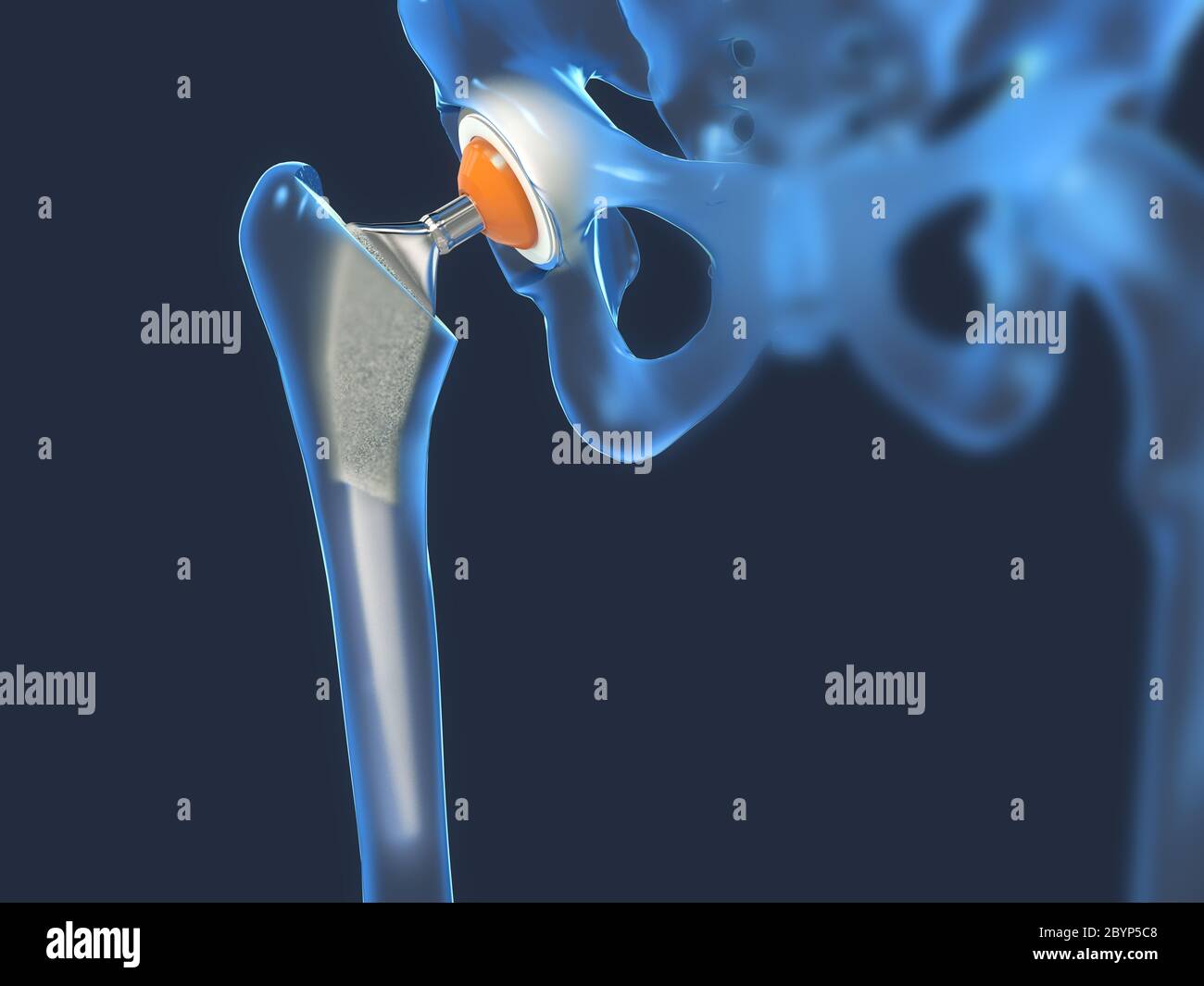 Function of a hip joint implant or hip prosthesis in frontal view - 3d illustration Stock Photohttps://www.alamy.com/image-license-details/?v=1https://www.alamy.com/function-of-a-hip-joint-implant-or-hip-prosthesis-in-frontal-view-3d-illustration-image361290280.html
RF2BYP5C8–Function of a hip joint implant or hip prosthesis in frontal view - 3d illustration
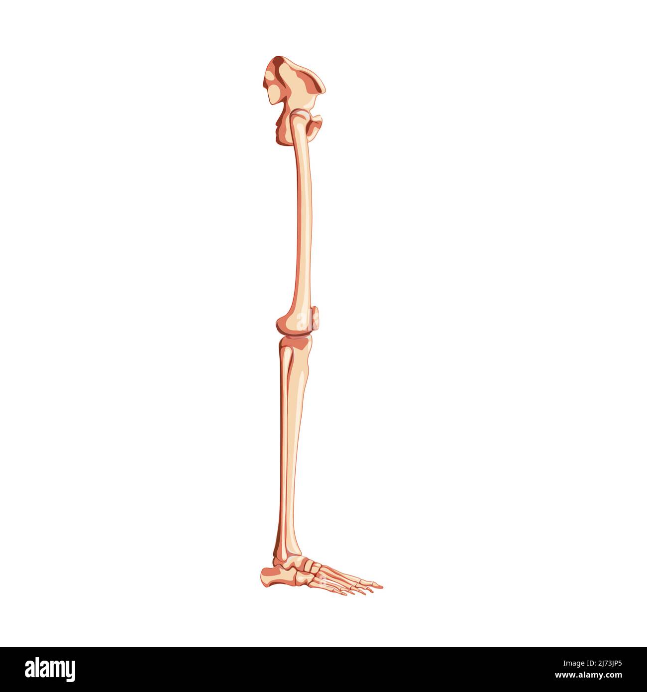 Human Pelvis with leg Skeleton side view with hip bone, thighs, foot, femur, knee, tibia. Anatomically correct 3D realistic flat natural color concept Vector illustration isolated on white background Stock Vectorhttps://www.alamy.com/image-license-details/?v=1https://www.alamy.com/human-pelvis-with-leg-skeleton-side-view-with-hip-bone-thighs-foot-femur-knee-tibia-anatomically-correct-3d-realistic-flat-natural-color-concept-vector-illustration-isolated-on-white-background-image469063117.html
RF2J73JP5–Human Pelvis with leg Skeleton side view with hip bone, thighs, foot, femur, knee, tibia. Anatomically correct 3D realistic flat natural color concept Vector illustration isolated on white background
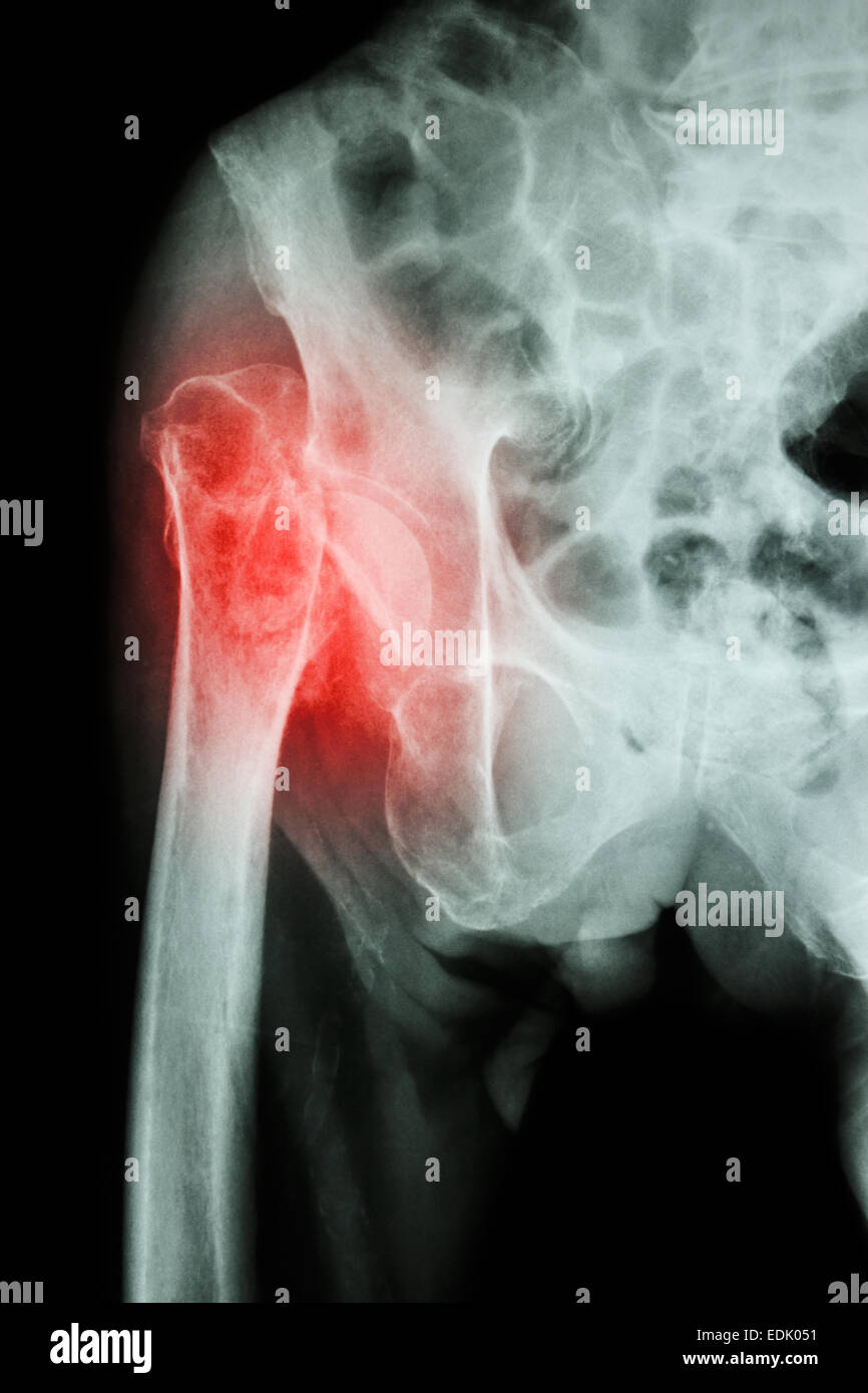 X-ray pelvis & hip joint : Fracture head of femur (thigh bone) Stock Photohttps://www.alamy.com/image-license-details/?v=1https://www.alamy.com/stock-photo-x-ray-pelvis-hip-joint-fracture-head-of-femur-thigh-bone-77249229.html
RFEDK051–X-ray pelvis & hip joint : Fracture head of femur (thigh bone)
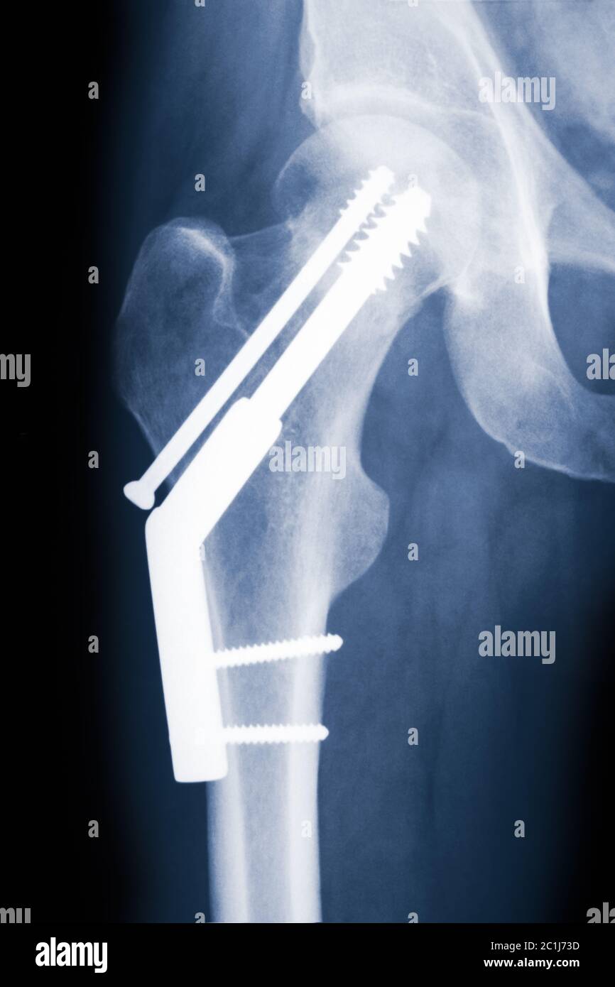 Hip X-ray showing fixation of a fractured hip with a dynamic hip screw and anti-rotation screw. Stock Photohttps://www.alamy.com/image-license-details/?v=1https://www.alamy.com/hip-x-ray-showing-fixation-of-a-fractured-hip-with-a-dynamic-hip-screw-and-anti-rotation-screw-image362433105.html
RF2C1J73D–Hip X-ray showing fixation of a fractured hip with a dynamic hip screw and anti-rotation screw.
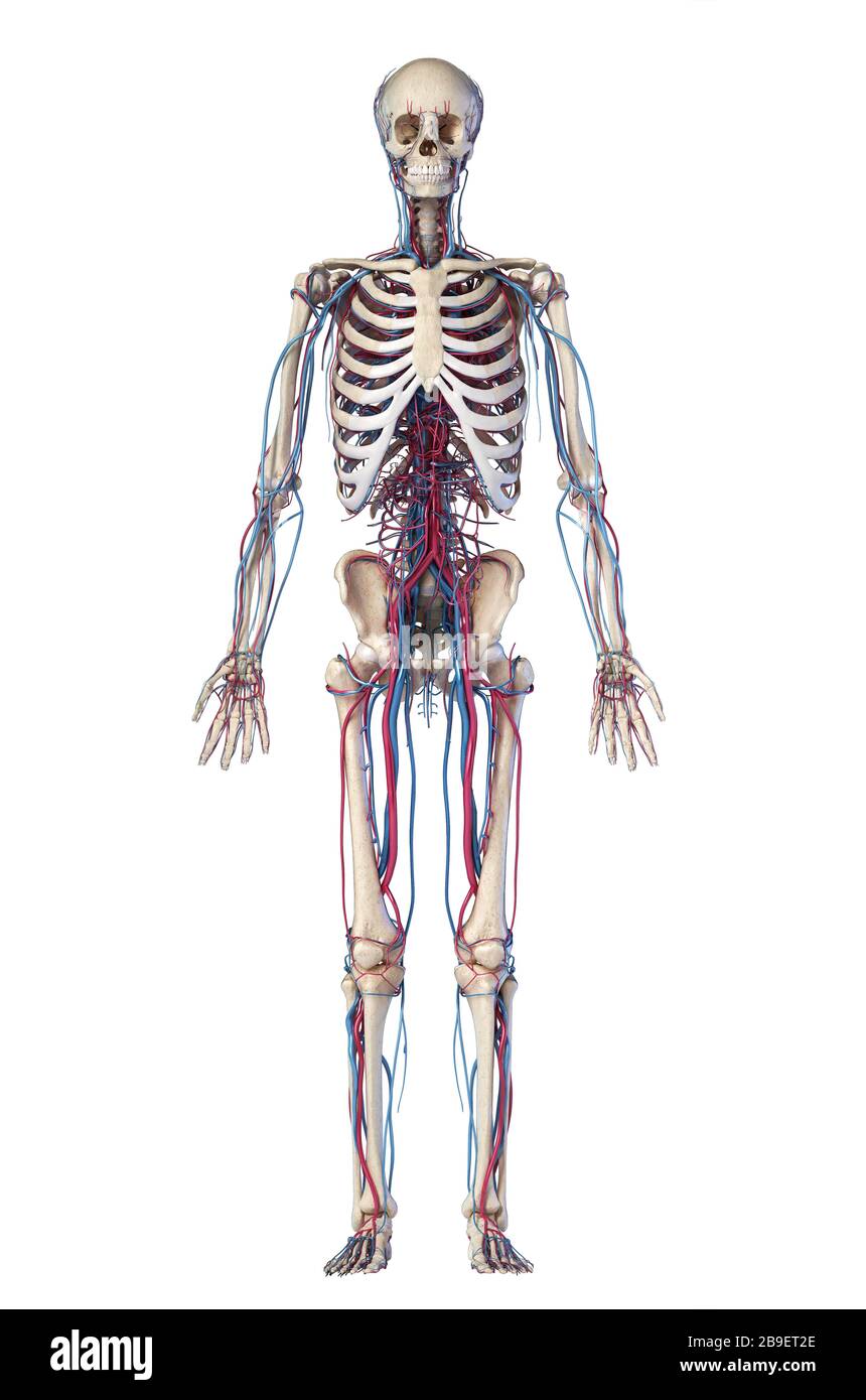 Anatomy of human skeleton with veins and arteries. Front view on black background. Stock Photohttps://www.alamy.com/image-license-details/?v=1https://www.alamy.com/anatomy-of-human-skeleton-with-veins-and-arteries-front-view-on-black-background-image350065478.html
RF2B9ET2E–Anatomy of human skeleton with veins and arteries. Front view on black background.
 Bone system: the bony joint of the hip. Stock Photohttps://www.alamy.com/image-license-details/?v=1https://www.alamy.com/bone-system-the-bony-joint-of-the-hip-image476923618.html
RF2JKWMXA–Bone system: the bony joint of the hip.
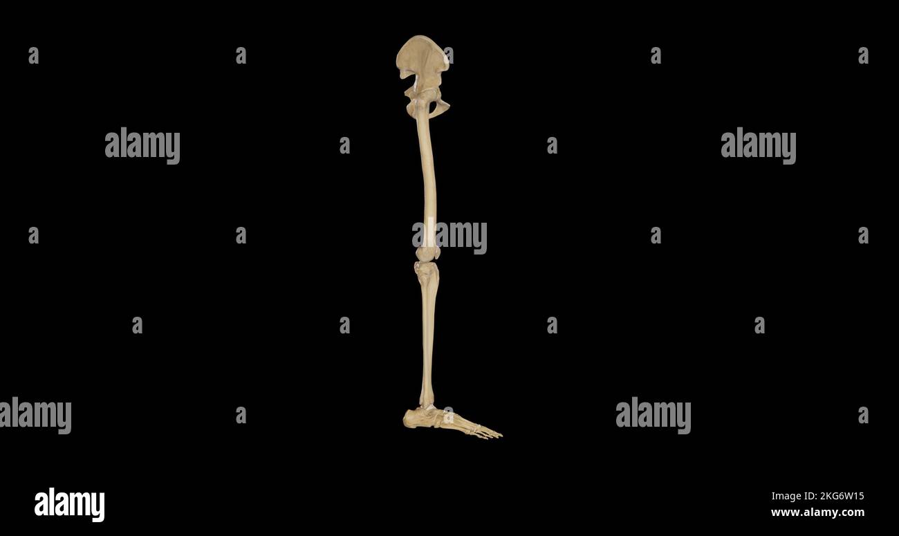 Bones of Right Lower Limb - Lateral View Stock Photohttps://www.alamy.com/image-license-details/?v=1https://www.alamy.com/bones-of-right-lower-limb-lateral-view-image491876145.html
RF2KG6W15–Bones of Right Lower Limb - Lateral View
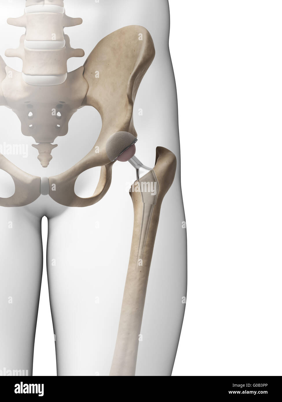 3d rendered illustration of a hip replacement Stock Photohttps://www.alamy.com/image-license-details/?v=1https://www.alamy.com/stock-photo-3d-rendered-illustration-of-a-hip-replacement-103506670.html
RMG0B3PP–3d rendered illustration of a hip replacement
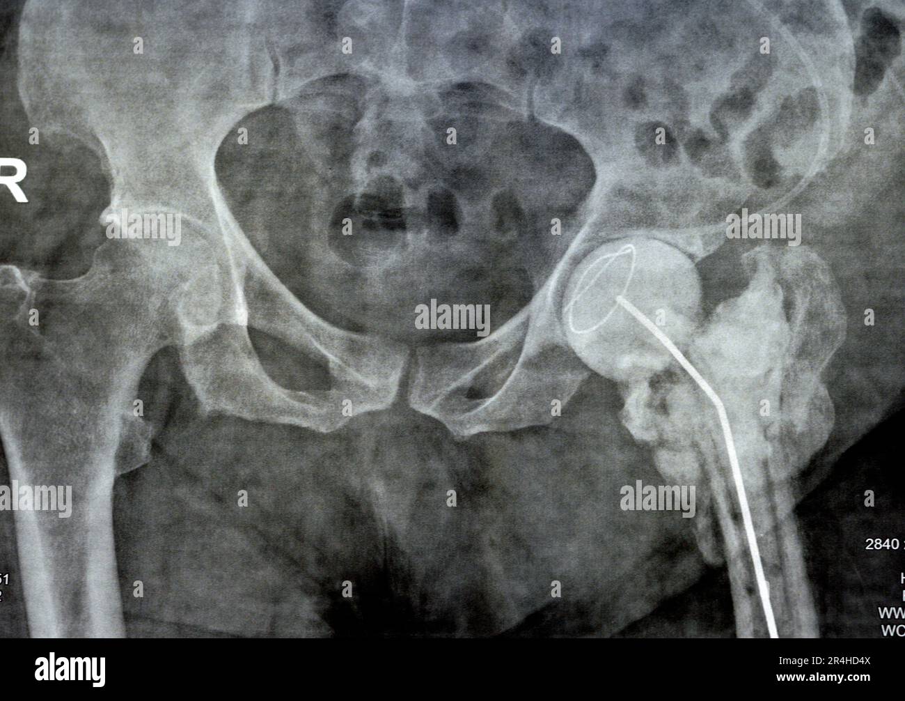 Plain X ray hip joint show left trans cervical fracture of the head of femur with temporary antibiotic loader spacer antibiotic-loaded bone cement aft Stock Photohttps://www.alamy.com/image-license-details/?v=1https://www.alamy.com/plain-x-ray-hip-joint-show-left-trans-cervical-fracture-of-the-head-of-femur-with-temporary-antibiotic-loader-spacer-antibiotic-loaded-bone-cement-aft-image553573914.html
RF2R4HD4X–Plain X ray hip joint show left trans cervical fracture of the head of femur with temporary antibiotic loader spacer antibiotic-loaded bone cement aft
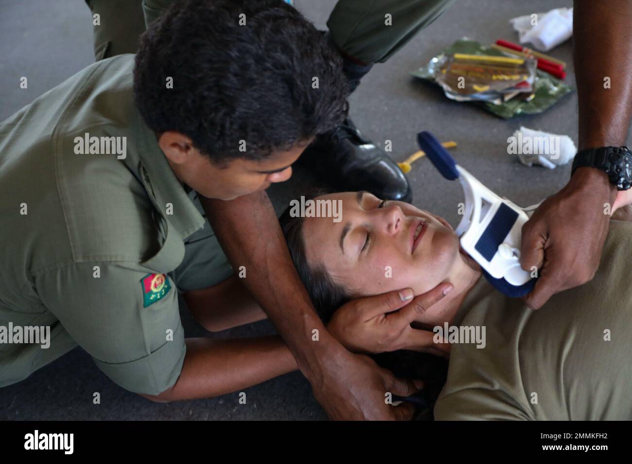 Tactical Combat Care Course skills testing during Exercise Cartwheel 2022 began with scenario one, led by Maj Rena White, 402nd Forward Resuscitative Surgical Team (FRST}Commander, and patient 1st LT Margret McDonell, Nurse Anesthetist, 402nd FRST, who simulated being involved in a motor vehicle accident with a head injury and femur fracture. Private Lusiana Likuomole, Territorial Force Brigade, Fiji Army, left, and classmates must Identify each concern while receiving little verbal guidance and the patient’s symptoms to pass. Exercise Cartwheel is a multilateral military-to-military training Stock Photohttps://www.alamy.com/image-license-details/?v=1https://www.alamy.com/tactical-combat-care-course-skills-testing-during-exercise-cartwheel-2022-began-with-scenario-one-led-by-maj-rena-white-402nd-forward-resuscitative-surgical-team-frstcommander-and-patient-1st-lt-margret-mcdonell-nurse-anesthetist-402nd-frst-who-simulated-being-involved-in-a-motor-vehicle-accident-with-a-head-injury-and-femur-fracture-private-lusiana-likuomole-territorial-force-brigade-fiji-army-left-and-classmates-must-identify-each-concern-while-receiving-little-verbal-guidance-and-the-patients-symptoms-to-pass-exercise-cartwheel-is-a-multilateral-military-to-military-training-image511823118.html
RM2MMKFH2–Tactical Combat Care Course skills testing during Exercise Cartwheel 2022 began with scenario one, led by Maj Rena White, 402nd Forward Resuscitative Surgical Team (FRST}Commander, and patient 1st LT Margret McDonell, Nurse Anesthetist, 402nd FRST, who simulated being involved in a motor vehicle accident with a head injury and femur fracture. Private Lusiana Likuomole, Territorial Force Brigade, Fiji Army, left, and classmates must Identify each concern while receiving little verbal guidance and the patient’s symptoms to pass. Exercise Cartwheel is a multilateral military-to-military training
 Head, Neck, and Trochanters of the Right Femur, shattered by a Conoidal Musket Ball, and successfully excised. Possibly William H. Bell, photographer (American, 1830 - 1910) about 1864–1865 Portrait of a portion of a femur bone suspended on a rod attached to a wooden base. The specimen is held together with metal screws, wires, and pins. (Recto, mount) upper center, handwritten in pencil: '13' Lower center, handwritten in faded pencil: '53' (Verso, mount) center, printed on paper affixed to mount: 'Surgeon General's Office. / ARMY MEDICAL MUSEUM. / SPECIMEN NO. 3375. Head, Neck, and Trochanter Stock Photohttps://www.alamy.com/image-license-details/?v=1https://www.alamy.com/head-neck-and-trochanters-of-the-right-femur-shattered-by-a-conoidal-musket-ball-and-successfully-excised-possibly-william-h-bell-photographer-american-1830-1910-about-18641865-portrait-of-a-portion-of-a-femur-bone-suspended-on-a-rod-attached-to-a-wooden-base-the-specimen-is-held-together-with-metal-screws-wires-and-pins-recto-mount-upper-center-handwritten-in-pencil-13-lower-center-handwritten-in-faded-pencil-53-verso-mount-center-printed-on-paper-affixed-to-mount-surgeon-generals-office-army-medical-museum-specimen-no-3375-head-neck-and-trochanter-image599990121.html
RM2WT3WEH–Head, Neck, and Trochanters of the Right Femur, shattered by a Conoidal Musket Ball, and successfully excised. Possibly William H. Bell, photographer (American, 1830 - 1910) about 1864–1865 Portrait of a portion of a femur bone suspended on a rod attached to a wooden base. The specimen is held together with metal screws, wires, and pins. (Recto, mount) upper center, handwritten in pencil: '13' Lower center, handwritten in faded pencil: '53' (Verso, mount) center, printed on paper affixed to mount: 'Surgeon General's Office. / ARMY MEDICAL MUSEUM. / SPECIMEN NO. 3375. Head, Neck, and Trochanter
 cave of the discovery of the head of a femur of a young Neanderthal man. Abruzzo, Italy, Europe Stock Photohttps://www.alamy.com/image-license-details/?v=1https://www.alamy.com/cave-of-the-discovery-of-the-head-of-a-femur-of-a-young-neanderthal-man-abruzzo-italy-europe-image350829614.html
RM2BANJN2–cave of the discovery of the head of a femur of a young Neanderthal man. Abruzzo, Italy, Europe
 Human skulls in the catacombs of Paris, France Stock Photohttps://www.alamy.com/image-license-details/?v=1https://www.alamy.com/human-skulls-in-the-catacombs-of-paris-france-image243069225.html
RMT3CN7N–Human skulls in the catacombs of Paris, France
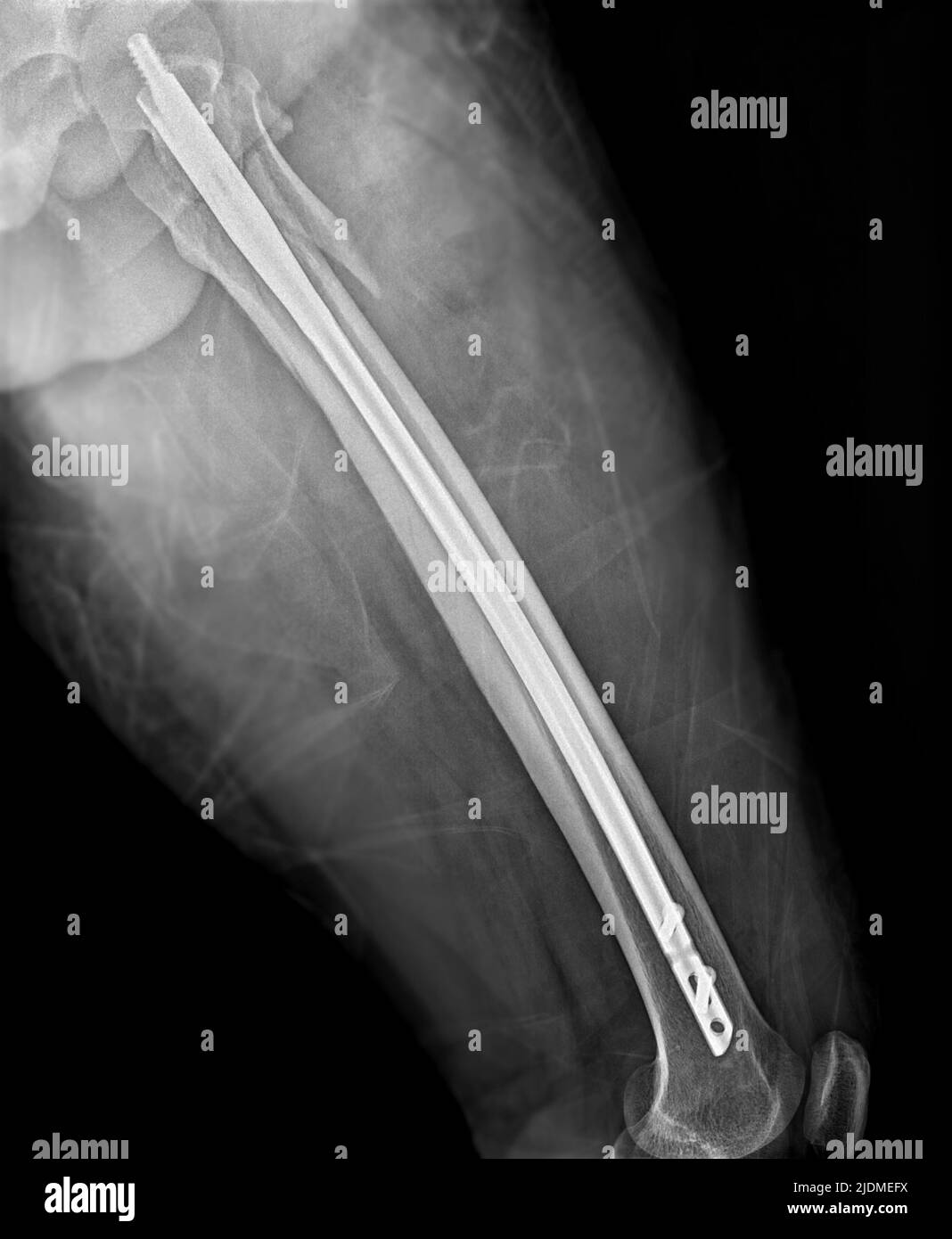 X-ray femur bone Fracture After Total Hip and femur Replacement. Stock Photohttps://www.alamy.com/image-license-details/?v=1https://www.alamy.com/x-ray-femur-bone-fracture-after-total-hip-and-femur-replacement-image473120926.html
RF2JDMEFX–X-ray femur bone Fracture After Total Hip and femur Replacement.
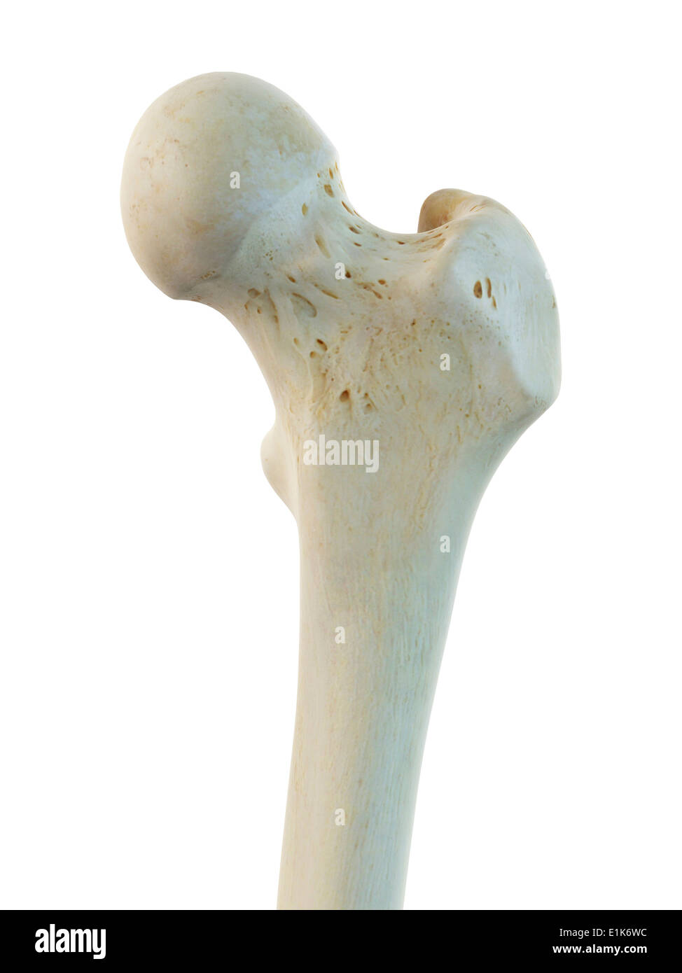 Human femur head computer artwork. Stock Photohttps://www.alamy.com/image-license-details/?v=1https://www.alamy.com/human-femur-head-computer-artwork-image69878632.html
RFE1K6WC–Human femur head computer artwork.
 Human skull isolated on white background Stock Vectorhttps://www.alamy.com/image-license-details/?v=1https://www.alamy.com/human-skull-isolated-on-white-background-image543507728.html
RF2PG6WJ8–Human skull isolated on white background
 3d rendered medically accurate illustration of a horse skeleton Stock Photohttps://www.alamy.com/image-license-details/?v=1https://www.alamy.com/3d-rendered-medically-accurate-illustration-of-a-horse-skeleton-image258153258.html
RFTYYW36–3d rendered medically accurate illustration of a horse skeleton
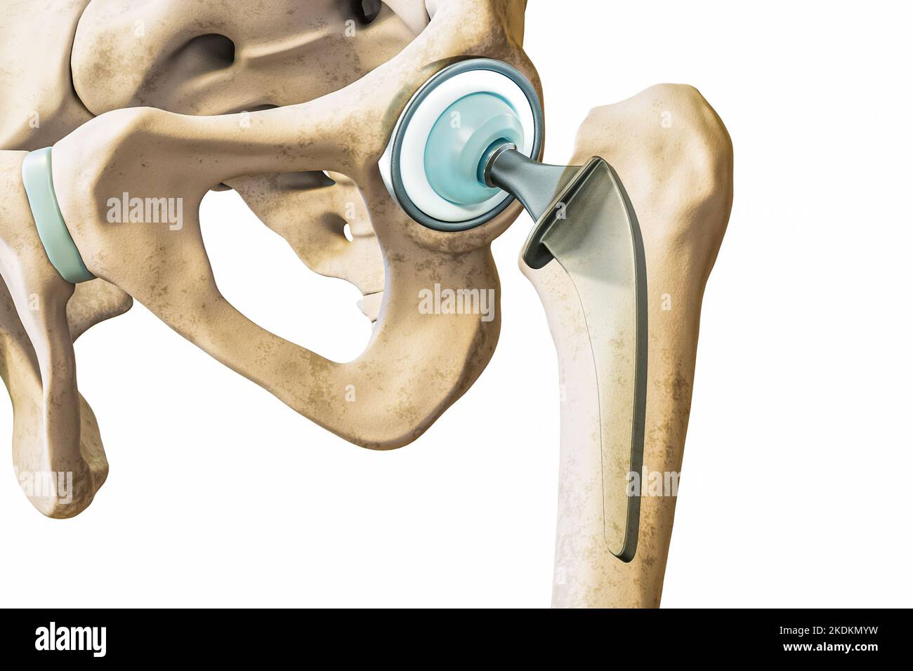 Hip prosthesis or implant isolated on white background close-up. Hip joint or femoral head replacement 3D rendering illustration. Medicine, medical an Stock Photohttps://www.alamy.com/image-license-details/?v=1https://www.alamy.com/hip-prosthesis-or-implant-isolated-on-white-background-close-up-hip-joint-or-femoral-head-replacement-3d-rendering-illustration-medicine-medical-an-image490314381.html
RF2KDKMYW–Hip prosthesis or implant isolated on white background close-up. Hip joint or femoral head replacement 3D rendering illustration. Medicine, medical an
 Silver Water Beetle (Hydrophilus piceus). Dorsal view. In a pond dipping identification tray. Showing body sections, head, thorax, elytra, legs. Stock Photohttps://www.alamy.com/image-license-details/?v=1https://www.alamy.com/silver-water-beetle-hydrophilus-piceus-dorsal-view-in-a-pond-dipping-identification-tray-showing-body-sections-head-thorax-elytra-legs-image329739292.html
RM2A4CWR8–Silver Water Beetle (Hydrophilus piceus). Dorsal view. In a pond dipping identification tray. Showing body sections, head, thorax, elytra, legs.
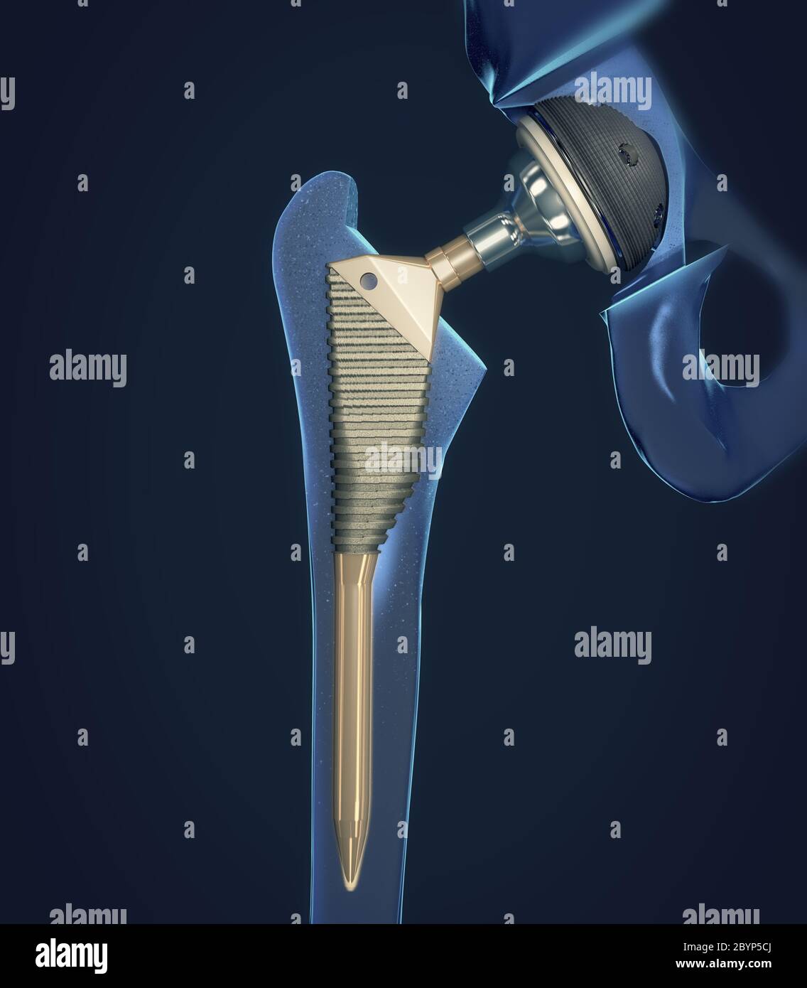 Function of a hip joint implant or hip prosthesis in frontal view - 3d illustration Stock Photohttps://www.alamy.com/image-license-details/?v=1https://www.alamy.com/function-of-a-hip-joint-implant-or-hip-prosthesis-in-frontal-view-3d-illustration-image361290290.html
RF2BYP5CJ–Function of a hip joint implant or hip prosthesis in frontal view - 3d illustration
 Antique Medical Illustration of Femur circa 1881 Stock Photohttps://www.alamy.com/image-license-details/?v=1https://www.alamy.com/stock-photo-antique-medical-illustration-of-femur-circa-1881-37154926.html
RFC4CFDJ–Antique Medical Illustration of Femur circa 1881
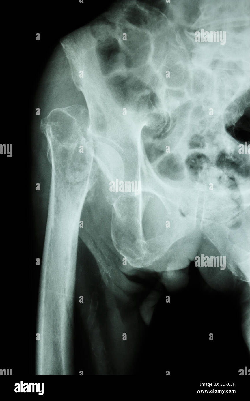 X-ray pelvis & hip joint : Fracture head of femur (thigh bone) Stock Photohttps://www.alamy.com/image-license-details/?v=1https://www.alamy.com/stock-photo-x-ray-pelvis-hip-joint-fracture-head-of-femur-thigh-bone-77249245.html
RFEDK05H–X-ray pelvis & hip joint : Fracture head of femur (thigh bone)
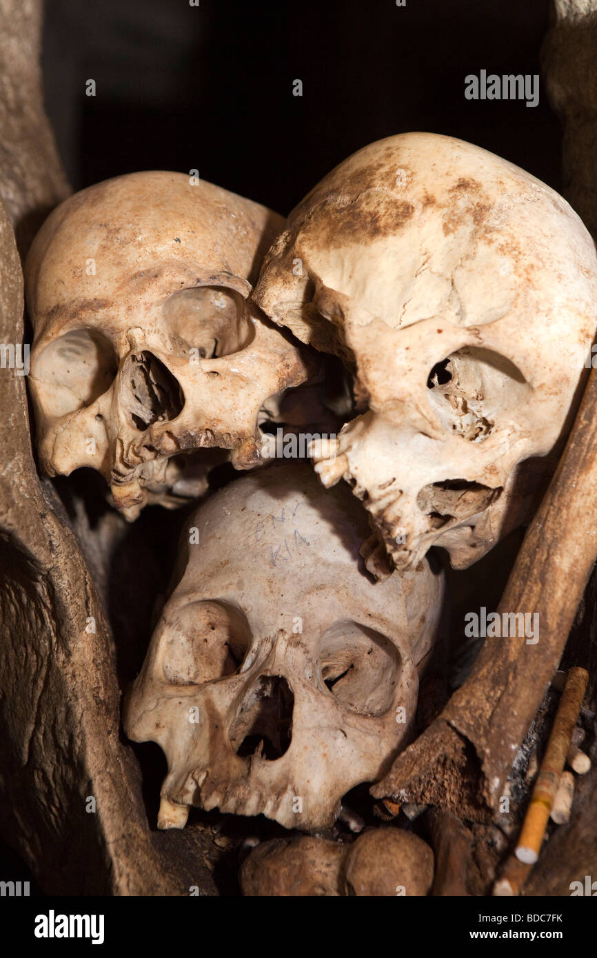 Indonesia Sulawesi Tana Toraja Londa village burial cave skulls and femur in rock cleft Stock Photohttps://www.alamy.com/image-license-details/?v=1https://www.alamy.com/stock-photo-indonesia-sulawesi-tana-toraja-londa-village-burial-cave-skulls-and-25470247.html
RMBDC7FK–Indonesia Sulawesi Tana Toraja Londa village burial cave skulls and femur in rock cleft
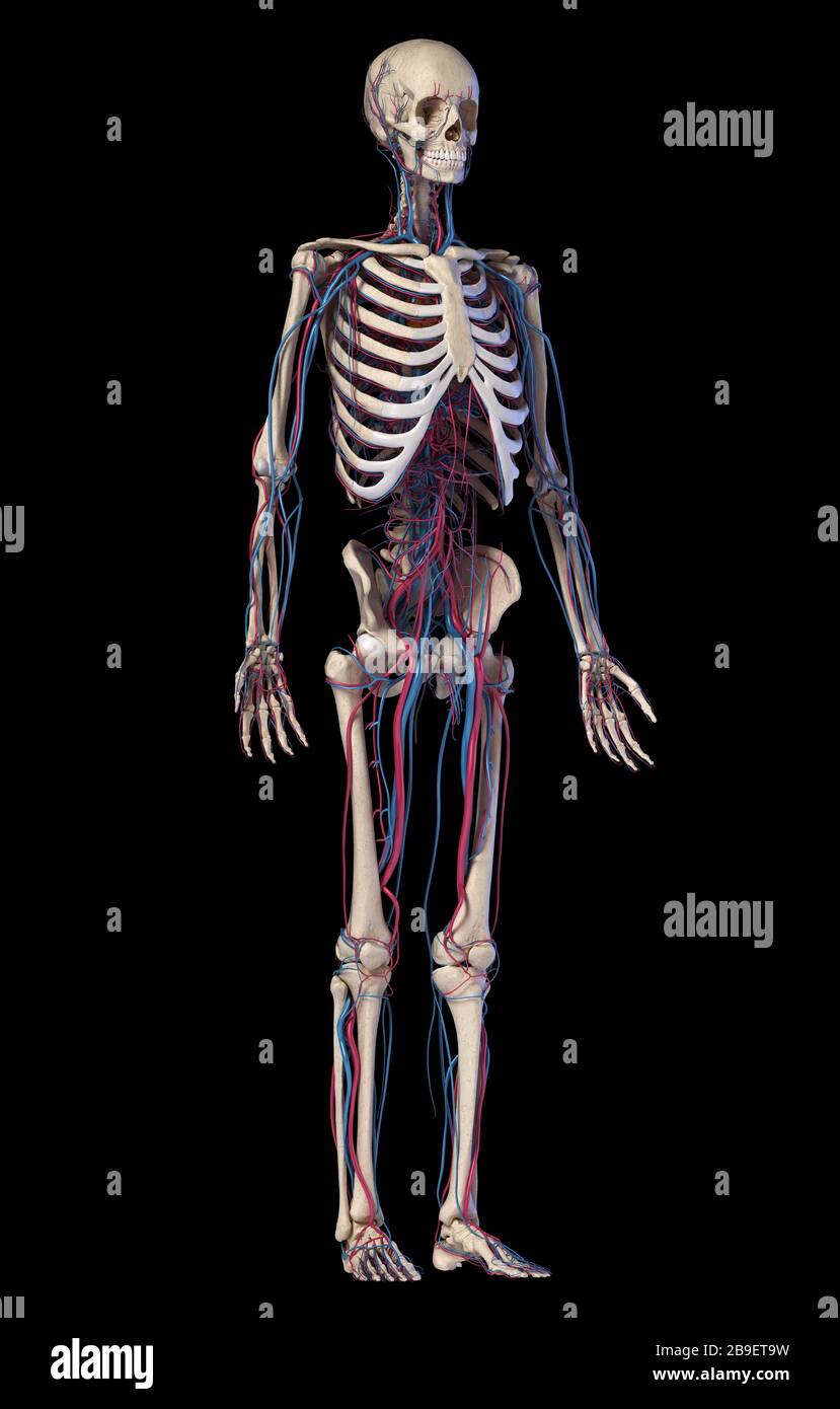 Human skeleton with veins and arteries. Front perspective view on black background. Stock Photohttps://www.alamy.com/image-license-details/?v=1https://www.alamy.com/human-skeleton-with-veins-and-arteries-front-perspective-view-on-black-background-image350065685.html
RF2B9ET9W–Human skeleton with veins and arteries. Front perspective view on black background.
