Meiosis ii Stock Photos and Images
 Meiosis II telophase II, illustration. Each daughter cell is haploid (has half the total number of chromosomes of the original cell). Stock Photohttps://www.alamy.com/image-license-details/?v=1https://www.alamy.com/meiosis-ii-telophase-ii-illustration-each-daughter-cell-is-haploid-has-half-the-total-number-of-chromosomes-of-the-original-cell-image618634143.html
RF2XXD64F–Meiosis II telophase II, illustration. Each daughter cell is haploid (has half the total number of chromosomes of the original cell).
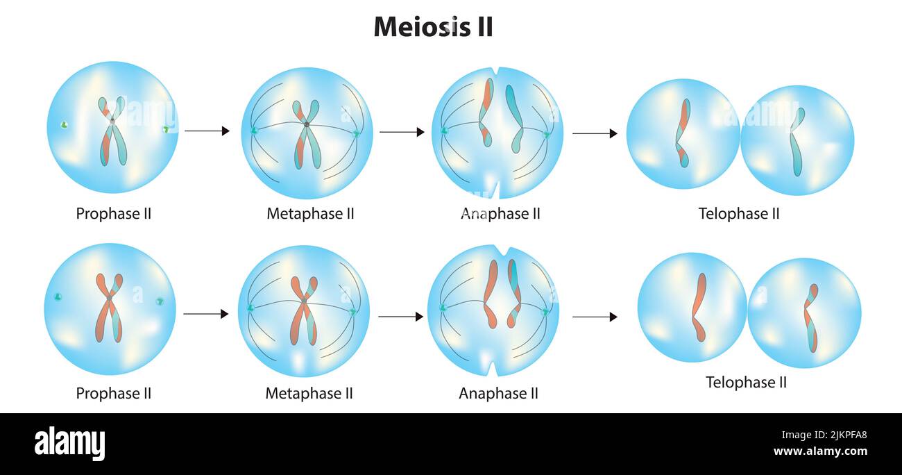 Meiosis ii stages Stock Photohttps://www.alamy.com/image-license-details/?v=1https://www.alamy.com/meiosis-ii-stages-image476853392.html
RF2JKPFA8–Meiosis ii stages
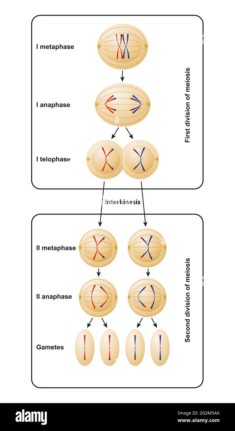 Division of meiosis. Meiosis is divided into meiosis I and meiosis II Stock Photohttps://www.alamy.com/image-license-details/?v=1https://www.alamy.com/division-of-meiosis-meiosis-is-divided-into-meiosis-i-and-meiosis-ii-image432546434.html
RF2G3M5AX–Division of meiosis. Meiosis is divided into meiosis I and meiosis II
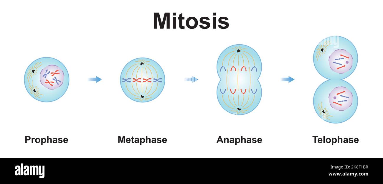 Scientific Designing of Mitosis Phases (Cell Division). Colorful Symbols. Vector Illustration. Stock Vectorhttps://www.alamy.com/image-license-details/?v=1https://www.alamy.com/scientific-designing-of-mitosis-phases-cell-division-colorful-symbols-vector-illustration-image487137947.html
RF2K8F1BR–Scientific Designing of Mitosis Phases (Cell Division). Colorful Symbols. Vector Illustration.
 Archive image from page 229 of Cytology, with special reference to. Cytology, with special reference to the metazoan nucleus cytologywithspec00agar 0 Year: 1920 Syngamy Aggregate Diplocgstis Certain of the Rhodophyceae. (Nemalicnes) Syngamy II Meiosis Syngamy Pteridophyla Meiosis Stock Photohttps://www.alamy.com/image-license-details/?v=1https://www.alamy.com/archive-image-from-page-229-of-cytology-with-special-reference-to-cytology-with-special-reference-to-the-metazoan-nucleus-cytologywithspec00agar-0-year-1920-syngamy-aggregate-diplocgstis-certain-of-the-rhodophyceae-nemalicnes-syngamy-ii-meiosis-syngamy-pteridophyla-meiosis-image259482615.html
RMW24CM7–Archive image from page 229 of Cytology, with special reference to. Cytology, with special reference to the metazoan nucleus cytologywithspec00agar 0 Year: 1920 Syngamy Aggregate Diplocgstis Certain of the Rhodophyceae. (Nemalicnes) Syngamy II Meiosis Syngamy Pteridophyla Meiosis
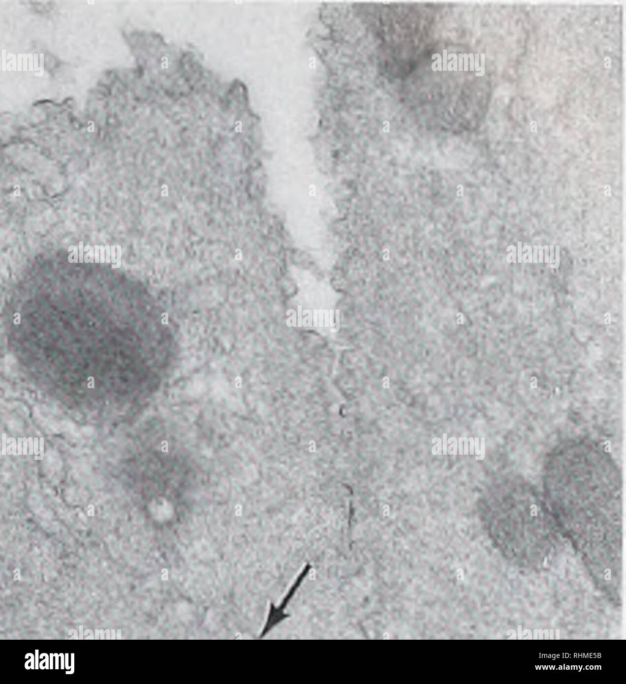 . The Biological bulletin. Biology; Zoology; Biology; Marine Biology. FERTILIZATION PROCESSES IN OYSTER EGGS 211 I * '. N b Figure 15. Polar bodies formed in eggs treated with cytochalasin B during the period of meiosis I (a) and meiosis II (b). Usually only polar bodies developing at meiosis II possess chromatin that becomes organized into a nucleus, (a) Nucleus (N) in a polar body formed after inhibition of the first polar body, (b) Chromatin of polar bodies that form at the first meiotic division may be surrounded by cisternae (arrows), however, and a nuclear envelope usually fails to form. Stock Photohttps://www.alamy.com/image-license-details/?v=1https://www.alamy.com/the-biological-bulletin-biology-zoology-biology-marine-biology-fertilization-processes-in-oyster-eggs-211-i-n-b-figure-15-polar-bodies-formed-in-eggs-treated-with-cytochalasin-b-during-the-period-of-meiosis-i-a-and-meiosis-ii-b-usually-only-polar-bodies-developing-at-meiosis-ii-possess-chromatin-that-becomes-organized-into-a-nucleus-a-nucleus-n-in-a-polar-body-formed-after-inhibition-of-the-first-polar-body-b-chromatin-of-polar-bodies-that-form-at-the-first-meiotic-division-may-be-surrounded-by-cisternae-arrows-however-and-a-nuclear-envelope-usually-fails-to-form-image234634103.html
RMRHME5B–. The Biological bulletin. Biology; Zoology; Biology; Marine Biology. FERTILIZATION PROCESSES IN OYSTER EGGS 211 I * '. N b Figure 15. Polar bodies formed in eggs treated with cytochalasin B during the period of meiosis I (a) and meiosis II (b). Usually only polar bodies developing at meiosis II possess chromatin that becomes organized into a nucleus, (a) Nucleus (N) in a polar body formed after inhibition of the first polar body, (b) Chromatin of polar bodies that form at the first meiotic division may be surrounded by cisternae (arrows), however, and a nuclear envelope usually fails to form.
 . Cytology, with special reference to the metazoan nucleus. Cells. Syngamy II Meiosis Syngojny Pteridophyla V Syngamy Phaneroga rns. Please note that these images are extracted from scanned page images that may have been digitally enhanced for readability - coloration and appearance of these illustrations may not perfectly resemble the original work.. Agar, Wilfred Eade, 1882-. London, Macmillan Stock Photohttps://www.alamy.com/image-license-details/?v=1https://www.alamy.com/cytology-with-special-reference-to-the-metazoan-nucleus-cells-syngamy-ii-meiosis-syngojny-pteridophyla-v-syngamy-phaneroga-rns-please-note-that-these-images-are-extracted-from-scanned-page-images-that-may-have-been-digitally-enhanced-for-readability-coloration-and-appearance-of-these-illustrations-may-not-perfectly-resemble-the-original-work-agar-wilfred-eade-1882-london-macmillan-image216167666.html
RMPFK81P–. Cytology, with special reference to the metazoan nucleus. Cells. Syngamy II Meiosis Syngojny Pteridophyla V Syngamy Phaneroga rns. Please note that these images are extracted from scanned page images that may have been digitally enhanced for readability - coloration and appearance of these illustrations may not perfectly resemble the original work.. Agar, Wilfred Eade, 1882-. London, Macmillan
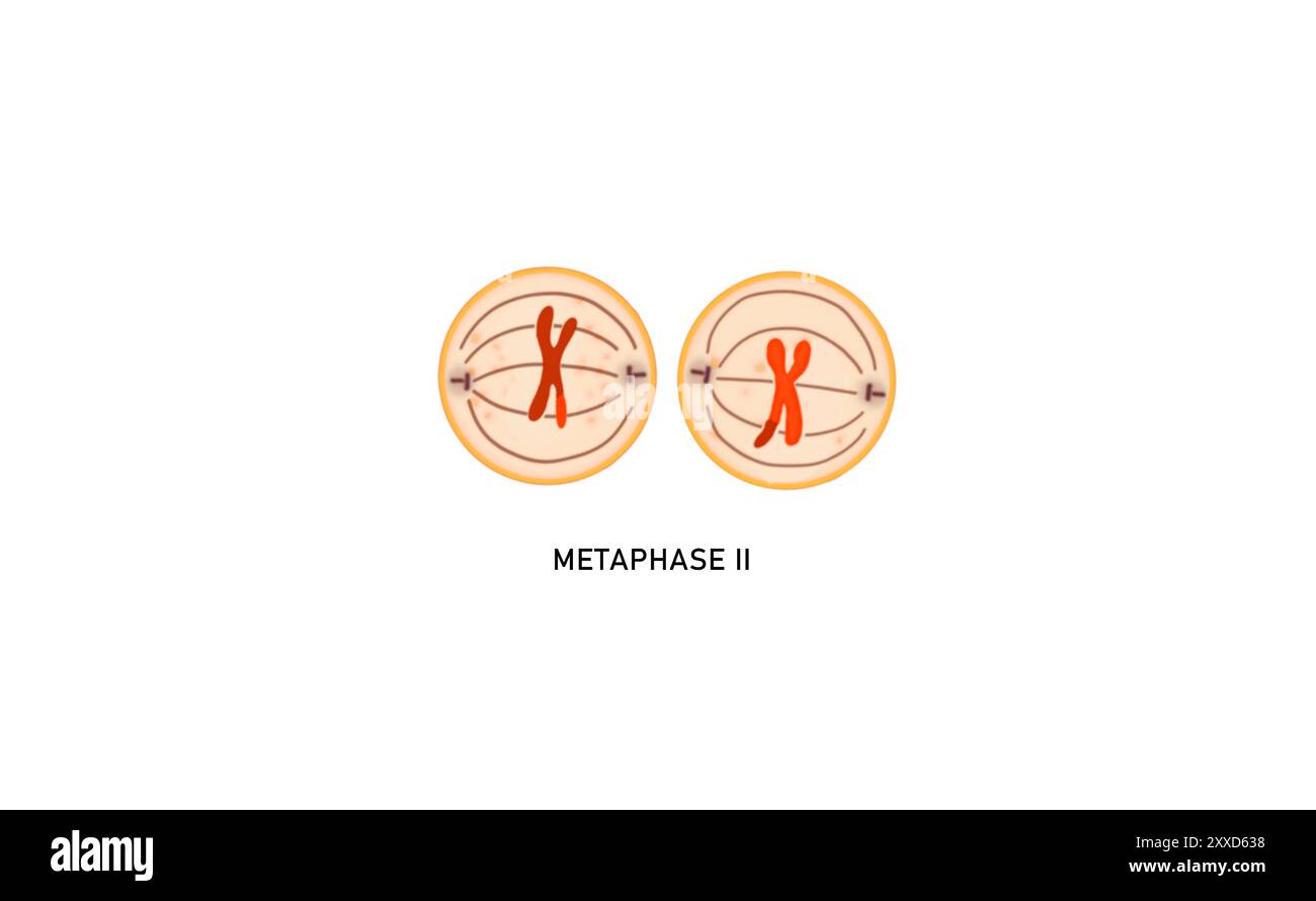 Meiosis II metaphase II, illustration. During metaphase II the centromeres of the paired chromatids align along the equatorial plate in both cells. Stock Photohttps://www.alamy.com/image-license-details/?v=1https://www.alamy.com/meiosis-ii-metaphase-ii-illustration-during-metaphase-ii-the-centromeres-of-the-paired-chromatids-align-along-the-equatorial-plate-in-both-cells-image618634108.html
RF2XXD638–Meiosis II metaphase II, illustration. During metaphase II the centromeres of the paired chromatids align along the equatorial plate in both cells.
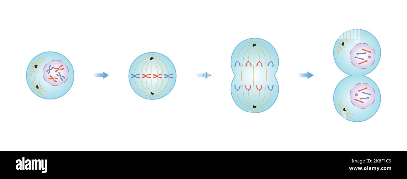 Scientific Designing of Mitosis Phases (Cell Division). Colorful Symbols. Vector Illustration. Stock Vectorhttps://www.alamy.com/image-license-details/?v=1https://www.alamy.com/scientific-designing-of-mitosis-phases-cell-division-colorful-symbols-vector-illustration-image487137961.html
RF2K8F1C9–Scientific Designing of Mitosis Phases (Cell Division). Colorful Symbols. Vector Illustration.
 Archive image from page 20 of Cytological studies of five interspecific. Cytological studies of five interspecific hybrids of Crepis leontodontoides cytologicalstudi65aver Year: 1930 c d Fig. 13. Meiosis in F, C. leontodontoides-marschalli. a, I—M, with four bivalents; b, I-A, showing' three large marscluilli chromo- somes ; c, II-A, after a 6-3 distribution at I-A; d, II-T, five large and two small nuclei. Mr A Ac 7£ Fig. 14. Somatic metaphases. a, of C aurea; b, of F (7. leontodontoides-aurea. Stock Photohttps://www.alamy.com/image-license-details/?v=1https://www.alamy.com/archive-image-from-page-20-of-cytological-studies-of-five-interspecific-cytological-studies-of-five-interspecific-hybrids-of-crepis-leontodontoides-cytologicalstudi65aver-year-1930-c-d-fig-13-meiosis-in-f-c-leontodontoides-marschalli-a-im-with-four-bivalents-b-i-a-showing-three-large-marscluilli-chromo-somes-c-ii-a-after-a-6-3-distribution-at-i-a-d-ii-t-five-large-and-two-small-nuclei-mr-a-ac-7-fig-14-somatic-metaphases-a-of-c-aurea-b-of-f-7-leontodontoides-aurea-image259443089.html
RMW22J8H–Archive image from page 20 of Cytological studies of five interspecific. Cytological studies of five interspecific hybrids of Crepis leontodontoides cytologicalstudi65aver Year: 1930 c d Fig. 13. Meiosis in F, C. leontodontoides-marschalli. a, I—M, with four bivalents; b, I-A, showing' three large marscluilli chromo- somes ; c, II-A, after a 6-3 distribution at I-A; d, II-T, five large and two small nuclei. Mr A Ac 7£ Fig. 14. Somatic metaphases. a, of C aurea; b, of F (7. leontodontoides-aurea.
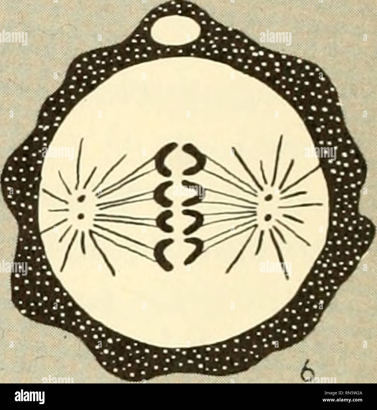 . Animal growth and development. Embryology; Growth; Biology; Growth; Embryology; Animals -- growth & development. 0. Fig. 26. Fertilization (after Wilson). (I) Sperm contacts egg. (2) Sperm head and midpiece enter egg through fertilization cone; egg nucleus pro- ceeds through meiosis II. (3) Sperm nucleus enlarges; midpiece constructs spindle and asters; meiosis II of egg completes to yield polar body and functional, haplold egg nucleus. (4) Sperm and egg nuclei migrate toward each other across midpiece spindle; nuclear membranes break down. (5) Chromosomes dispose themselves upon mitotic Stock Photohttps://www.alamy.com/image-license-details/?v=1https://www.alamy.com/animal-growth-and-development-embryology-growth-biology-growth-embryology-animals-growth-amp-development-0-fig-26-fertilization-after-wilson-i-sperm-contacts-egg-2-sperm-head-and-midpiece-enter-egg-through-fertilization-cone-egg-nucleus-pro-ceeds-through-meiosis-ii-3-sperm-nucleus-enlarges-midpiece-constructs-spindle-and-asters-meiosis-ii-of-egg-completes-to-yield-polar-body-and-functional-haplold-egg-nucleus-4-sperm-and-egg-nuclei-migrate-toward-each-other-across-midpiece-spindle-nuclear-membranes-break-down-5-chromosomes-dispose-themselves-upon-mitotic-image236771986.html
RMRN5W2A–. Animal growth and development. Embryology; Growth; Biology; Growth; Embryology; Animals -- growth & development. 0. Fig. 26. Fertilization (after Wilson). (I) Sperm contacts egg. (2) Sperm head and midpiece enter egg through fertilization cone; egg nucleus pro- ceeds through meiosis II. (3) Sperm nucleus enlarges; midpiece constructs spindle and asters; meiosis II of egg completes to yield polar body and functional, haplold egg nucleus. (4) Sperm and egg nuclei migrate toward each other across midpiece spindle; nuclear membranes break down. (5) Chromosomes dispose themselves upon mitotic
 . Cytology, with special reference to the metazoan nucleus. Cells. Syngamy II Meiosis Syngamy Pteridophyla Meiosis. V Syngamy Phanerogam*. Please note that these images are extracted from scanned page images that may have been digitally enhanced for readability - coloration and appearance of these illustrations may not perfectly resemble the original work.. Agar, W. E. (Wilfred Eade), 1882-1951. London, Macmillan and Co. , limited Stock Photohttps://www.alamy.com/image-license-details/?v=1https://www.alamy.com/cytology-with-special-reference-to-the-metazoan-nucleus-cells-syngamy-ii-meiosis-syngamy-pteridophyla-meiosis-v-syngamy-phanerogam-please-note-that-these-images-are-extracted-from-scanned-page-images-that-may-have-been-digitally-enhanced-for-readability-coloration-and-appearance-of-these-illustrations-may-not-perfectly-resemble-the-original-work-agar-w-e-wilfred-eade-1882-1951-london-macmillan-and-co-limited-image216168311.html
RMPFK8TR–. Cytology, with special reference to the metazoan nucleus. Cells. Syngamy II Meiosis Syngamy Pteridophyla Meiosis. V Syngamy Phanerogam*. Please note that these images are extracted from scanned page images that may have been digitally enhanced for readability - coloration and appearance of these illustrations may not perfectly resemble the original work.. Agar, W. E. (Wilfred Eade), 1882-1951. London, Macmillan and Co. , limited
 Meiosis II anaphase II, illustration. In this phase of meiosis II there is a simultaneous splitting of the centromere of each chromosome and the sister chromatids are pulled away towards the opposite poles. Stock Photohttps://www.alamy.com/image-license-details/?v=1https://www.alamy.com/meiosis-ii-anaphase-ii-illustration-in-this-phase-of-meiosis-ii-there-is-a-simultaneous-splitting-of-the-centromere-of-each-chromosome-and-the-sister-chromatids-are-pulled-away-towards-the-opposite-poles-image618634070.html
RF2XXD61X–Meiosis II anaphase II, illustration. In this phase of meiosis II there is a simultaneous splitting of the centromere of each chromosome and the sister chromatids are pulled away towards the opposite poles.
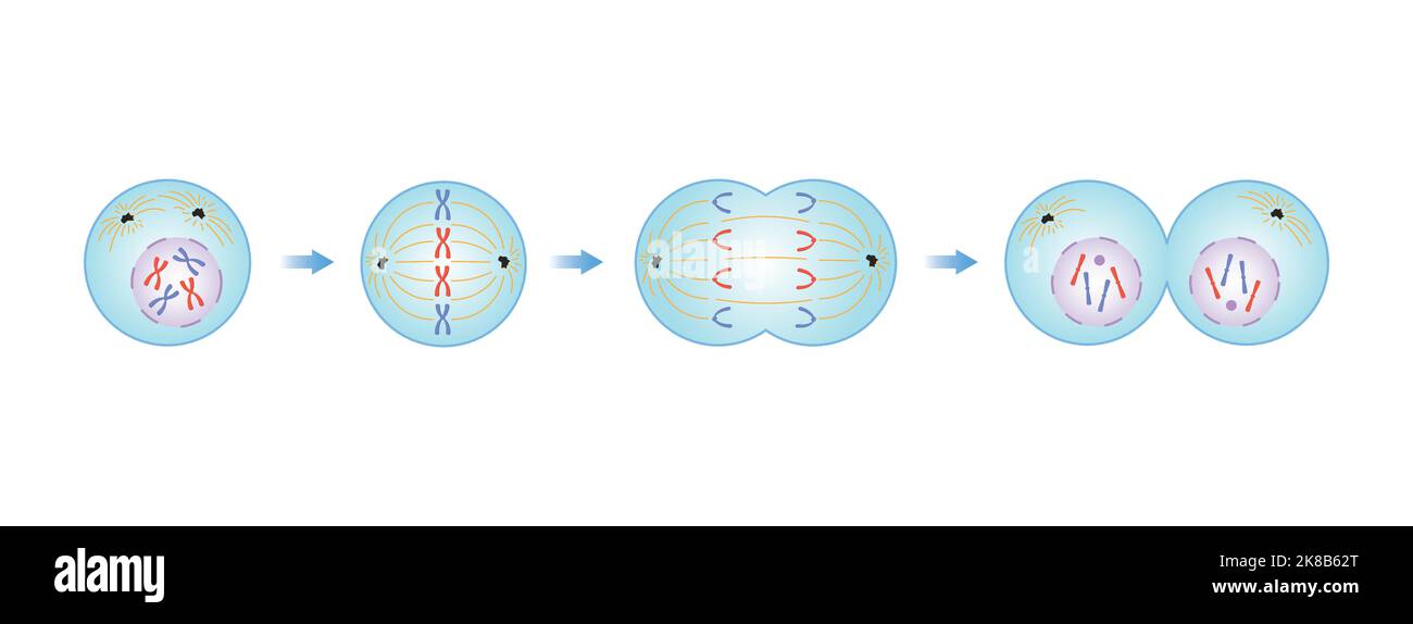 Scientific Designing of Mitosis Phases (Cell Division). Colorful Symbols. Vector Illustration. Stock Vectorhttps://www.alamy.com/image-license-details/?v=1https://www.alamy.com/scientific-designing-of-mitosis-phases-cell-division-colorful-symbols-vector-illustration-image487053808.html
RF2K8B62T–Scientific Designing of Mitosis Phases (Cell Division). Colorful Symbols. Vector Illustration.
 Archive image from page 54 of Cytology, with special reference to. Cytology, with special reference to the metazoan nucleus cytologywithspec00agar Year: 1920 II MEIOSIS IN LEPIDOSIREN 39 conjugation of the chromosomes. Hence the word synizesis was proposed for the contraction, and syndesis for the chromosome conjugation. Stock Photohttps://www.alamy.com/image-license-details/?v=1https://www.alamy.com/archive-image-from-page-54-of-cytology-with-special-reference-to-cytology-with-special-reference-to-the-metazoan-nucleus-cytologywithspec00agar-year-1920-ii-meiosis-in-lepidosiren-39-conjugation-of-the-chromosomes-hence-the-word-synizesis-was-proposed-for-the-contraction-and-syndesis-for-the-chromosome-conjugation-image259285630.html
RMW1RDD2–Archive image from page 54 of Cytology, with special reference to. Cytology, with special reference to the metazoan nucleus cytologywithspec00agar Year: 1920 II MEIOSIS IN LEPIDOSIREN 39 conjugation of the chromosomes. Hence the word synizesis was proposed for the contraction, and syndesis for the chromosome conjugation.
 . Cytology, with special reference to the metazoan nucleus. Cells. Syngamy II Meiosis Syngojny Pteridophyla V Syngamy Phaneroga rns. Please note that these images are extracted from scanned page images that may have been digitally enhanced for readability - coloration and appearance of these illustrations may not perfectly resemble the original work.. Agar, Wilfred Eade, 1882-. London, Macmillan Stock Photohttps://www.alamy.com/image-license-details/?v=1https://www.alamy.com/cytology-with-special-reference-to-the-metazoan-nucleus-cells-syngamy-ii-meiosis-syngojny-pteridophyla-v-syngamy-phaneroga-rns-please-note-that-these-images-are-extracted-from-scanned-page-images-that-may-have-been-digitally-enhanced-for-readability-coloration-and-appearance-of-these-illustrations-may-not-perfectly-resemble-the-original-work-agar-wilfred-eade-1882-london-macmillan-image231789264.html
RMRD2WG0–. Cytology, with special reference to the metazoan nucleus. Cells. Syngamy II Meiosis Syngojny Pteridophyla V Syngamy Phaneroga rns. Please note that these images are extracted from scanned page images that may have been digitally enhanced for readability - coloration and appearance of these illustrations may not perfectly resemble the original work.. Agar, Wilfred Eade, 1882-. London, Macmillan
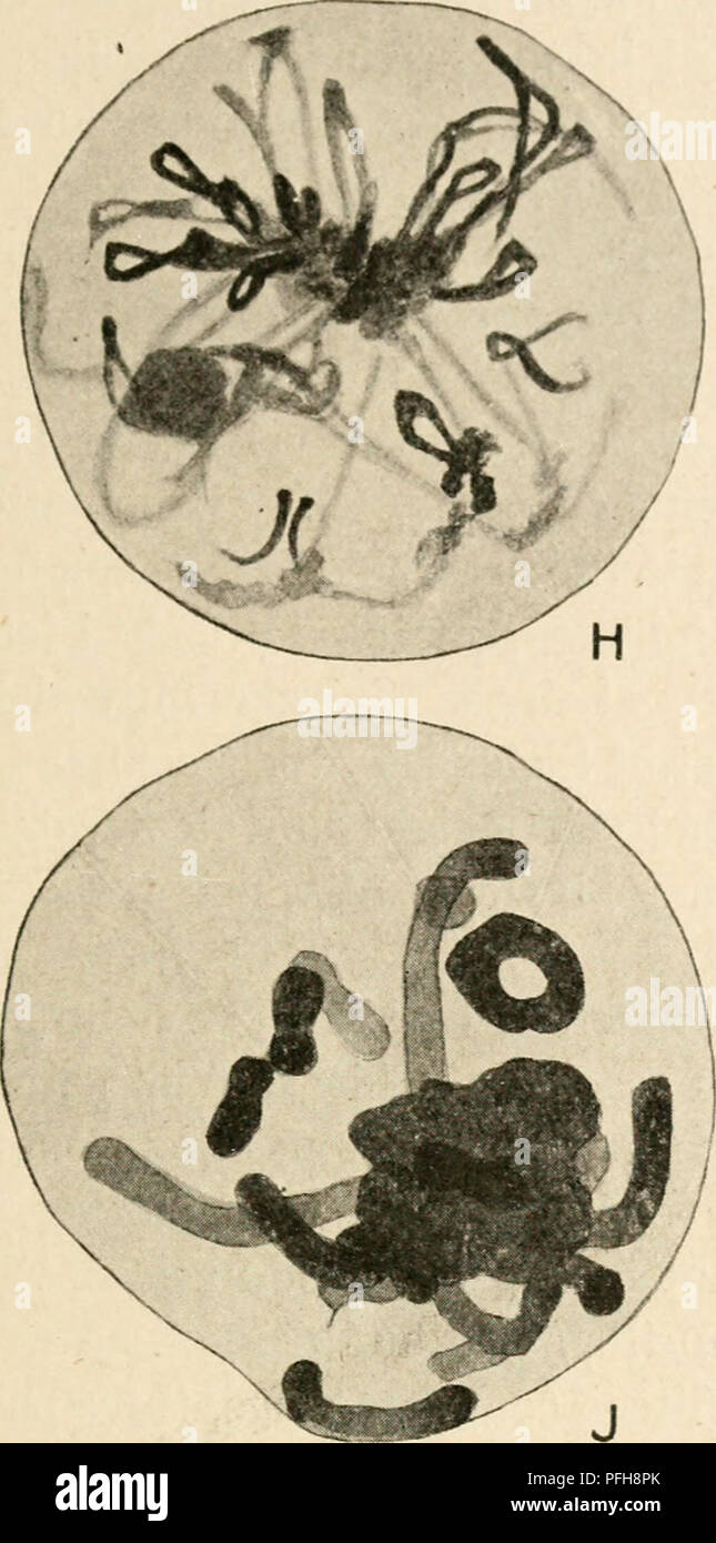 . Cytology, with special reference to the metazoan nucleus. Cells. II MEIOSIS IN LEPIDOSIREN 39 conjugation of the chromosomes. Hence the word synizesis was proposed for the contraction, and syndesis for the chromosome conjugation.. Please note that these images are extracted from scanned page images that may have been digitally enhanced for readability - coloration and appearance of these illustrations may not perfectly resemble the original work.. Agar, Wilfred Eade, 1882-. London, Macmillan Stock Photohttps://www.alamy.com/image-license-details/?v=1https://www.alamy.com/cytology-with-special-reference-to-the-metazoan-nucleus-cells-ii-meiosis-in-lepidosiren-39-conjugation-of-the-chromosomes-hence-the-word-synizesis-was-proposed-for-the-contraction-and-syndesis-for-the-chromosome-conjugation-please-note-that-these-images-are-extracted-from-scanned-page-images-that-may-have-been-digitally-enhanced-for-readability-coloration-and-appearance-of-these-illustrations-may-not-perfectly-resemble-the-original-work-agar-wilfred-eade-1882-london-macmillan-image216124347.html
RMPFH8PK–. Cytology, with special reference to the metazoan nucleus. Cells. II MEIOSIS IN LEPIDOSIREN 39 conjugation of the chromosomes. Hence the word synizesis was proposed for the contraction, and syndesis for the chromosome conjugation.. Please note that these images are extracted from scanned page images that may have been digitally enhanced for readability - coloration and appearance of these illustrations may not perfectly resemble the original work.. Agar, Wilfred Eade, 1882-. London, Macmillan
 Meiosis II prophase II, illustration. During prophase II the chromosomes condense and a new set of spindle fibres form. The chromosomes begin moving towards the equator of the cell. Stock Photohttps://www.alamy.com/image-license-details/?v=1https://www.alamy.com/meiosis-ii-prophase-ii-illustration-during-prophase-ii-the-chromosomes-condense-and-a-new-set-of-spindle-fibres-form-the-chromosomes-begin-moving-towards-the-equator-of-the-cell-image618634126.html
RF2XXD63X–Meiosis II prophase II, illustration. During prophase II the chromosomes condense and a new set of spindle fibres form. The chromosomes begin moving towards the equator of the cell.
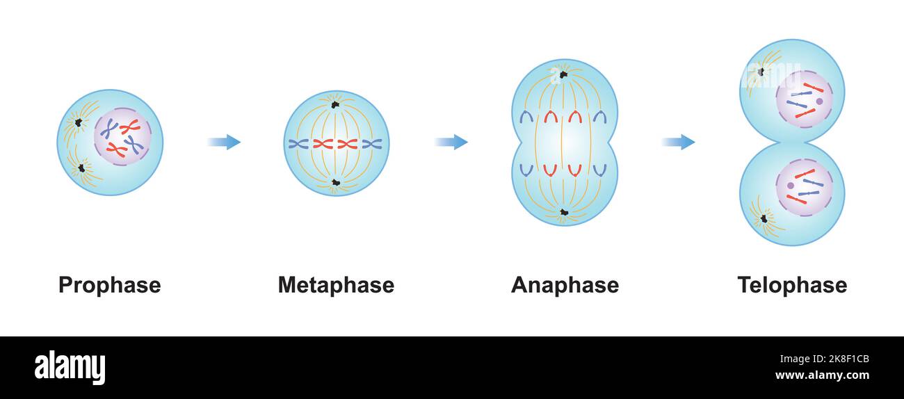 Scientific Designing of Mitosis Phases (Cell Division). Colorful Symbols. Vector Illustration. Stock Vectorhttps://www.alamy.com/image-license-details/?v=1https://www.alamy.com/scientific-designing-of-mitosis-phases-cell-division-colorful-symbols-vector-illustration-image487137963.html
RF2K8F1CB–Scientific Designing of Mitosis Phases (Cell Division). Colorful Symbols. Vector Illustration.
 Archive image from page 76 of Cytology, with special reference to. Cytology, with special reference to the metazoan nucleus cytologywithspec00agar Year: 1920 II MEIOSIS IN THE FEMALE 6i germinal vesicles with diffuse chromosomes are to be found, and it is evident that the bivalents resulting from syjidesis condense continuously ' :.f Stock Photohttps://www.alamy.com/image-license-details/?v=1https://www.alamy.com/archive-image-from-page-76-of-cytology-with-special-reference-to-cytology-with-special-reference-to-the-metazoan-nucleus-cytologywithspec00agar-year-1920-ii-meiosis-in-the-female-6i-germinal-vesicles-with-diffuse-chromosomes-are-to-be-found-and-it-is-evident-that-the-bivalents-resulting-from-syjidesis-condense-continuously-f-image259293587.html
RMW1RRH7–Archive image from page 76 of Cytology, with special reference to. Cytology, with special reference to the metazoan nucleus cytologywithspec00agar Year: 1920 II MEIOSIS IN THE FEMALE 6i germinal vesicles with diffuse chromosomes are to be found, and it is evident that the bivalents resulting from syjidesis condense continuously ' :.f
 . Cytology, with special reference to the metazoan nucleus. Cells. Syngamy II Meiosis Syngamy Pteridophyla Meiosis. V Syngamy Phanerogam*. Please note that these images are extracted from scanned page images that may have been digitally enhanced for readability - coloration and appearance of these illustrations may not perfectly resemble the original work.. Agar, W. E. (Wilfred Eade), 1882-1951. London, Macmillan and Co. , limited Stock Photohttps://www.alamy.com/image-license-details/?v=1https://www.alamy.com/cytology-with-special-reference-to-the-metazoan-nucleus-cells-syngamy-ii-meiosis-syngamy-pteridophyla-meiosis-v-syngamy-phanerogam-please-note-that-these-images-are-extracted-from-scanned-page-images-that-may-have-been-digitally-enhanced-for-readability-coloration-and-appearance-of-these-illustrations-may-not-perfectly-resemble-the-original-work-agar-w-e-wilfred-eade-1882-1951-london-macmillan-and-co-limited-image231777679.html
RMRD2AP7–. Cytology, with special reference to the metazoan nucleus. Cells. Syngamy II Meiosis Syngamy Pteridophyla Meiosis. V Syngamy Phanerogam*. Please note that these images are extracted from scanned page images that may have been digitally enhanced for readability - coloration and appearance of these illustrations may not perfectly resemble the original work.. Agar, W. E. (Wilfred Eade), 1882-1951. London, Macmillan and Co. , limited
 . Cytology, with special reference to the metazoan nucleus. Cells. Syngamy Aggregate Diplocgstis Certain of the Rhodophyceae. (Nemalicnes). Syngamy II Meiosis Syngamy Pteridophyla Meiosis. Please note that these images are extracted from scanned page images that may have been digitally enhanced for readability - coloration and appearance of these illustrations may not perfectly resemble the original work.. Agar, W. E. (Wilfred Eade), 1882-1951. London, Macmillan and Co. , limited Stock Photohttps://www.alamy.com/image-license-details/?v=1https://www.alamy.com/cytology-with-special-reference-to-the-metazoan-nucleus-cells-syngamy-aggregate-diplocgstis-certain-of-the-rhodophyceae-nemalicnes-syngamy-ii-meiosis-syngamy-pteridophyla-meiosis-please-note-that-these-images-are-extracted-from-scanned-page-images-that-may-have-been-digitally-enhanced-for-readability-coloration-and-appearance-of-these-illustrations-may-not-perfectly-resemble-the-original-work-agar-w-e-wilfred-eade-1882-1951-london-macmillan-and-co-limited-image216168314.html
RMPFK8TX–. Cytology, with special reference to the metazoan nucleus. Cells. Syngamy Aggregate Diplocgstis Certain of the Rhodophyceae. (Nemalicnes). Syngamy II Meiosis Syngamy Pteridophyla Meiosis. Please note that these images are extracted from scanned page images that may have been digitally enhanced for readability - coloration and appearance of these illustrations may not perfectly resemble the original work.. Agar, W. E. (Wilfred Eade), 1882-1951. London, Macmillan and Co. , limited
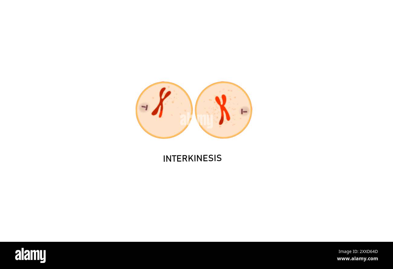 Meiosis I interkinesis, illustration. Interkinesis or interphase II is a period of rest that cells of some species enter between meiosis I and meiosis II. Stock Photohttps://www.alamy.com/image-license-details/?v=1https://www.alamy.com/meiosis-i-interkinesis-illustration-interkinesis-or-interphase-ii-is-a-period-of-rest-that-cells-of-some-species-enter-between-meiosis-i-and-meiosis-ii-image618634141.html
RF2XXD64D–Meiosis I interkinesis, illustration. Interkinesis or interphase II is a period of rest that cells of some species enter between meiosis I and meiosis II.
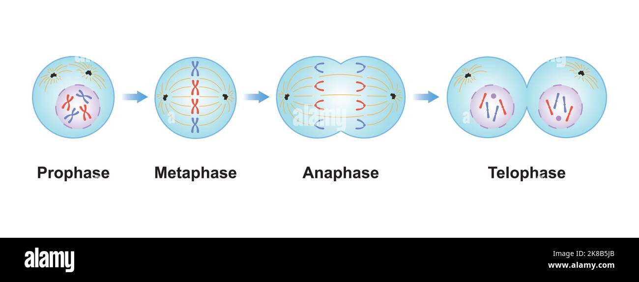 Scientific Designing of Mitosis Phases (Cell Division). Colorful Symbols. Vector Illustration. Stock Vectorhttps://www.alamy.com/image-license-details/?v=1https://www.alamy.com/scientific-designing-of-mitosis-phases-cell-division-colorful-symbols-vector-illustration-image487053459.html
RF2K8B5JB–Scientific Designing of Mitosis Phases (Cell Division). Colorful Symbols. Vector Illustration.
 Archive image from page 74 of Cytology, with special reference to. Cytology, with special reference to the metazoan nucleus cytologywithspec00agaruoft Year: 1920 II MEIOSIS IN THE FEMALE 59 The particular problems raised by the germinal vesicle stage in oogenesis are : (i) The continuity of the chromosomes throughout this period. (2) The relation between the chromosomes and the nucleoli. (â 3) The connection between the peculiar germinal vesicle stage and the synchronous enormous growth of the cytoplasm of the egg, together with the formation of yolk. (4) Does any comparable stage occur in sp Stock Photohttps://www.alamy.com/image-license-details/?v=1https://www.alamy.com/archive-image-from-page-74-of-cytology-with-special-reference-to-cytology-with-special-reference-to-the-metazoan-nucleus-cytologywithspec00agaruoft-year-1920-ii-meiosis-in-the-female-59-the-particular-problems-raised-by-the-germinal-vesicle-stage-in-oogenesis-are-i-the-continuity-of-the-chromosomes-throughout-this-period-2-the-relation-between-the-chromosomes-and-the-nucleoli-3-the-connection-between-the-peculiar-germinal-vesicle-stage-and-the-synchronous-enormous-growth-of-the-cytoplasm-of-the-egg-together-with-the-formation-of-yolk-4-does-any-comparable-stage-occur-in-sp-image259292686.html
RMW1RPD2–Archive image from page 74 of Cytology, with special reference to. Cytology, with special reference to the metazoan nucleus cytologywithspec00agaruoft Year: 1920 II MEIOSIS IN THE FEMALE 59 The particular problems raised by the germinal vesicle stage in oogenesis are : (i) The continuity of the chromosomes throughout this period. (2) The relation between the chromosomes and the nucleoli. (â 3) The connection between the peculiar germinal vesicle stage and the synchronous enormous growth of the cytoplasm of the egg, together with the formation of yolk. (4) Does any comparable stage occur in sp
 . Cytology, with special reference to the metazoan nucleus. Cells. II MEIOSIS IN LEPIDOSIREN 39 conjugation of the chromosomes. Hence the word synizesis was proposed for the contraction, and syndesis for the chromosome conjugation.. Please note that these images are extracted from scanned page images that may have been digitally enhanced for readability - coloration and appearance of these illustrations may not perfectly resemble the original work.. Agar, Wilfred Eade, 1882-. London, Macmillan Stock Photohttps://www.alamy.com/image-license-details/?v=1https://www.alamy.com/cytology-with-special-reference-to-the-metazoan-nucleus-cells-ii-meiosis-in-lepidosiren-39-conjugation-of-the-chromosomes-hence-the-word-synizesis-was-proposed-for-the-contraction-and-syndesis-for-the-chromosome-conjugation-please-note-that-these-images-are-extracted-from-scanned-page-images-that-may-have-been-digitally-enhanced-for-readability-coloration-and-appearance-of-these-illustrations-may-not-perfectly-resemble-the-original-work-agar-wilfred-eade-1882-london-macmillan-image231777978.html
RMRD2B4X–. Cytology, with special reference to the metazoan nucleus. Cells. II MEIOSIS IN LEPIDOSIREN 39 conjugation of the chromosomes. Hence the word synizesis was proposed for the contraction, and syndesis for the chromosome conjugation.. Please note that these images are extracted from scanned page images that may have been digitally enhanced for readability - coloration and appearance of these illustrations may not perfectly resemble the original work.. Agar, Wilfred Eade, 1882-. London, Macmillan
 . Cytology, with special reference to the metazoan nucleus. Cells. II MEIOSIS IN THE FEMALE 6i germinal vesicles with diffuse chromosomes are to be found, and it is evident that the bivalents resulting from syjidesis condense continuously. '^^ ^:^.f^. Please note that these images are extracted from scanned page images that may have been digitally enhanced for readability - coloration and appearance of these illustrations may not perfectly resemble the original work.. Agar, Wilfred Eade, 1882-. London, Macmillan Stock Photohttps://www.alamy.com/image-license-details/?v=1https://www.alamy.com/cytology-with-special-reference-to-the-metazoan-nucleus-cells-ii-meiosis-in-the-female-6i-germinal-vesicles-with-diffuse-chromosomes-are-to-be-found-and-it-is-evident-that-the-bivalents-resulting-from-syjidesis-condense-continuously-f-please-note-that-these-images-are-extracted-from-scanned-page-images-that-may-have-been-digitally-enhanced-for-readability-coloration-and-appearance-of-these-illustrations-may-not-perfectly-resemble-the-original-work-agar-wilfred-eade-1882-london-macmillan-image216168294.html
RMPFK8T6–. Cytology, with special reference to the metazoan nucleus. Cells. II MEIOSIS IN THE FEMALE 6i germinal vesicles with diffuse chromosomes are to be found, and it is evident that the bivalents resulting from syjidesis condense continuously. '^^ ^:^.f^. Please note that these images are extracted from scanned page images that may have been digitally enhanced for readability - coloration and appearance of these illustrations may not perfectly resemble the original work.. Agar, Wilfred Eade, 1882-. London, Macmillan
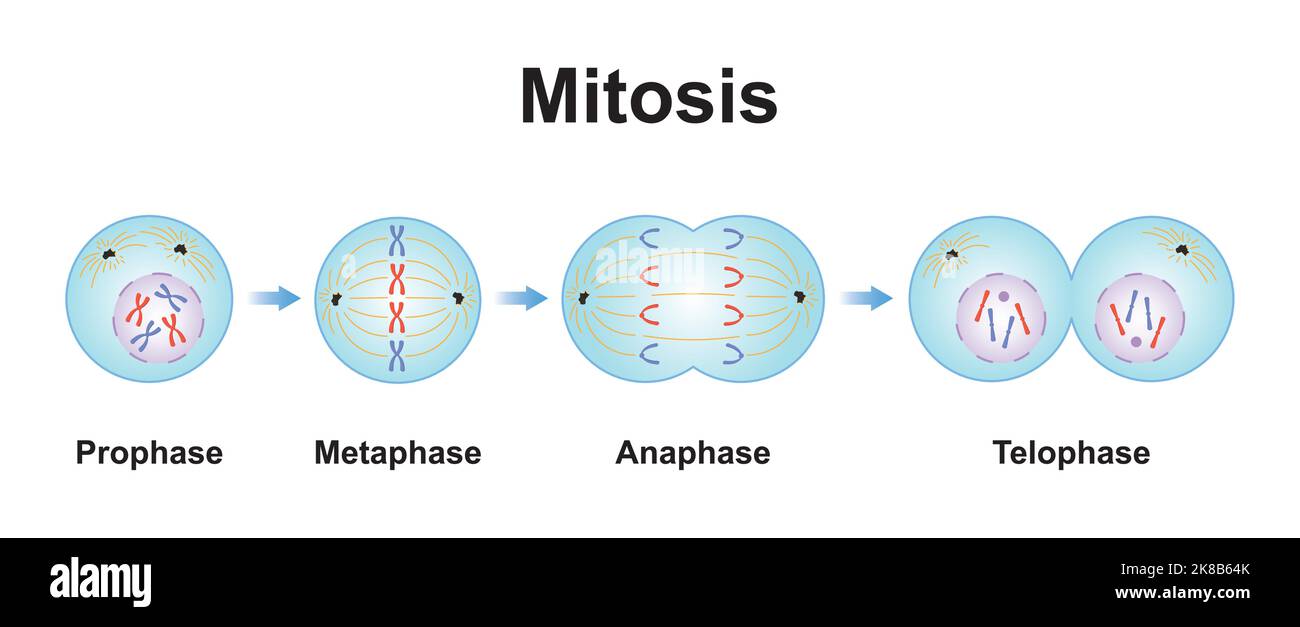 Scientific Designing of Mitosis Phases (Cell Division). Colorful Symbols. Vector Illustration. Stock Vectorhttps://www.alamy.com/image-license-details/?v=1https://www.alamy.com/scientific-designing-of-mitosis-phases-cell-division-colorful-symbols-vector-illustration-image487053859.html
RF2K8B64K–Scientific Designing of Mitosis Phases (Cell Division). Colorful Symbols. Vector Illustration.
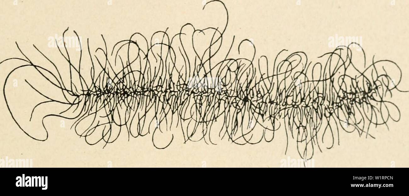 Archive image from page 74 of Cytology, with special reference to. Cytology, with special reference to the metazoan nucleus cytologywithspec00agar Year: 1920 II MEIOSIS IN THE FEMALE 59 The particular problems raised by the germinal vesicle stage in oogenesis are : (i) The continuity of the chromosomes throughout this period. (2) The relation between the chromosomes and the nucleoli. (3) The connection between the pecuhar germinal vesicle stage and the synchronous enormous growth of the cytoplasm of the egg, together with the formation of yolk. (4) Does any comparable stage occur in spermatog Stock Photohttps://www.alamy.com/image-license-details/?v=1https://www.alamy.com/archive-image-from-page-74-of-cytology-with-special-reference-to-cytology-with-special-reference-to-the-metazoan-nucleus-cytologywithspec00agar-year-1920-ii-meiosis-in-the-female-59-the-particular-problems-raised-by-the-germinal-vesicle-stage-in-oogenesis-are-i-the-continuity-of-the-chromosomes-throughout-this-period-2-the-relation-between-the-chromosomes-and-the-nucleoli-3-the-connection-between-the-pecuhar-germinal-vesicle-stage-and-the-synchronous-enormous-growth-of-the-cytoplasm-of-the-egg-together-with-the-formation-of-yolk-4-does-any-comparable-stage-occur-in-spermatog-image259292677.html
RMW1RPCN–Archive image from page 74 of Cytology, with special reference to. Cytology, with special reference to the metazoan nucleus cytologywithspec00agar Year: 1920 II MEIOSIS IN THE FEMALE 59 The particular problems raised by the germinal vesicle stage in oogenesis are : (i) The continuity of the chromosomes throughout this period. (2) The relation between the chromosomes and the nucleoli. (3) The connection between the pecuhar germinal vesicle stage and the synchronous enormous growth of the cytoplasm of the egg, together with the formation of yolk. (4) Does any comparable stage occur in spermatog
 . Cytology, with special reference to the metazoan nucleus. Cells. II MEIOSIS IN THE FEMALE 6i germinal vesicles with diffuse chromosomes are to be found, and it is evident that the bivalents resulting from syjidesis condense continuously. '^^ ^:^.f^. Please note that these images are extracted from scanned page images that may have been digitally enhanced for readability - coloration and appearance of these illustrations may not perfectly resemble the original work.. Agar, Wilfred Eade, 1882-. London, Macmillan Stock Photohttps://www.alamy.com/image-license-details/?v=1https://www.alamy.com/cytology-with-special-reference-to-the-metazoan-nucleus-cells-ii-meiosis-in-the-female-6i-germinal-vesicles-with-diffuse-chromosomes-are-to-be-found-and-it-is-evident-that-the-bivalents-resulting-from-syjidesis-condense-continuously-f-please-note-that-these-images-are-extracted-from-scanned-page-images-that-may-have-been-digitally-enhanced-for-readability-coloration-and-appearance-of-these-illustrations-may-not-perfectly-resemble-the-original-work-agar-wilfred-eade-1882-london-macmillan-image231777629.html
RMRD2AMD–. Cytology, with special reference to the metazoan nucleus. Cells. II MEIOSIS IN THE FEMALE 6i germinal vesicles with diffuse chromosomes are to be found, and it is evident that the bivalents resulting from syjidesis condense continuously. '^^ ^:^.f^. Please note that these images are extracted from scanned page images that may have been digitally enhanced for readability - coloration and appearance of these illustrations may not perfectly resemble the original work.. Agar, Wilfred Eade, 1882-. London, Macmillan
 . Cytology, with special reference to the metazoan nucleus. Cells. Syngamy II Meiosis Syngojny Pteridophyla V Syngamy Phaneroga rns. Meiosis Syngamy Metazoa and certain of the Rhodophyceae. (Fllclls) Lastrea Pseudomas vancristala A'. Please note that these images are extracted from scanned page images that may have been digitally enhanced for readability - coloration and appearance of these illustrations may not perfectly resemble the original work.. Agar, Wilfred Eade, 1882-. London, Macmillan Stock Photohttps://www.alamy.com/image-license-details/?v=1https://www.alamy.com/cytology-with-special-reference-to-the-metazoan-nucleus-cells-syngamy-ii-meiosis-syngojny-pteridophyla-v-syngamy-phaneroga-rns-meiosis-syngamy-metazoa-and-certain-of-the-rhodophyceae-fllclls-lastrea-pseudomas-vancristala-a-please-note-that-these-images-are-extracted-from-scanned-page-images-that-may-have-been-digitally-enhanced-for-readability-coloration-and-appearance-of-these-illustrations-may-not-perfectly-resemble-the-original-work-agar-wilfred-eade-1882-london-macmillan-image216167655.html
RMPFK81B–. Cytology, with special reference to the metazoan nucleus. Cells. Syngamy II Meiosis Syngojny Pteridophyla V Syngamy Phaneroga rns. Meiosis Syngamy Metazoa and certain of the Rhodophyceae. (Fllclls) Lastrea Pseudomas vancristala A'. Please note that these images are extracted from scanned page images that may have been digitally enhanced for readability - coloration and appearance of these illustrations may not perfectly resemble the original work.. Agar, Wilfred Eade, 1882-. London, Macmillan
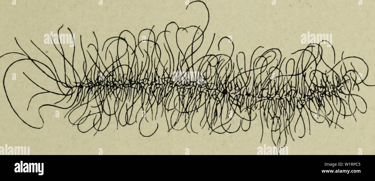 Archive image from page 74 of Cytology, with special reference to. Cytology, with special reference to the metazoan nucleus cytologywithspec00agar 0 Year: 1920 II MEIOSIS IN THE FEMALE 59 The particular problems raised by the germinal vesicle stage in oogenesis are : (1) The continuity of the chromosomes throughout this period. (2) The relation between the chromosomes and the nucleoli. (3) The connection between the peculiar germinal vesicle stage and the synchronous enormous growth of the cytoplasm of the egg, together with the formation of yolk. (4) Does any comparable stage occur in sperma Stock Photohttps://www.alamy.com/image-license-details/?v=1https://www.alamy.com/archive-image-from-page-74-of-cytology-with-special-reference-to-cytology-with-special-reference-to-the-metazoan-nucleus-cytologywithspec00agar-0-year-1920-ii-meiosis-in-the-female-59-the-particular-problems-raised-by-the-germinal-vesicle-stage-in-oogenesis-are-1-the-continuity-of-the-chromosomes-throughout-this-period-2-the-relation-between-the-chromosomes-and-the-nucleoli-3-the-connection-between-the-peculiar-germinal-vesicle-stage-and-the-synchronous-enormous-growth-of-the-cytoplasm-of-the-egg-together-with-the-formation-of-yolk-4-does-any-comparable-stage-occur-in-sperma-image259292661.html
RMW1RPC5–Archive image from page 74 of Cytology, with special reference to. Cytology, with special reference to the metazoan nucleus cytologywithspec00agar 0 Year: 1920 II MEIOSIS IN THE FEMALE 59 The particular problems raised by the germinal vesicle stage in oogenesis are : (1) The continuity of the chromosomes throughout this period. (2) The relation between the chromosomes and the nucleoli. (3) The connection between the peculiar germinal vesicle stage and the synchronous enormous growth of the cytoplasm of the egg, together with the formation of yolk. (4) Does any comparable stage occur in sperma
 . Cytology, with special reference to the metazoan nucleus. Cells. Syngamy Aggregate Diplocgstis Certain of the Rhodophyceae. (Nemalicnes). Syngamy II Meiosis Syngamy Pteridophyla Meiosis. Please note that these images are extracted from scanned page images that may have been digitally enhanced for readability - coloration and appearance of these illustrations may not perfectly resemble the original work.. Agar, W. E. (Wilfred Eade), 1882-1951. London, Macmillan and Co. , limited Stock Photohttps://www.alamy.com/image-license-details/?v=1https://www.alamy.com/cytology-with-special-reference-to-the-metazoan-nucleus-cells-syngamy-aggregate-diplocgstis-certain-of-the-rhodophyceae-nemalicnes-syngamy-ii-meiosis-syngamy-pteridophyla-meiosis-please-note-that-these-images-are-extracted-from-scanned-page-images-that-may-have-been-digitally-enhanced-for-readability-coloration-and-appearance-of-these-illustrations-may-not-perfectly-resemble-the-original-work-agar-w-e-wilfred-eade-1882-1951-london-macmillan-and-co-limited-image231777686.html
RMRD2APE–. Cytology, with special reference to the metazoan nucleus. Cells. Syngamy Aggregate Diplocgstis Certain of the Rhodophyceae. (Nemalicnes). Syngamy II Meiosis Syngamy Pteridophyla Meiosis. Please note that these images are extracted from scanned page images that may have been digitally enhanced for readability - coloration and appearance of these illustrations may not perfectly resemble the original work.. Agar, W. E. (Wilfred Eade), 1882-1951. London, Macmillan and Co. , limited
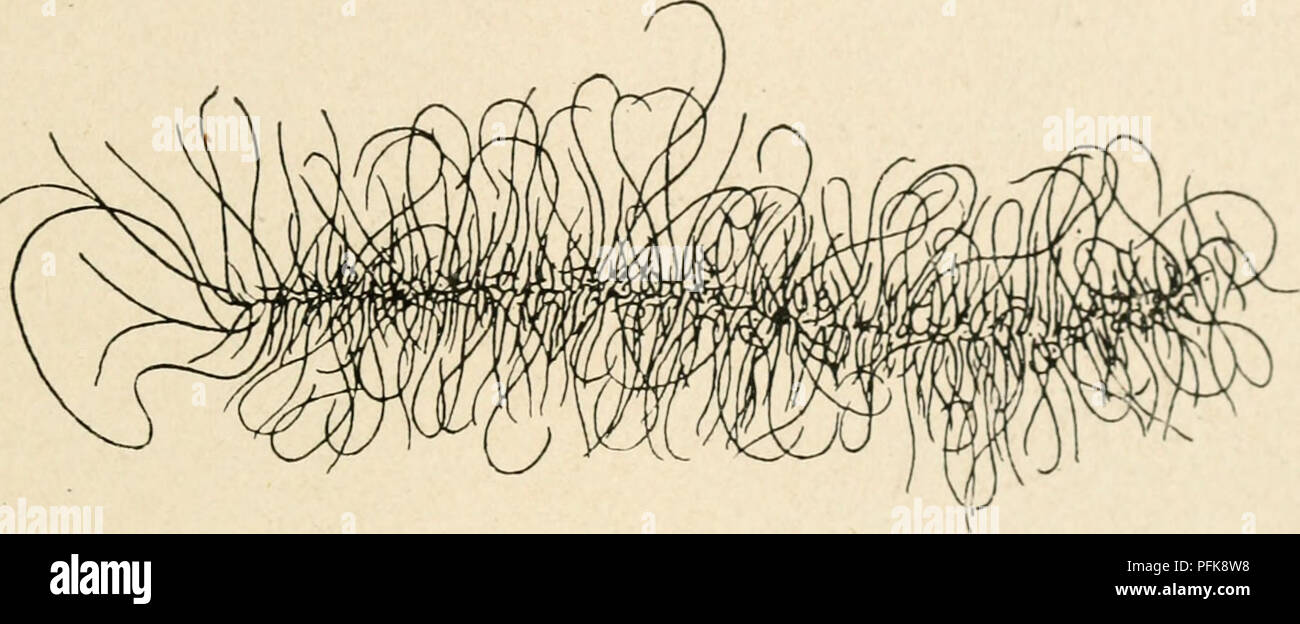 . Cytology, with special reference to the metazoan nucleus. Cells. II MEIOSIS IN THE FEMALE 59 The particular problems raised by the germinal vesicle stage in oogenesis are : (i) The continuity of the chromosomes throughout this period. (2) The relation between the chromosomes and the nucleoli. (3) The connection between the pecuhar germinal vesicle stage and the synchronous enormous growth of the cytoplasm of the egg, together with the formation of yolk. (4) Does any comparable stage occur in spermatogenesis ? (i) The Continuity of the Chromosomes The conditions in the germinal vesicle have b Stock Photohttps://www.alamy.com/image-license-details/?v=1https://www.alamy.com/cytology-with-special-reference-to-the-metazoan-nucleus-cells-ii-meiosis-in-the-female-59-the-particular-problems-raised-by-the-germinal-vesicle-stage-in-oogenesis-are-i-the-continuity-of-the-chromosomes-throughout-this-period-2-the-relation-between-the-chromosomes-and-the-nucleoli-3-the-connection-between-the-pecuhar-germinal-vesicle-stage-and-the-synchronous-enormous-growth-of-the-cytoplasm-of-the-egg-together-with-the-formation-of-yolk-4-does-any-comparable-stage-occur-in-spermatogenesis-i-the-continuity-of-the-chromosomes-the-conditions-in-the-germinal-vesicle-have-b-image216168324.html
RMPFK8W8–. Cytology, with special reference to the metazoan nucleus. Cells. II MEIOSIS IN THE FEMALE 59 The particular problems raised by the germinal vesicle stage in oogenesis are : (i) The continuity of the chromosomes throughout this period. (2) The relation between the chromosomes and the nucleoli. (3) The connection between the pecuhar germinal vesicle stage and the synchronous enormous growth of the cytoplasm of the egg, together with the formation of yolk. (4) Does any comparable stage occur in spermatogenesis ? (i) The Continuity of the Chromosomes The conditions in the germinal vesicle have b
 Archive image from page 78 of Cytology, with special reference to. Cytology, with special reference to the metazoan nucleus cytologywithspec00agar Year: 1920 II MEIOSIS IN THE FEMALE 63 nature (amphinucleoli), consisting of a plastin groundwork (plasmosome), covered or impregnated with chromatin or a chromatin-Uke substance ; or the two constituents may be separate, so that the nucleolus consists of two parts, a chromatin and a plastin portion. These remarks refer especially to the main nucleolus, which persists right through the growth period. In many animals the secondary nucleoli which dev Stock Photohttps://www.alamy.com/image-license-details/?v=1https://www.alamy.com/archive-image-from-page-78-of-cytology-with-special-reference-to-cytology-with-special-reference-to-the-metazoan-nucleus-cytologywithspec00agar-year-1920-ii-meiosis-in-the-female-63-nature-amphinucleoli-consisting-of-a-plastin-groundwork-plasmosome-covered-or-impregnated-with-chromatin-or-a-chromatin-uke-substance-or-the-two-constituents-may-be-separate-so-that-the-nucleolus-consists-of-two-parts-a-chromatin-and-a-plastin-portion-these-remarks-refer-especially-to-the-main-nucleolus-which-persists-right-through-the-growth-period-in-many-animals-the-secondary-nucleoli-which-dev-image259294278.html
RMW1RTDX–Archive image from page 78 of Cytology, with special reference to. Cytology, with special reference to the metazoan nucleus cytologywithspec00agar Year: 1920 II MEIOSIS IN THE FEMALE 63 nature (amphinucleoli), consisting of a plastin groundwork (plasmosome), covered or impregnated with chromatin or a chromatin-Uke substance ; or the two constituents may be separate, so that the nucleolus consists of two parts, a chromatin and a plastin portion. These remarks refer especially to the main nucleolus, which persists right through the growth period. In many animals the secondary nucleoli which dev
 . Cytology, with special reference to the metazoan nucleus. Cells. Syngamy II Meiosis Syngojny Pteridophyla V Syngamy Phaneroga rns. Meiosis Syngamy Metazoa and certain of the Rhodophyceae. (Fllclls) Lastrea Pseudomas vancristala A'. Please note that these images are extracted from scanned page images that may have been digitally enhanced for readability - coloration and appearance of these illustrations may not perfectly resemble the original work.. Agar, Wilfred Eade, 1882-. London, Macmillan Stock Photohttps://www.alamy.com/image-license-details/?v=1https://www.alamy.com/cytology-with-special-reference-to-the-metazoan-nucleus-cells-syngamy-ii-meiosis-syngojny-pteridophyla-v-syngamy-phaneroga-rns-meiosis-syngamy-metazoa-and-certain-of-the-rhodophyceae-fllclls-lastrea-pseudomas-vancristala-a-please-note-that-these-images-are-extracted-from-scanned-page-images-that-may-have-been-digitally-enhanced-for-readability-coloration-and-appearance-of-these-illustrations-may-not-perfectly-resemble-the-original-work-agar-wilfred-eade-1882-london-macmillan-image231789255.html
RMRD2WFK–. Cytology, with special reference to the metazoan nucleus. Cells. Syngamy II Meiosis Syngojny Pteridophyla V Syngamy Phaneroga rns. Meiosis Syngamy Metazoa and certain of the Rhodophyceae. (Fllclls) Lastrea Pseudomas vancristala A'. Please note that these images are extracted from scanned page images that may have been digitally enhanced for readability - coloration and appearance of these illustrations may not perfectly resemble the original work.. Agar, Wilfred Eade, 1882-. London, Macmillan
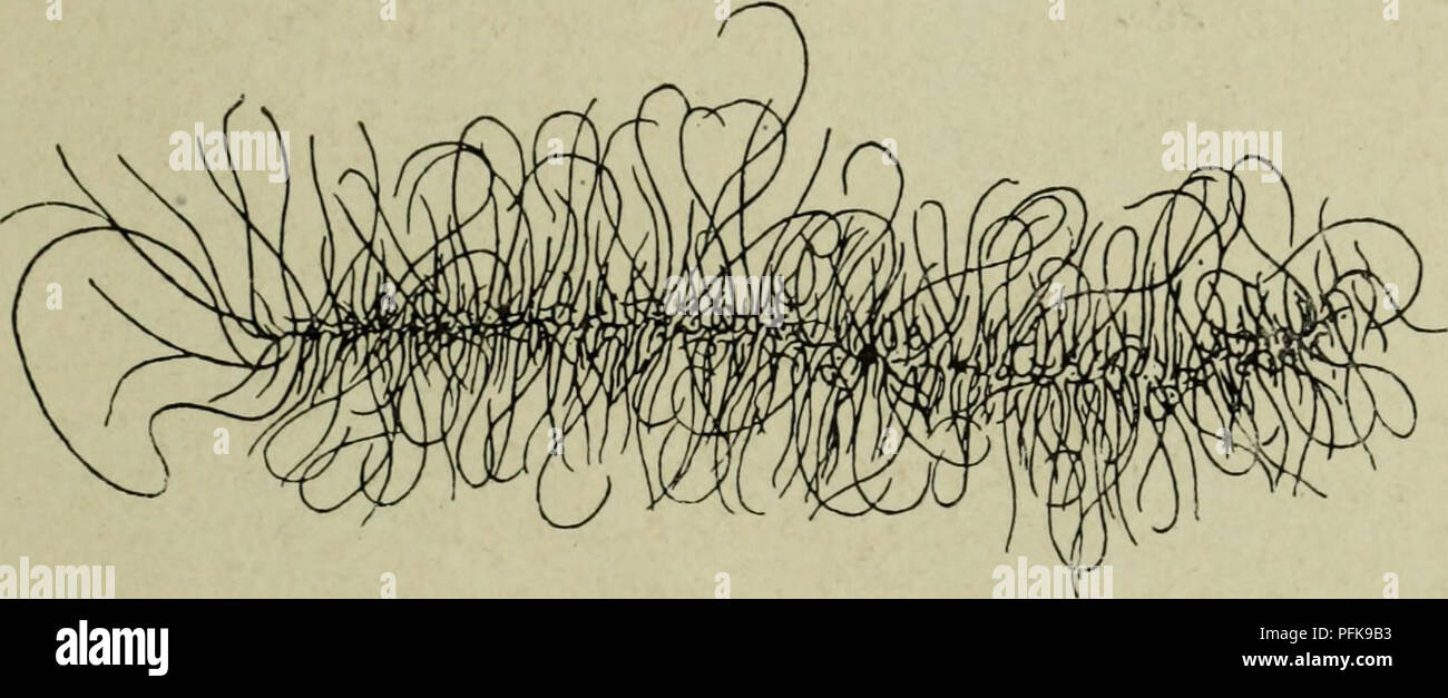 . Cytology, with special reference to the metazoan nucleus. Cells. II MEIOSIS IN THE FEMALE 59 The particular problems raised by the germinal vesicle stage in oogenesis are : (1) The continuity of the chromosomes throughout this period. (2) The relation between the chromosomes and the nucleoli. (3) The connection between the peculiar germinal vesicle stage and the synchronous enormous growth of the cytoplasm of the egg, together with the formation of yolk. (4) Does any comparable stage occur in spermatogenesis ? (1) The Continuity of the Chromosomes The conditions in the germinal vesicle have Stock Photohttps://www.alamy.com/image-license-details/?v=1https://www.alamy.com/cytology-with-special-reference-to-the-metazoan-nucleus-cells-ii-meiosis-in-the-female-59-the-particular-problems-raised-by-the-germinal-vesicle-stage-in-oogenesis-are-1-the-continuity-of-the-chromosomes-throughout-this-period-2-the-relation-between-the-chromosomes-and-the-nucleoli-3-the-connection-between-the-peculiar-germinal-vesicle-stage-and-the-synchronous-enormous-growth-of-the-cytoplasm-of-the-egg-together-with-the-formation-of-yolk-4-does-any-comparable-stage-occur-in-spermatogenesis-1-the-continuity-of-the-chromosomes-the-conditions-in-the-germinal-vesicle-have-image216168711.html
RMPFK9B3–. Cytology, with special reference to the metazoan nucleus. Cells. II MEIOSIS IN THE FEMALE 59 The particular problems raised by the germinal vesicle stage in oogenesis are : (1) The continuity of the chromosomes throughout this period. (2) The relation between the chromosomes and the nucleoli. (3) The connection between the peculiar germinal vesicle stage and the synchronous enormous growth of the cytoplasm of the egg, together with the formation of yolk. (4) Does any comparable stage occur in spermatogenesis ? (1) The Continuity of the Chromosomes The conditions in the germinal vesicle have
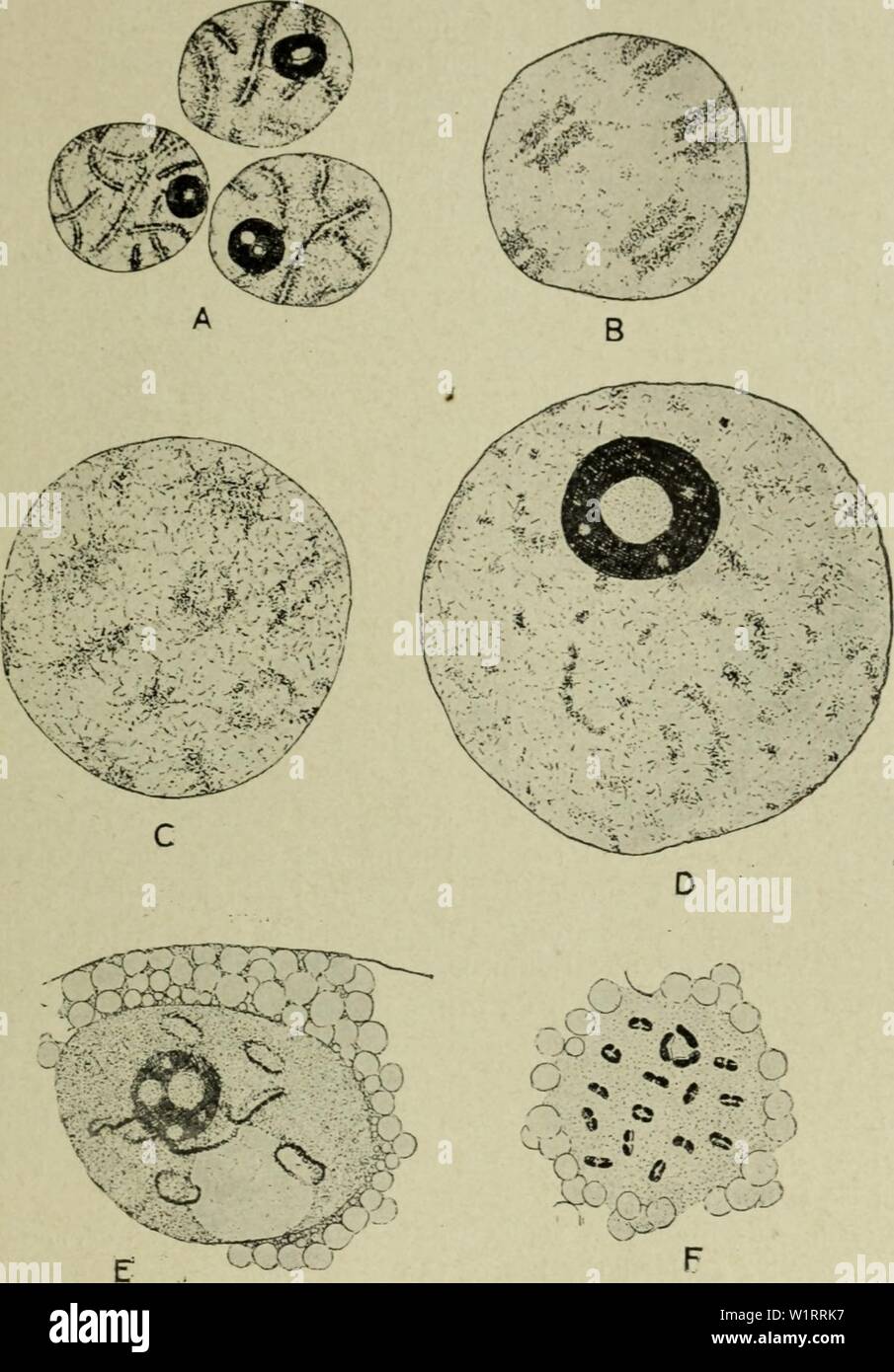 Archive image from page 76 of Cytology, with special reference to. Cytology, with special reference to the metazoan nucleus cytologywithspec00agar 0 Year: 1920 ii MEIOSIS IN THE FEMALE 6l germinal vesicles with diffuse chromosomes are to be found, and it is evident that the bivalents resulting from syndesis condense continuously Fig. 25. The chromosomes during the oogenesis of Diaptomus castor from the pachytene stage (A), through the germinal vesicle stage (B-E) to the condensation of the definitive bivalents (F). (Matschek, A.Z., 1910.) into the definitive bivalents of metaphase I. Anima Stock Photohttps://www.alamy.com/image-license-details/?v=1https://www.alamy.com/archive-image-from-page-76-of-cytology-with-special-reference-to-cytology-with-special-reference-to-the-metazoan-nucleus-cytologywithspec00agar-0-year-1920-ii-meiosis-in-the-female-6l-germinal-vesicles-with-diffuse-chromosomes-are-to-be-found-and-it-is-evident-that-the-bivalents-resulting-from-syndesis-condense-continuously-fig-25-the-chromosomes-during-the-oogenesis-of-diaptomus-castor-from-the-pachytene-stage-a-through-the-germinal-vesicle-stage-b-e-to-the-condensation-of-the-definitive-bivalents-f-matschek-az-1910-into-the-definitive-bivalents-of-metaphase-i-anima-image259293643.html
RMW1RRK7–Archive image from page 76 of Cytology, with special reference to. Cytology, with special reference to the metazoan nucleus cytologywithspec00agar 0 Year: 1920 ii MEIOSIS IN THE FEMALE 6l germinal vesicles with diffuse chromosomes are to be found, and it is evident that the bivalents resulting from syndesis condense continuously Fig. 25. The chromosomes during the oogenesis of Diaptomus castor from the pachytene stage (A), through the germinal vesicle stage (B-E) to the condensation of the definitive bivalents (F). (Matschek, A.Z., 1910.) into the definitive bivalents of metaphase I. Anima
 . An atlas of the fertilization and karyokinesis of the ovum. Ovum; Fertilization (Biology); Meiosis; Embryology -- Echinodermata. ATLAS OF FERTILIZATION. PLATE II. ^. Please note that these images are extracted from scanned page images that may have been digitally enhanced for readability - coloration and appearance of these illustrations may not perfectly resemble the original work.. Wilson, Edmund B. (Edmund Beecher), 1856-1939; Leaming, Edward, 1861-1916. New York, London, Pub. for the Columbia university press by Macmillan and co. Stock Photohttps://www.alamy.com/image-license-details/?v=1https://www.alamy.com/an-atlas-of-the-fertilization-and-karyokinesis-of-the-ovum-ovum-fertilization-biology-meiosis-embryology-echinodermata-atlas-of-fertilization-plate-ii-please-note-that-these-images-are-extracted-from-scanned-page-images-that-may-have-been-digitally-enhanced-for-readability-coloration-and-appearance-of-these-illustrations-may-not-perfectly-resemble-the-original-work-wilson-edmund-b-edmund-beecher-1856-1939-leaming-edward-1861-1916-new-york-london-pub-for-the-columbia-university-press-by-macmillan-and-co-image235402222.html
RMRJYDX6–. An atlas of the fertilization and karyokinesis of the ovum. Ovum; Fertilization (Biology); Meiosis; Embryology -- Echinodermata. ATLAS OF FERTILIZATION. PLATE II. ^. Please note that these images are extracted from scanned page images that may have been digitally enhanced for readability - coloration and appearance of these illustrations may not perfectly resemble the original work.. Wilson, Edmund B. (Edmund Beecher), 1856-1939; Leaming, Edward, 1861-1916. New York, London, Pub. for the Columbia university press by Macmillan and co.
 . Cytology, with special reference to the metazoan nucleus. Cells. II MEIOSIS 65 (4) Does any Stage comparable to the Germinal Vesicle occur in Spermatogenesis ? The undoubted correlation between the pecuhar conditions of the germinal vesicle and the long duration of the growth period in the female meiotic phase, and probably also with the deposition of yolk, makes it. /^"^^. Please note that these images are extracted from scanned page images that may have been digitally enhanced for readability - coloration and appearance of these illustrations may not perfectly resemble the original wo Stock Photohttps://www.alamy.com/image-license-details/?v=1https://www.alamy.com/cytology-with-special-reference-to-the-metazoan-nucleus-cells-ii-meiosis-65-4-does-any-stage-comparable-to-the-germinal-vesicle-occur-in-spermatogenesis-the-undoubted-correlation-between-the-pecuhar-conditions-of-the-germinal-vesicle-and-the-long-duration-of-the-growth-period-in-the-female-meiotic-phase-and-probably-also-with-the-deposition-of-yolk-makes-it-quot-please-note-that-these-images-are-extracted-from-scanned-page-images-that-may-have-been-digitally-enhanced-for-readability-coloration-and-appearance-of-these-illustrations-may-not-perfectly-resemble-the-original-wo-image216168245.html
RMPFK8PD–. Cytology, with special reference to the metazoan nucleus. Cells. II MEIOSIS 65 (4) Does any Stage comparable to the Germinal Vesicle occur in Spermatogenesis ? The undoubted correlation between the pecuhar conditions of the germinal vesicle and the long duration of the growth period in the female meiotic phase, and probably also with the deposition of yolk, makes it. /^"^^. Please note that these images are extracted from scanned page images that may have been digitally enhanced for readability - coloration and appearance of these illustrations may not perfectly resemble the original wo
 Archive image from page 66 of Cytology, with special reference to. Cytology, with special reference to the metazoan nucleus cytologywithspec00agar Year: 1920 II MEIOSIS IN ASCARIS 51 By the later stage shown in Fig. 20, C, synizesis has completely dis- appeared, and all the chromatin is in the form of a long, doubly split thread (only one split is visible in the plane of the figure). As there are really two bivalent chromosomes present, these must be joined tempor- FiG. 20. end view, and therefore its quadruple constitution is revealed. F, metaphase I. ; G, H, anaphase I. ; I, preparation Stock Photohttps://www.alamy.com/image-license-details/?v=1https://www.alamy.com/archive-image-from-page-66-of-cytology-with-special-reference-to-cytology-with-special-reference-to-the-metazoan-nucleus-cytologywithspec00agar-year-1920-ii-meiosis-in-ascaris-51-by-the-later-stage-shown-in-fig-20-c-synizesis-has-completely-dis-appeared-and-all-the-chromatin-is-in-the-form-of-a-long-doubly-split-thread-only-one-split-is-visible-in-the-plane-of-the-figure-as-there-are-really-two-bivalent-chromosomes-present-these-must-be-joined-tempor-fig-20-end-view-and-therefore-its-quadruple-constitution-is-revealed-f-metaphase-i-g-h-anaphase-i-i-preparation-image259289460.html
RMW1RJ9T–Archive image from page 66 of Cytology, with special reference to. Cytology, with special reference to the metazoan nucleus cytologywithspec00agar Year: 1920 II MEIOSIS IN ASCARIS 51 By the later stage shown in Fig. 20, C, synizesis has completely dis- appeared, and all the chromatin is in the form of a long, doubly split thread (only one split is visible in the plane of the figure). As there are really two bivalent chromosomes present, these must be joined tempor- FiG. 20. end view, and therefore its quadruple constitution is revealed. F, metaphase I. ; G, H, anaphase I. ; I, preparation
 . An atlas of the fertilization and karyokinesis of the ovum. Ovum; Fertilization (Biology); Meiosis; Embryology -- Echinodermata. ATLAS OF FERTILIZATION. PLATE II.. i. ' >L, /"'. Please note that these images are extracted from scanned page images that may have been digitally enhanced for readability - coloration and appearance of these illustrations may not perfectly resemble the original work.. Wilson, Edmund B. (Edmund Beecher), 1856-1939; Leaming, Edward, 1861-1916. New York, London, Pub. for the Columbia university press by Macmillan and co. Stock Photohttps://www.alamy.com/image-license-details/?v=1https://www.alamy.com/an-atlas-of-the-fertilization-and-karyokinesis-of-the-ovum-ovum-fertilization-biology-meiosis-embryology-echinodermata-atlas-of-fertilization-plate-ii-i-gtl-quot-please-note-that-these-images-are-extracted-from-scanned-page-images-that-may-have-been-digitally-enhanced-for-readability-coloration-and-appearance-of-these-illustrations-may-not-perfectly-resemble-the-original-work-wilson-edmund-b-edmund-beecher-1856-1939-leaming-edward-1861-1916-new-york-london-pub-for-the-columbia-university-press-by-macmillan-and-co-image235402263.html
RMRJYDYK–. An atlas of the fertilization and karyokinesis of the ovum. Ovum; Fertilization (Biology); Meiosis; Embryology -- Echinodermata. ATLAS OF FERTILIZATION. PLATE II.. i. ' >L, /"'. Please note that these images are extracted from scanned page images that may have been digitally enhanced for readability - coloration and appearance of these illustrations may not perfectly resemble the original work.. Wilson, Edmund B. (Edmund Beecher), 1856-1939; Leaming, Edward, 1861-1916. New York, London, Pub. for the Columbia university press by Macmillan and co.
 . Cytology, with special reference to the metazoan nucleus. Cells; Cytology. II MEIOSIS IN THE FEMALE 59 The particular problems raised by the germinal vesicle stage in oogenesis are : (i) The continuity of the chromosomes throughout this period. (2) The relation between the chromosomes and the nucleoli. (â 3) The connection between the peculiar germinal vesicle stage and the synchronous enormous growth of the cytoplasm of the egg, together with the formation of yolk. (4) Does any comparable stage occur in spermatogenesis ? (i) The Continuity of the Chromosomes The conditions in the germinal v Stock Photohttps://www.alamy.com/image-license-details/?v=1https://www.alamy.com/cytology-with-special-reference-to-the-metazoan-nucleus-cells-cytology-ii-meiosis-in-the-female-59-the-particular-problems-raised-by-the-germinal-vesicle-stage-in-oogenesis-are-i-the-continuity-of-the-chromosomes-throughout-this-period-2-the-relation-between-the-chromosomes-and-the-nucleoli-3-the-connection-between-the-peculiar-germinal-vesicle-stage-and-the-synchronous-enormous-growth-of-the-cytoplasm-of-the-egg-together-with-the-formation-of-yolk-4-does-any-comparable-stage-occur-in-spermatogenesis-i-the-continuity-of-the-chromosomes-the-conditions-in-the-germinal-v-image216168101.html
RMPFK8H9–. Cytology, with special reference to the metazoan nucleus. Cells; Cytology. II MEIOSIS IN THE FEMALE 59 The particular problems raised by the germinal vesicle stage in oogenesis are : (i) The continuity of the chromosomes throughout this period. (2) The relation between the chromosomes and the nucleoli. (â 3) The connection between the peculiar germinal vesicle stage and the synchronous enormous growth of the cytoplasm of the egg, together with the formation of yolk. (4) Does any comparable stage occur in spermatogenesis ? (i) The Continuity of the Chromosomes The conditions in the germinal v
 . Cytology, with special reference to the metazoan nucleus. Cells. II MEIOSIS 65 (4) Does any Stage comparable to the Germinal Vesicle occur in Spermatogenesis ? The undoubted correlation between the pecuhar conditions of the germinal vesicle and the long duration of the growth period in the female meiotic phase, and probably also with the deposition of yolk, makes it. /^"^^. Please note that these images are extracted from scanned page images that may have been digitally enhanced for readability - coloration and appearance of these illustrations may not perfectly resemble the original wo Stock Photohttps://www.alamy.com/image-license-details/?v=1https://www.alamy.com/cytology-with-special-reference-to-the-metazoan-nucleus-cells-ii-meiosis-65-4-does-any-stage-comparable-to-the-germinal-vesicle-occur-in-spermatogenesis-the-undoubted-correlation-between-the-pecuhar-conditions-of-the-germinal-vesicle-and-the-long-duration-of-the-growth-period-in-the-female-meiotic-phase-and-probably-also-with-the-deposition-of-yolk-makes-it-quot-please-note-that-these-images-are-extracted-from-scanned-page-images-that-may-have-been-digitally-enhanced-for-readability-coloration-and-appearance-of-these-illustrations-may-not-perfectly-resemble-the-original-wo-image231777550.html
RMRD2AHJ–. Cytology, with special reference to the metazoan nucleus. Cells. II MEIOSIS 65 (4) Does any Stage comparable to the Germinal Vesicle occur in Spermatogenesis ? The undoubted correlation between the pecuhar conditions of the germinal vesicle and the long duration of the growth period in the female meiotic phase, and probably also with the deposition of yolk, makes it. /^"^^. Please note that these images are extracted from scanned page images that may have been digitally enhanced for readability - coloration and appearance of these illustrations may not perfectly resemble the original wo
 . Cytology, with special reference to the metazoan nucleus. Cells. II MEIOSIS IN THE FEMALE 63 nature (amphinucleoli), consisting of a plastin groundwork (plasmosome), covered or impregnated with chromatin or a chromatin-Uke substance ; or the two constituents may be separate, so that the nucleolus consists of two parts, a chromatin and a plastin portion. These remarks refer especially to the main nucleolus, which persists right through the growth period. In many animals the secondary nucleoli which develop later. Please note that these images are extracted from scanned page images that may ha Stock Photohttps://www.alamy.com/image-license-details/?v=1https://www.alamy.com/cytology-with-special-reference-to-the-metazoan-nucleus-cells-ii-meiosis-in-the-female-63-nature-amphinucleoli-consisting-of-a-plastin-groundwork-plasmosome-covered-or-impregnated-with-chromatin-or-a-chromatin-uke-substance-or-the-two-constituents-may-be-separate-so-that-the-nucleolus-consists-of-two-parts-a-chromatin-and-a-plastin-portion-these-remarks-refer-especially-to-the-main-nucleolus-which-persists-right-through-the-growth-period-in-many-animals-the-secondary-nucleoli-which-develop-later-please-note-that-these-images-are-extracted-from-scanned-page-images-that-may-ha-image216168271.html
RMPFK8RB–. Cytology, with special reference to the metazoan nucleus. Cells. II MEIOSIS IN THE FEMALE 63 nature (amphinucleoli), consisting of a plastin groundwork (plasmosome), covered or impregnated with chromatin or a chromatin-Uke substance ; or the two constituents may be separate, so that the nucleolus consists of two parts, a chromatin and a plastin portion. These remarks refer especially to the main nucleolus, which persists right through the growth period. In many animals the secondary nucleoli which develop later. Please note that these images are extracted from scanned page images that may ha
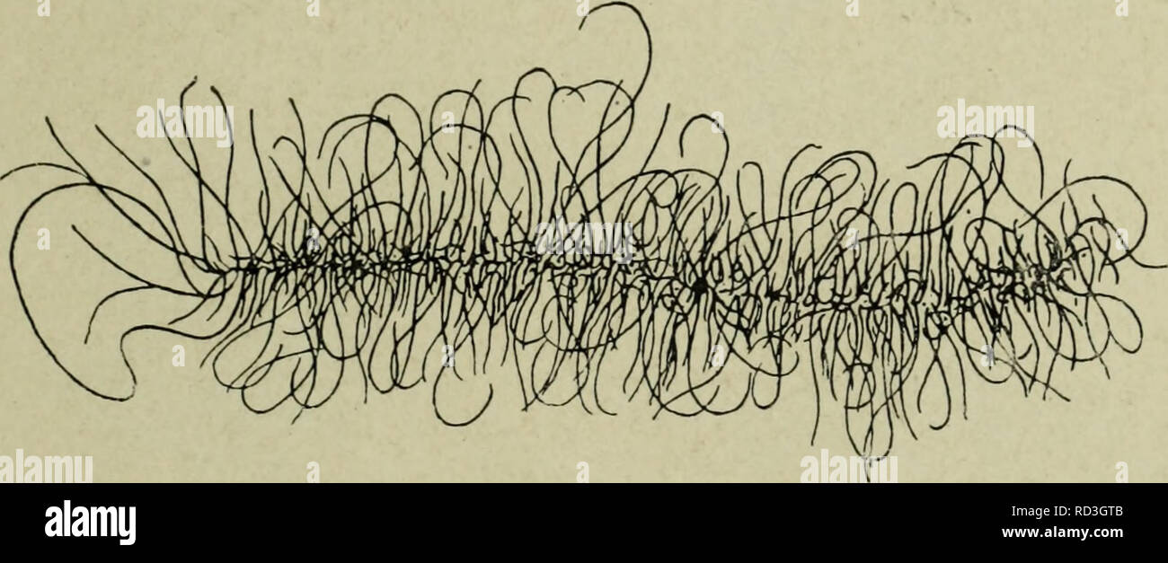 . Cytology, with special reference to the metazoan nucleus. Cells. II MEIOSIS IN THE FEMALE 59 The particular problems raised by the germinal vesicle stage in oogenesis are : (1) The continuity of the chromosomes throughout this period. (2) The relation between the chromosomes and the nucleoli. (3) The connection between the peculiar germinal vesicle stage and the synchronous enormous growth of the cytoplasm of the egg, together with the formation of yolk. (4) Does any comparable stage occur in spermatogenesis ? (1) The Continuity of the Chromosomes The conditions in the germinal vesicle have Stock Photohttps://www.alamy.com/image-license-details/?v=1https://www.alamy.com/cytology-with-special-reference-to-the-metazoan-nucleus-cells-ii-meiosis-in-the-female-59-the-particular-problems-raised-by-the-germinal-vesicle-stage-in-oogenesis-are-1-the-continuity-of-the-chromosomes-throughout-this-period-2-the-relation-between-the-chromosomes-and-the-nucleoli-3-the-connection-between-the-peculiar-germinal-vesicle-stage-and-the-synchronous-enormous-growth-of-the-cytoplasm-of-the-egg-together-with-the-formation-of-yolk-4-does-any-comparable-stage-occur-in-spermatogenesis-1-the-continuity-of-the-chromosomes-the-conditions-in-the-germinal-vesicle-have-image231804395.html
RMRD3GTB–. Cytology, with special reference to the metazoan nucleus. Cells. II MEIOSIS IN THE FEMALE 59 The particular problems raised by the germinal vesicle stage in oogenesis are : (1) The continuity of the chromosomes throughout this period. (2) The relation between the chromosomes and the nucleoli. (3) The connection between the peculiar germinal vesicle stage and the synchronous enormous growth of the cytoplasm of the egg, together with the formation of yolk. (4) Does any comparable stage occur in spermatogenesis ? (1) The Continuity of the Chromosomes The conditions in the germinal vesicle have
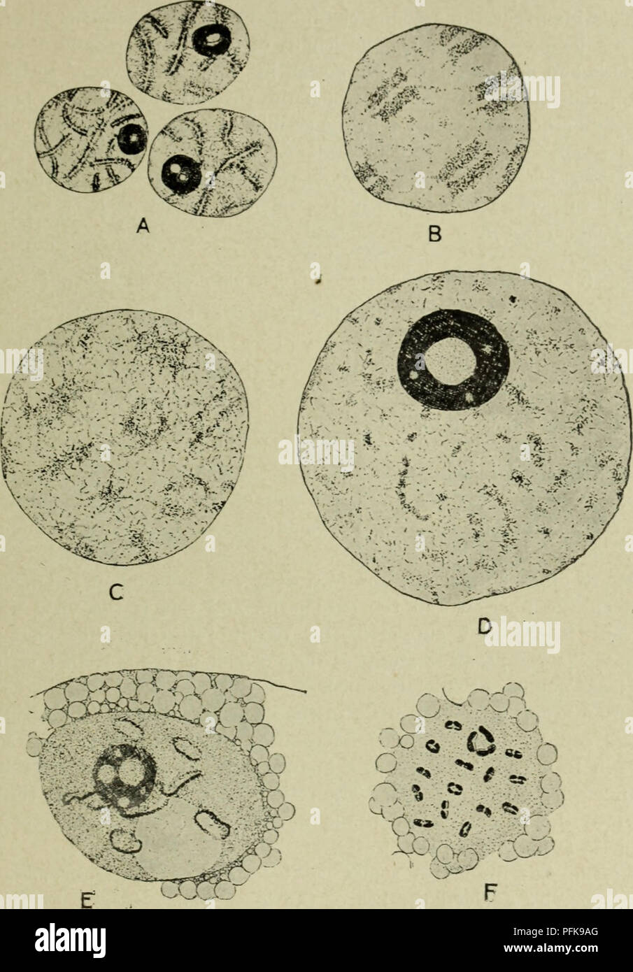 . Cytology, with special reference to the metazoan nucleus. Cells. ii MEIOSIS IN THE FEMALE 6l germinal vesicles with diffuse chromosomes are to be found, and it is evident that the bivalents resulting from syndesis condense continuously. Fig. 25. The chromosomes during the oogenesis of Diaptomus castor from the pachytene stage (A), through the germinal vesicle stage (B-E) to the condensation of the definitive bivalents (F). (Matschek, A.Z., 1910.) into the definitive bivalents of metaphase I. Animals, however, which are carrying egg-sacs contain in their oviducts oocytes with well-developed g Stock Photohttps://www.alamy.com/image-license-details/?v=1https://www.alamy.com/cytology-with-special-reference-to-the-metazoan-nucleus-cells-ii-meiosis-in-the-female-6l-germinal-vesicles-with-diffuse-chromosomes-are-to-be-found-and-it-is-evident-that-the-bivalents-resulting-from-syndesis-condense-continuously-fig-25-the-chromosomes-during-the-oogenesis-of-diaptomus-castor-from-the-pachytene-stage-a-through-the-germinal-vesicle-stage-b-e-to-the-condensation-of-the-definitive-bivalents-f-matschek-az-1910-into-the-definitive-bivalents-of-metaphase-i-animals-however-which-are-carrying-egg-sacs-contain-in-their-oviducts-oocytes-with-well-developed-g-image216168696.html
RMPFK9AG–. Cytology, with special reference to the metazoan nucleus. Cells. ii MEIOSIS IN THE FEMALE 6l germinal vesicles with diffuse chromosomes are to be found, and it is evident that the bivalents resulting from syndesis condense continuously. Fig. 25. The chromosomes during the oogenesis of Diaptomus castor from the pachytene stage (A), through the germinal vesicle stage (B-E) to the condensation of the definitive bivalents (F). (Matschek, A.Z., 1910.) into the definitive bivalents of metaphase I. Animals, however, which are carrying egg-sacs contain in their oviducts oocytes with well-developed g
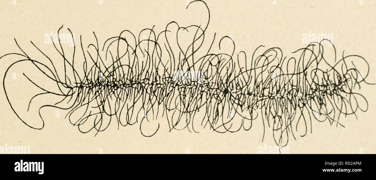 . Cytology, with special reference to the metazoan nucleus. Cells. II MEIOSIS IN THE FEMALE 59 The particular problems raised by the germinal vesicle stage in oogenesis are : (i) The continuity of the chromosomes throughout this period. (2) The relation between the chromosomes and the nucleoli. (3) The connection between the pecuhar germinal vesicle stage and the synchronous enormous growth of the cytoplasm of the egg, together with the formation of yolk. (4) Does any comparable stage occur in spermatogenesis ? (i) The Continuity of the Chromosomes The conditions in the germinal vesicle have b Stock Photohttps://www.alamy.com/image-license-details/?v=1https://www.alamy.com/cytology-with-special-reference-to-the-metazoan-nucleus-cells-ii-meiosis-in-the-female-59-the-particular-problems-raised-by-the-germinal-vesicle-stage-in-oogenesis-are-i-the-continuity-of-the-chromosomes-throughout-this-period-2-the-relation-between-the-chromosomes-and-the-nucleoli-3-the-connection-between-the-pecuhar-germinal-vesicle-stage-and-the-synchronous-enormous-growth-of-the-cytoplasm-of-the-egg-together-with-the-formation-of-yolk-4-does-any-comparable-stage-occur-in-spermatogenesis-i-the-continuity-of-the-chromosomes-the-conditions-in-the-germinal-vesicle-have-b-image231777692.html
RMRD2APM–. Cytology, with special reference to the metazoan nucleus. Cells. II MEIOSIS IN THE FEMALE 59 The particular problems raised by the germinal vesicle stage in oogenesis are : (i) The continuity of the chromosomes throughout this period. (2) The relation between the chromosomes and the nucleoli. (3) The connection between the pecuhar germinal vesicle stage and the synchronous enormous growth of the cytoplasm of the egg, together with the formation of yolk. (4) Does any comparable stage occur in spermatogenesis ? (i) The Continuity of the Chromosomes The conditions in the germinal vesicle have b
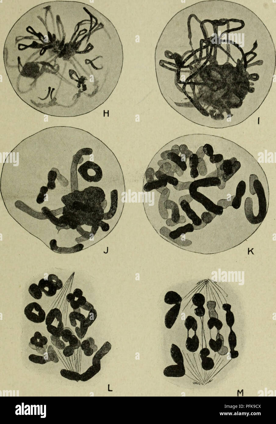 . Cytology, with special reference to the metazoan nucleus. Cells. II MEIOSIS IN LEPIDOSIREN 39 conjugation of the chromosomes. Hence the word symzesis was proposed for the contraction, and syndesis for the chromosome conjugation.. Fig. 16a. Meiosis in Lepidosiren (male . (Agar, Q.J.M.S., 1911.) H, diplotene stage and beginning of synizesis; I, synizesis further advanced ; J, synizesis breaking up; K, diakinesis, showing that the bivalents which were formed in the zygotene stage are completely resolved into their univalent constituents; L, immediate prophase of the meiotic division, showing th Stock Photohttps://www.alamy.com/image-license-details/?v=1https://www.alamy.com/cytology-with-special-reference-to-the-metazoan-nucleus-cells-ii-meiosis-in-lepidosiren-39-conjugation-of-the-chromosomes-hence-the-word-symzesis-was-proposed-for-the-contraction-and-syndesis-for-the-chromosome-conjugation-fig-16a-meiosis-in-lepidosiren-male-agar-qjms-1911-h-diplotene-stage-and-beginning-of-synizesis-i-synizesis-further-advanced-j-synizesis-breaking-up-k-diakinesis-showing-that-the-bivalents-which-were-formed-in-the-zygotene-stage-are-completely-resolved-into-their-univalent-constituents-l-immediate-prophase-of-the-meiotic-division-showing-th-image216168762.html
RMPFK9CX–. Cytology, with special reference to the metazoan nucleus. Cells. II MEIOSIS IN LEPIDOSIREN 39 conjugation of the chromosomes. Hence the word symzesis was proposed for the contraction, and syndesis for the chromosome conjugation.. Fig. 16a. Meiosis in Lepidosiren (male . (Agar, Q.J.M.S., 1911.) H, diplotene stage and beginning of synizesis; I, synizesis further advanced ; J, synizesis breaking up; K, diakinesis, showing that the bivalents which were formed in the zygotene stage are completely resolved into their univalent constituents; L, immediate prophase of the meiotic division, showing th
 . Cytology, with special reference to the metazoan nucleus. Cells; Cytology. II MEIOSIS IN THE FEMALE 59 The particular problems raised by the germinal vesicle stage in oogenesis are : (i) The continuity of the chromosomes throughout this period. (2) The relation between the chromosomes and the nucleoli. (â 3) The connection between the peculiar germinal vesicle stage and the synchronous enormous growth of the cytoplasm of the egg, together with the formation of yolk. (4) Does any comparable stage occur in spermatogenesis ? (i) The Continuity of the Chromosomes The conditions in the germinal v Stock Photohttps://www.alamy.com/image-license-details/?v=1https://www.alamy.com/cytology-with-special-reference-to-the-metazoan-nucleus-cells-cytology-ii-meiosis-in-the-female-59-the-particular-problems-raised-by-the-germinal-vesicle-stage-in-oogenesis-are-i-the-continuity-of-the-chromosomes-throughout-this-period-2-the-relation-between-the-chromosomes-and-the-nucleoli-3-the-connection-between-the-peculiar-germinal-vesicle-stage-and-the-synchronous-enormous-growth-of-the-cytoplasm-of-the-egg-together-with-the-formation-of-yolk-4-does-any-comparable-stage-occur-in-spermatogenesis-i-the-continuity-of-the-chromosomes-the-conditions-in-the-germinal-v-image231777234.html
RMRD2A6A–. Cytology, with special reference to the metazoan nucleus. Cells; Cytology. II MEIOSIS IN THE FEMALE 59 The particular problems raised by the germinal vesicle stage in oogenesis are : (i) The continuity of the chromosomes throughout this period. (2) The relation between the chromosomes and the nucleoli. (â 3) The connection between the peculiar germinal vesicle stage and the synchronous enormous growth of the cytoplasm of the egg, together with the formation of yolk. (4) Does any comparable stage occur in spermatogenesis ? (i) The Continuity of the Chromosomes The conditions in the germinal v
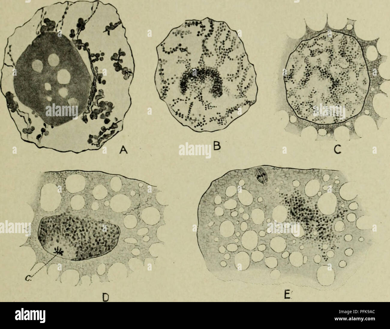 . Cytology, with special reference to the metazoan nucleus. Cells. ii MEIOSIS IN THE FEMALE 63 nature (amphinucleoli), consisting of a plastin groundwork (plasmosome), covered or impregnated with chromatin or a chromatin-like substance ; or the two constituents may be separate, so that the nucleolus consists of two parts, a chromatin and a plastin portion. These remarks refer especially to the main nucleolus, which persists right through the growth period. In many animals the secondary nucleoli which develop later. Fig. "26. Showing the fate of the nucleolus in the oogenesis of Daphnia pu Stock Photohttps://www.alamy.com/image-license-details/?v=1https://www.alamy.com/cytology-with-special-reference-to-the-metazoan-nucleus-cells-ii-meiosis-in-the-female-63-nature-amphinucleoli-consisting-of-a-plastin-groundwork-plasmosome-covered-or-impregnated-with-chromatin-or-a-chromatin-like-substance-or-the-two-constituents-may-be-separate-so-that-the-nucleolus-consists-of-two-parts-a-chromatin-and-a-plastin-portion-these-remarks-refer-especially-to-the-main-nucleolus-which-persists-right-through-the-growth-period-in-many-animals-the-secondary-nucleoli-which-develop-later-fig-quot26-showing-the-fate-of-the-nucleolus-in-the-oogenesis-of-daphnia-pu-image216168692.html
RMPFK9AC–. Cytology, with special reference to the metazoan nucleus. Cells. ii MEIOSIS IN THE FEMALE 63 nature (amphinucleoli), consisting of a plastin groundwork (plasmosome), covered or impregnated with chromatin or a chromatin-like substance ; or the two constituents may be separate, so that the nucleolus consists of two parts, a chromatin and a plastin portion. These remarks refer especially to the main nucleolus, which persists right through the growth period. In many animals the secondary nucleoli which develop later. Fig. "26. Showing the fate of the nucleolus in the oogenesis of Daphnia pu
 . Cytology, with special reference to the metazoan nucleus. Cells. ii MEIOSIS IN THE FEMALE 63 nature (amphinucleoli), consisting of a plastin groundwork (plasmosome), covered or impregnated with chromatin or a chromatin-like substance ; or the two constituents may be separate, so that the nucleolus consists of two parts, a chromatin and a plastin portion. These remarks refer especially to the main nucleolus, which persists right through the growth period. In many animals the secondary nucleoli which develop later. Fig. "26. Showing the fate of the nucleolus in the oogenesis of Daphnia pu Stock Photohttps://www.alamy.com/image-license-details/?v=1https://www.alamy.com/cytology-with-special-reference-to-the-metazoan-nucleus-cells-ii-meiosis-in-the-female-63-nature-amphinucleoli-consisting-of-a-plastin-groundwork-plasmosome-covered-or-impregnated-with-chromatin-or-a-chromatin-like-substance-or-the-two-constituents-may-be-separate-so-that-the-nucleolus-consists-of-two-parts-a-chromatin-and-a-plastin-portion-these-remarks-refer-especially-to-the-main-nucleolus-which-persists-right-through-the-growth-period-in-many-animals-the-secondary-nucleoli-which-develop-later-fig-quot26-showing-the-fate-of-the-nucleolus-in-the-oogenesis-of-daphnia-pu-image231790372.html
RMRD2XYG–. Cytology, with special reference to the metazoan nucleus. Cells. ii MEIOSIS IN THE FEMALE 63 nature (amphinucleoli), consisting of a plastin groundwork (plasmosome), covered or impregnated with chromatin or a chromatin-like substance ; or the two constituents may be separate, so that the nucleolus consists of two parts, a chromatin and a plastin portion. These remarks refer especially to the main nucleolus, which persists right through the growth period. In many animals the secondary nucleoli which develop later. Fig. "26. Showing the fate of the nucleolus in the oogenesis of Daphnia pu
 . Cytology, with special reference to the metazoan nucleus. Cells. ii MEIOSIS 65 (4) Does any Stage comparable to the Germinal Vesicle occur in Spermatogenesis ? The undoubted correlation between the peculiar conditions of the germinal vesicle and the long duration of the growth period in the female meiotic phase, and probably also with the deposition of yolk, makes it. Fig. 28. Germinal vesicle-like stages in spermatogenesis. A-G, Notodromas monacha (after Schmalz, A.Z., 1912) ; H-N, Scolopendra heros (after Blackman, B.M.C.Z.H., 1905). In both cases the principal stages between the pachytene Stock Photohttps://www.alamy.com/image-license-details/?v=1https://www.alamy.com/cytology-with-special-reference-to-the-metazoan-nucleus-cells-ii-meiosis-65-4-does-any-stage-comparable-to-the-germinal-vesicle-occur-in-spermatogenesis-the-undoubted-correlation-between-the-peculiar-conditions-of-the-germinal-vesicle-and-the-long-duration-of-the-growth-period-in-the-female-meiotic-phase-and-probably-also-with-the-deposition-of-yolk-makes-it-fig-28-germinal-vesicle-like-stages-in-spermatogenesis-a-g-notodromas-monacha-after-schmalz-az-1912-h-n-scolopendra-heros-after-blackman-bmczh-1905-in-both-cases-the-principal-stages-between-the-pachytene-image216168677.html
RMPFK99W–. Cytology, with special reference to the metazoan nucleus. Cells. ii MEIOSIS 65 (4) Does any Stage comparable to the Germinal Vesicle occur in Spermatogenesis ? The undoubted correlation between the peculiar conditions of the germinal vesicle and the long duration of the growth period in the female meiotic phase, and probably also with the deposition of yolk, makes it. Fig. 28. Germinal vesicle-like stages in spermatogenesis. A-G, Notodromas monacha (after Schmalz, A.Z., 1912) ; H-N, Scolopendra heros (after Blackman, B.M.C.Z.H., 1905). In both cases the principal stages between the pachytene
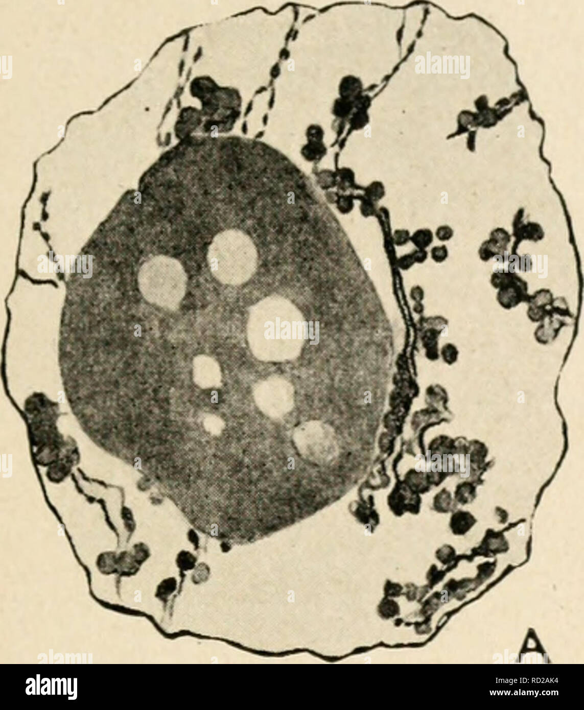 . Cytology, with special reference to the metazoan nucleus. Cells. II MEIOSIS IN THE FEMALE 63 nature (amphinucleoli), consisting of a plastin groundwork (plasmosome), covered or impregnated with chromatin or a chromatin-Uke substance ; or the two constituents may be separate, so that the nucleolus consists of two parts, a chromatin and a plastin portion. These remarks refer especially to the main nucleolus, which persists right through the growth period. In many animals the secondary nucleoli which develop later. Please note that these images are extracted from scanned page images that may ha Stock Photohttps://www.alamy.com/image-license-details/?v=1https://www.alamy.com/cytology-with-special-reference-to-the-metazoan-nucleus-cells-ii-meiosis-in-the-female-63-nature-amphinucleoli-consisting-of-a-plastin-groundwork-plasmosome-covered-or-impregnated-with-chromatin-or-a-chromatin-uke-substance-or-the-two-constituents-may-be-separate-so-that-the-nucleolus-consists-of-two-parts-a-chromatin-and-a-plastin-portion-these-remarks-refer-especially-to-the-main-nucleolus-which-persists-right-through-the-growth-period-in-many-animals-the-secondary-nucleoli-which-develop-later-please-note-that-these-images-are-extracted-from-scanned-page-images-that-may-ha-image231777592.html
RMRD2AK4–. Cytology, with special reference to the metazoan nucleus. Cells. II MEIOSIS IN THE FEMALE 63 nature (amphinucleoli), consisting of a plastin groundwork (plasmosome), covered or impregnated with chromatin or a chromatin-Uke substance ; or the two constituents may be separate, so that the nucleolus consists of two parts, a chromatin and a plastin portion. These remarks refer especially to the main nucleolus, which persists right through the growth period. In many animals the secondary nucleoli which develop later. Please note that these images are extracted from scanned page images that may ha
 . Cytology, with special reference to the metazoan nucleus. Cells. II MEIOSIS IN ASCARIS 51 By the later stage shown in Fig. 20, C, synizesis has completely dis- appeared, and all the chromatin is in the form of a long, doubly split thread (only one split is visible in the plane of the figure). As there are really two bivalent chromosomes present, these must be joined tempor-. FiG. 20. end view, and therefore its quadruple constitution is revealed. F, metaphase I. ; G, H, anaphase I. ; I, preparation for second division ; J, metaphase II. ; K, anaphase II. ; L-M and N-P, condensation of the de Stock Photohttps://www.alamy.com/image-license-details/?v=1https://www.alamy.com/cytology-with-special-reference-to-the-metazoan-nucleus-cells-ii-meiosis-in-ascaris-51-by-the-later-stage-shown-in-fig-20-c-synizesis-has-completely-dis-appeared-and-all-the-chromatin-is-in-the-form-of-a-long-doubly-split-thread-only-one-split-is-visible-in-the-plane-of-the-figure-as-there-are-really-two-bivalent-chromosomes-present-these-must-be-joined-tempor-fig-20-end-view-and-therefore-its-quadruple-constitution-is-revealed-f-metaphase-i-g-h-anaphase-i-i-preparation-for-second-division-j-metaphase-ii-k-anaphase-ii-l-m-and-n-p-condensation-of-the-de-image216168377.html
RMPFK8Y5–. Cytology, with special reference to the metazoan nucleus. Cells. II MEIOSIS IN ASCARIS 51 By the later stage shown in Fig. 20, C, synizesis has completely dis- appeared, and all the chromatin is in the form of a long, doubly split thread (only one split is visible in the plane of the figure). As there are really two bivalent chromosomes present, these must be joined tempor-. FiG. 20. end view, and therefore its quadruple constitution is revealed. F, metaphase I. ; G, H, anaphase I. ; I, preparation for second division ; J, metaphase II. ; K, anaphase II. ; L-M and N-P, condensation of the de
 . An atlas of the fertilization and karyokinesis of the ovum. Ovum; Fertilization (Biology); Meiosis; Embryology -- Echinodermata. V',i'i// â ^â f'jif^^- '"^'Ml^-^ Fig. XIII. âThe segmentation- or cleavage-nucleus (looo diameters). A. The " pause " (thirty minutes). The two germ-nuclei have fused imlistinguishably to form a cleavage-nucleus, traversed by a chromatin network. Some of the chromatin in the form of rings. The astral rays are much shorter and more granular. In the centre of each aster is a small group of granules. B. Initial stage in the formation of the karyokinetic Stock Photohttps://www.alamy.com/image-license-details/?v=1https://www.alamy.com/an-atlas-of-the-fertilization-and-karyokinesis-of-the-ovum-ovum-fertilization-biology-meiosis-embryology-echinodermata-vii-fjif-quotml-fig-xiii-the-segmentation-or-cleavage-nucleus-looo-diameters-a-the-quot-pause-quot-thirty-minutes-the-two-germ-nuclei-have-fused-imlistinguishably-to-form-a-cleavage-nucleus-traversed-by-a-chromatin-network-some-of-the-chromatin-in-the-form-of-rings-the-astral-rays-are-much-shorter-and-more-granular-in-the-centre-of-each-aster-is-a-small-group-of-granules-b-initial-stage-in-the-formation-of-the-karyokinetic-image235391389.html
RMRJY039–. An atlas of the fertilization and karyokinesis of the ovum. Ovum; Fertilization (Biology); Meiosis; Embryology -- Echinodermata. V',i'i// â ^â f'jif^^- '"^'Ml^-^ Fig. XIII. âThe segmentation- or cleavage-nucleus (looo diameters). A. The " pause " (thirty minutes). The two germ-nuclei have fused imlistinguishably to form a cleavage-nucleus, traversed by a chromatin network. Some of the chromatin in the form of rings. The astral rays are much shorter and more granular. In the centre of each aster is a small group of granules. B. Initial stage in the formation of the karyokinetic
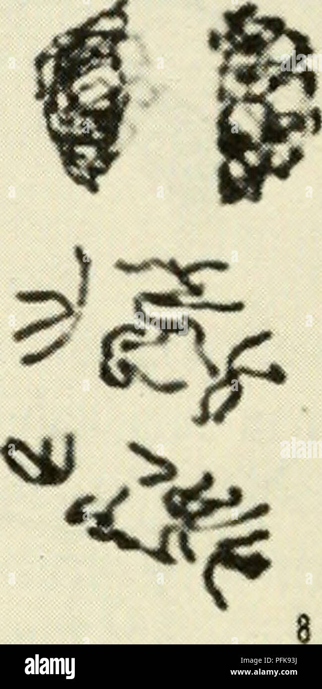 . Cytology. Cytology. '^f I / ^7 if'/ ,' '' ^ ^ A â ^ '^^h' 'A-. -t ^ ^ 11 ^ Figure 5-7. Stages of Meiosis in Microsporocytes of Podophyllum: (1) zygotene; (2) pachytene; (3) diplotene; (4) diakinesis; (5) metaphase I; (6) anaphase I; (7) telophase I; (8) prophase II; (9) metaphase II; (10) anaphase II; (11) early telophase II; (12) late telophase II. (Courtesy of Miss N. Gabriele MLihling, Montana State University.) 128 / CHAPTER 5. Please note that these images are extracted from scanned page images that may have been digitally enhanced for readability - coloration and appearance of these Stock Photohttps://www.alamy.com/image-license-details/?v=1https://www.alamy.com/cytology-cytology-f-i-7-if-a-h-a-t-11-figure-5-7-stages-of-meiosis-in-microsporocytes-of-podophyllum-1-zygotene-2-pachytene-3-diplotene-4-diakinesis-5-metaphase-i-6-anaphase-i-7-telophase-i-8-prophase-ii-9-metaphase-ii-10-anaphase-ii-11-early-telophase-ii-12-late-telophase-ii-courtesy-of-miss-n-gabriele-mlihling-montana-state-university-128-chapter-5-please-note-that-these-images-are-extracted-from-scanned-page-images-that-may-have-been-digitally-enhanced-for-readability-coloration-and-appearance-of-these-image216168502.html
RMPFK93J–. Cytology. Cytology. '^f I / ^7 if'/ ,' '' ^ ^ A â ^ '^^h' 'A-. -t ^ ^ 11 ^ Figure 5-7. Stages of Meiosis in Microsporocytes of Podophyllum: (1) zygotene; (2) pachytene; (3) diplotene; (4) diakinesis; (5) metaphase I; (6) anaphase I; (7) telophase I; (8) prophase II; (9) metaphase II; (10) anaphase II; (11) early telophase II; (12) late telophase II. (Courtesy of Miss N. Gabriele MLihling, Montana State University.) 128 / CHAPTER 5. Please note that these images are extracted from scanned page images that may have been digitally enhanced for readability - coloration and appearance of these
 . Cytology, with special reference to the metazoan nucleus. Cells. ii MEIOSIS IN THE FEMALE 6l germinal vesicles with diffuse chromosomes are to be found, and it is evident that the bivalents resulting from syndesis condense continuously. Fig. 25. The chromosomes during the oogenesis of Diaptomus castor from the pachytene stage (A), through the germinal vesicle stage (B-E) to the condensation of the definitive bivalents (F). (Matschek, A.Z., 1910.) into the definitive bivalents of metaphase I. Animals, however, which are carrying egg-sacs contain in their oviducts oocytes with well-developed g Stock Photohttps://www.alamy.com/image-license-details/?v=1https://www.alamy.com/cytology-with-special-reference-to-the-metazoan-nucleus-cells-ii-meiosis-in-the-female-6l-germinal-vesicles-with-diffuse-chromosomes-are-to-be-found-and-it-is-evident-that-the-bivalents-resulting-from-syndesis-condense-continuously-fig-25-the-chromosomes-during-the-oogenesis-of-diaptomus-castor-from-the-pachytene-stage-a-through-the-germinal-vesicle-stage-b-e-to-the-condensation-of-the-definitive-bivalents-f-matschek-az-1910-into-the-definitive-bivalents-of-metaphase-i-animals-however-which-are-carrying-egg-sacs-contain-in-their-oviducts-oocytes-with-well-developed-g-image231790381.html
RMRD2XYW–. Cytology, with special reference to the metazoan nucleus. Cells. ii MEIOSIS IN THE FEMALE 6l germinal vesicles with diffuse chromosomes are to be found, and it is evident that the bivalents resulting from syndesis condense continuously. Fig. 25. The chromosomes during the oogenesis of Diaptomus castor from the pachytene stage (A), through the germinal vesicle stage (B-E) to the condensation of the definitive bivalents (F). (Matschek, A.Z., 1910.) into the definitive bivalents of metaphase I. Animals, however, which are carrying egg-sacs contain in their oviducts oocytes with well-developed g
 . Cytology, with special reference to the metazoan nucleus. Cells. ii MEIOSIS IN ASCARIS 51 By the later stage shown in Fig. 20, C, synizesis has completely dis- appeared, and all the chromatin is in the form of a long, doubly split thread (only one split is visible in the plane of the figure). As there are really two bivalent chromosomes present, these must be joined tempor-. P Fig. 20. Meiosis in Ascaris megalocephala bivalens. (A-M, after Brauer, A .m.A., 1893 ; N-P, after O. Hertwig, A .m.A., 1890.) A, B, synizesis and syndesis ; C, D, E, formation of the definitive bivalents. In E the lef Stock Photohttps://www.alamy.com/image-license-details/?v=1https://www.alamy.com/cytology-with-special-reference-to-the-metazoan-nucleus-cells-ii-meiosis-in-ascaris-51-by-the-later-stage-shown-in-fig-20-c-synizesis-has-completely-dis-appeared-and-all-the-chromatin-is-in-the-form-of-a-long-doubly-split-thread-only-one-split-is-visible-in-the-plane-of-the-figure-as-there-are-really-two-bivalent-chromosomes-present-these-must-be-joined-tempor-p-fig-20-meiosis-in-ascaris-megalocephala-bivalens-a-m-after-brauer-a-ma-1893-n-p-after-o-hertwig-a-ma-1890-a-b-synizesis-and-syndesis-c-d-e-formation-of-the-definitive-bivalents-in-e-the-lef-image216168735.html
RMPFK9BY–. Cytology, with special reference to the metazoan nucleus. Cells. ii MEIOSIS IN ASCARIS 51 By the later stage shown in Fig. 20, C, synizesis has completely dis- appeared, and all the chromatin is in the form of a long, doubly split thread (only one split is visible in the plane of the figure). As there are really two bivalent chromosomes present, these must be joined tempor-. P Fig. 20. Meiosis in Ascaris megalocephala bivalens. (A-M, after Brauer, A .m.A., 1893 ; N-P, after O. Hertwig, A .m.A., 1890.) A, B, synizesis and syndesis ; C, D, E, formation of the definitive bivalents. In E the lef
 . Cytology, with special reference to the metazoan nucleus. Cells. II MEIOSIS IN LEPIDOSIREN 39 conjugation of the chromosomes. Hence the word symzesis was proposed for the contraction, and syndesis for the chromosome conjugation.. Fig. 16a. Meiosis in Lepidosiren (male . (Agar, Q.J.M.S., 1911.) H, diplotene stage and beginning of synizesis; I, synizesis further advanced ; J, synizesis breaking up; K, diakinesis, showing that the bivalents which were formed in the zygotene stage are completely resolved into their univalent constituents; L, immediate prophase of the meiotic division, showing th Stock Photohttps://www.alamy.com/image-license-details/?v=1https://www.alamy.com/cytology-with-special-reference-to-the-metazoan-nucleus-cells-ii-meiosis-in-lepidosiren-39-conjugation-of-the-chromosomes-hence-the-word-symzesis-was-proposed-for-the-contraction-and-syndesis-for-the-chromosome-conjugation-fig-16a-meiosis-in-lepidosiren-male-agar-qjms-1911-h-diplotene-stage-and-beginning-of-synizesis-i-synizesis-further-advanced-j-synizesis-breaking-up-k-diakinesis-showing-that-the-bivalents-which-were-formed-in-the-zygotene-stage-are-completely-resolved-into-their-univalent-constituents-l-immediate-prophase-of-the-meiotic-division-showing-th-image231804525.html
RMRD3H11–. Cytology, with special reference to the metazoan nucleus. Cells. II MEIOSIS IN LEPIDOSIREN 39 conjugation of the chromosomes. Hence the word symzesis was proposed for the contraction, and syndesis for the chromosome conjugation.. Fig. 16a. Meiosis in Lepidosiren (male . (Agar, Q.J.M.S., 1911.) H, diplotene stage and beginning of synizesis; I, synizesis further advanced ; J, synizesis breaking up; K, diakinesis, showing that the bivalents which were formed in the zygotene stage are completely resolved into their univalent constituents; L, immediate prophase of the meiotic division, showing th
 . Cytological studies of five interspecific hybrids of Crepis leontodontoides. Karyokinesis; Crepis. c d Fig. 13. Meiosis in F, C. leontodontoides-marschalli. a, I—M, with four bivalents; b, I-A, showing' three large marscluilli chromo- somes ; c, II-A, after a 6-3 distribution at I-A; d, II-T, five large and two small nuclei. Mr A Ac 7£ Fig. 14. Somatic metaphases. a, of C aurea; b, of F! (7. leontodontoides-aurea.. Please note that these images are extracted from scanned page images that may have been digitally enhanced for readability - coloration and appearance of these illustrations may Stock Photohttps://www.alamy.com/image-license-details/?v=1https://www.alamy.com/cytological-studies-of-five-interspecific-hybrids-of-crepis-leontodontoides-karyokinesis-crepis-c-d-fig-13-meiosis-in-f-c-leontodontoides-marschalli-a-im-with-four-bivalents-b-i-a-showing-three-large-marscluilli-chromo-somes-c-ii-a-after-a-6-3-distribution-at-i-a-d-ii-t-five-large-and-two-small-nuclei-mr-a-ac-7-fig-14-somatic-metaphases-a-of-c-aurea-b-of-f!-7-leontodontoides-aurea-please-note-that-these-images-are-extracted-from-scanned-page-images-that-may-have-been-digitally-enhanced-for-readability-coloration-and-appearance-of-these-illustrations-may-image216169728.html
RMPFKAKC–. Cytological studies of five interspecific hybrids of Crepis leontodontoides. Karyokinesis; Crepis. c d Fig. 13. Meiosis in F, C. leontodontoides-marschalli. a, I—M, with four bivalents; b, I-A, showing' three large marscluilli chromo- somes ; c, II-A, after a 6-3 distribution at I-A; d, II-T, five large and two small nuclei. Mr A Ac 7£ Fig. 14. Somatic metaphases. a, of C aurea; b, of F! (7. leontodontoides-aurea.. Please note that these images are extracted from scanned page images that may have been digitally enhanced for readability - coloration and appearance of these illustrations may
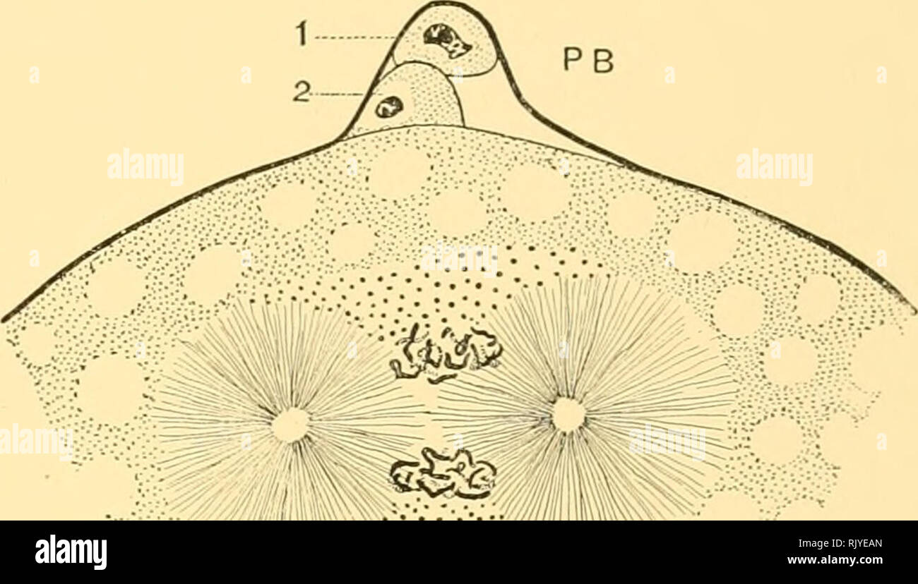 . An atlas of the fertilization and karyokinesis of the ovum. Ovum; Fertilization (Biology); Meiosis; Embryology -- Echinodermata. iiiii!ii^:;-;';>v-:V:Vfiijii^-.:^ B Fig. IV. l-ic IV — The two aerm-iiuclei in the egg of the gastropod Plerotrachea. Highly magnified, after Boveri. E, the egg-nucleus; S, the sperm-nucleus, each containing sixteen elongated chromosomes. PB, the polar bodies. The centrosome has divided into two to form an amphiaster. Its origm m this animal has not yet been determmed. i;. Later stage, showing the fully developed amphiaster. Above it lie the sixteen maternal c Stock Photohttps://www.alamy.com/image-license-details/?v=1https://www.alamy.com/an-atlas-of-the-fertilization-and-karyokinesis-of-the-ovum-ovum-fertilization-biology-meiosis-embryology-echinodermata-iiiii!ii-gtv-vvfiijii-b-fig-iv-l-ic-iv-the-two-aerm-iiuclei-in-the-egg-of-the-gastropod-plerotrachea-highly-magnified-after-boveri-e-the-egg-nucleus-s-the-sperm-nucleus-each-containing-sixteen-elongated-chromosomes-pb-the-polar-bodies-the-centrosome-has-divided-into-two-to-form-an-amphiaster-its-origm-m-this-animal-has-not-yet-been-determmed-i-later-stage-showing-the-fully-developed-amphiaster-above-it-lie-the-sixteen-maternal-c-image235402573.html
RMRJYEAN–. An atlas of the fertilization and karyokinesis of the ovum. Ovum; Fertilization (Biology); Meiosis; Embryology -- Echinodermata. iiiii!ii^:;-;';>v-:V:Vfiijii^-.:^ B Fig. IV. l-ic IV — The two aerm-iiuclei in the egg of the gastropod Plerotrachea. Highly magnified, after Boveri. E, the egg-nucleus; S, the sperm-nucleus, each containing sixteen elongated chromosomes. PB, the polar bodies. The centrosome has divided into two to form an amphiaster. Its origm m this animal has not yet been determmed. i;. Later stage, showing the fully developed amphiaster. Above it lie the sixteen maternal c
 . An atlas of the fertilization and karyokinesis of the ovum. Ovum; Fertilization (Biology); Meiosis; Embryology -- Echinodermata. Fi.:. III. Fig. III. — History of the germ-nuclei in the KYi^i'i.iX-wmm Ascaris megalocephaUi (after Boveri), highly magnified. A. Egg immediately after the formation of the second polar body, PB; E, the egg-nucleus, consisting of two branching chromosomes; S, the sperm-nucleus derived from the head of a spermatozoon that has entered the egg. B. Following stage, in which the egg-nucleus (E) and sperm-nucleus (S) have assumed the same size and structure. C. Later st Stock Photohttps://www.alamy.com/image-license-details/?v=1https://www.alamy.com/an-atlas-of-the-fertilization-and-karyokinesis-of-the-ovum-ovum-fertilization-biology-meiosis-embryology-echinodermata-fi-iii-fig-iii-history-of-the-germ-nuclei-in-the-kyiiiix-wmm-ascaris-megalocephaui-after-boveri-highly-magnified-a-egg-immediately-after-the-formation-of-the-second-polar-body-pb-e-the-egg-nucleus-consisting-of-two-branching-chromosomes-s-the-sperm-nucleus-derived-from-the-head-of-a-spermatozoon-that-has-entered-the-egg-b-following-stage-in-which-the-egg-nucleus-e-and-sperm-nucleus-s-have-assumed-the-same-size-and-structure-c-later-st-image235402599.html
RMRJYEBK–. An atlas of the fertilization and karyokinesis of the ovum. Ovum; Fertilization (Biology); Meiosis; Embryology -- Echinodermata. Fi.:. III. Fig. III. — History of the germ-nuclei in the KYi^i'i.iX-wmm Ascaris megalocephaUi (after Boveri), highly magnified. A. Egg immediately after the formation of the second polar body, PB; E, the egg-nucleus, consisting of two branching chromosomes; S, the sperm-nucleus derived from the head of a spermatozoon that has entered the egg. B. Following stage, in which the egg-nucleus (E) and sperm-nucleus (S) have assumed the same size and structure. C. Later st
 . Colchicine in agriculture, medicine, biology, and chemistry. Colchicine; Colchicine. 172 Colchicine that action during division leading up to niciosis creates octoploid or tetraploid pollen mother cellsJ'^ In contrast, activity dm ing meiotic divisions I and II creates tetraploid monads, and activity at division II only, diploid monads. Monadal formation is a special feature of the c-meiosis. The monads replace the usual tetrads of microspores forming at the close of a meiosis.-^' ^*^- ^-- "^^ Since archesporial divisions become regular c-mitoses, these are not described in great detai Stock Photohttps://www.alamy.com/image-license-details/?v=1https://www.alamy.com/colchicine-in-agriculture-medicine-biology-and-chemistry-colchicine-colchicine-172-colchicine-that-action-during-division-leading-up-to-niciosis-creates-octoploid-or-tetraploid-pollen-mother-cellsj-in-contrast-activity-dm-ing-meiotic-divisions-i-and-ii-creates-tetraploid-monads-and-activity-at-division-ii-only-diploid-monads-monadal-formation-is-a-special-feature-of-the-c-meiosis-the-monads-replace-the-usual-tetrads-of-microspores-forming-at-the-close-of-a-meiosis-quot-since-archesporial-divisions-become-regular-c-mitoses-these-are-not-described-in-great-detai-image232744757.html
RMREJC8N–. Colchicine in agriculture, medicine, biology, and chemistry. Colchicine; Colchicine. 172 Colchicine that action during division leading up to niciosis creates octoploid or tetraploid pollen mother cellsJ'^ In contrast, activity dm ing meiotic divisions I and II creates tetraploid monads, and activity at division II only, diploid monads. Monadal formation is a special feature of the c-meiosis. The monads replace the usual tetrads of microspores forming at the close of a meiosis.-^' ^*^- ^-- "^^ Since archesporial divisions become regular c-mitoses, these are not described in great detai
 . Cytology, with special reference to the metazoan nucleus. Cells. ii MEIOSIS 65 (4) Does any Stage comparable to the Germinal Vesicle occur in Spermatogenesis ? The undoubted correlation between the peculiar conditions of the germinal vesicle and the long duration of the growth period in the female meiotic phase, and probably also with the deposition of yolk, makes it. Fig. 28. Germinal vesicle-like stages in spermatogenesis. A-G, Notodromas monacha (after Schmalz, A.Z., 1912) ; H-N, Scolopendra heros (after Blackman, B.M.C.Z.H., 1905). In both cases the principal stages between the pachytene Stock Photohttps://www.alamy.com/image-license-details/?v=1https://www.alamy.com/cytology-with-special-reference-to-the-metazoan-nucleus-cells-ii-meiosis-65-4-does-any-stage-comparable-to-the-germinal-vesicle-occur-in-spermatogenesis-the-undoubted-correlation-between-the-peculiar-conditions-of-the-germinal-vesicle-and-the-long-duration-of-the-growth-period-in-the-female-meiotic-phase-and-probably-also-with-the-deposition-of-yolk-makes-it-fig-28-germinal-vesicle-like-stages-in-spermatogenesis-a-g-notodromas-monacha-after-schmalz-az-1912-h-n-scolopendra-heros-after-blackman-bmczh-1905-in-both-cases-the-principal-stages-between-the-pachytene-image231790348.html
RMRD2XXM–. Cytology, with special reference to the metazoan nucleus. Cells. ii MEIOSIS 65 (4) Does any Stage comparable to the Germinal Vesicle occur in Spermatogenesis ? The undoubted correlation between the peculiar conditions of the germinal vesicle and the long duration of the growth period in the female meiotic phase, and probably also with the deposition of yolk, makes it. Fig. 28. Germinal vesicle-like stages in spermatogenesis. A-G, Notodromas monacha (after Schmalz, A.Z., 1912) ; H-N, Scolopendra heros (after Blackman, B.M.C.Z.H., 1905). In both cases the principal stages between the pachytene
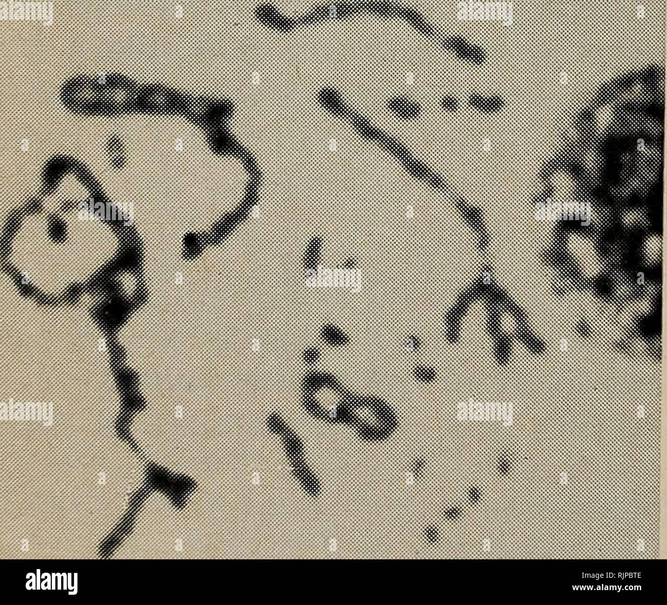 . The Australian zoologist. Zoology; Zoology; Zoology. Fig.. Fig. 3 Fig. 4 FIG. 1 — Cut-out representation of karyotype of diploid (left) and triploid A nobbi FIG. 2 — Photomicrograph of triploid mitotic figure. Bar represents lOu. FIG. 3 — Diakinesis of triploid individual. Most figures of this stage snowed even less organisation. FIG. 4 — Metaphase II of meiosis in triploid A. nobbi. c.f. Fig 2 for appearance of chromosomes. 306 Aust. Zool. 19(3), 1978. Please note that these images are extracted from scanned page images that may have been digitally enhanced for readability - coloration and Stock Photohttps://www.alamy.com/image-license-details/?v=1https://www.alamy.com/the-australian-zoologist-zoology-zoology-zoology-fig-fig-3-fig-4-fig-1-cut-out-representation-of-karyotype-of-diploid-left-and-triploid-a-nobbi-fig-2-photomicrograph-of-triploid-mitotic-figure-bar-represents-lou-fig-3-diakinesis-of-triploid-individual-most-figures-of-this-stage-snowed-even-less-organisation-fig-4-metaphase-ii-of-meiosis-in-triploid-a-nobbi-cf-fig-2-for-appearance-of-chromosomes-306-aust-zool-193-1978-please-note-that-these-images-are-extracted-from-scanned-page-images-that-may-have-been-digitally-enhanced-for-readability-coloration-and-image235290846.html
RMRJPBTE–. The Australian zoologist. Zoology; Zoology; Zoology. Fig.. Fig. 3 Fig. 4 FIG. 1 — Cut-out representation of karyotype of diploid (left) and triploid A nobbi FIG. 2 — Photomicrograph of triploid mitotic figure. Bar represents lOu. FIG. 3 — Diakinesis of triploid individual. Most figures of this stage snowed even less organisation. FIG. 4 — Metaphase II of meiosis in triploid A. nobbi. c.f. Fig 2 for appearance of chromosomes. 306 Aust. Zool. 19(3), 1978. Please note that these images are extracted from scanned page images that may have been digitally enhanced for readability - coloration and
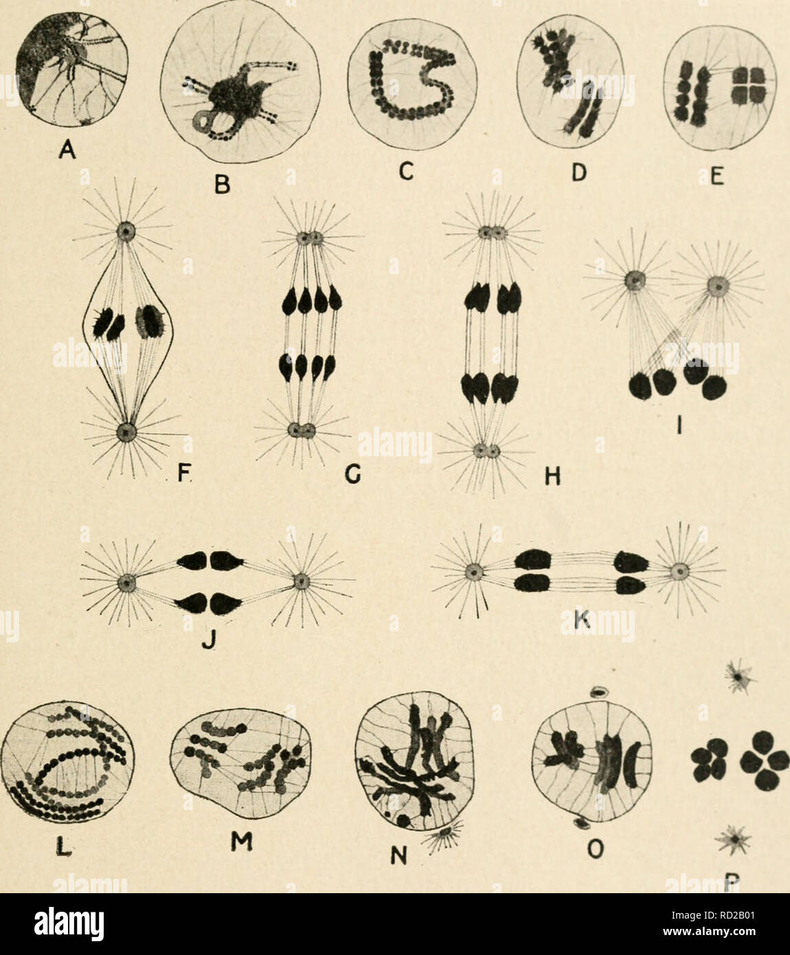 . Cytology, with special reference to the metazoan nucleus. Cells. II MEIOSIS IN ASCARIS 51 By the later stage shown in Fig. 20, C, synizesis has completely dis- appeared, and all the chromatin is in the form of a long, doubly split thread (only one split is visible in the plane of the figure). As there are really two bivalent chromosomes present, these must be joined tempor-. FiG. 20. end view, and therefore its quadruple constitution is revealed. F, metaphase I. ; G, H, anaphase I. ; I, preparation for second division ; J, metaphase II. ; K, anaphase II. ; L-M and N-P, condensation of the de Stock Photohttps://www.alamy.com/image-license-details/?v=1https://www.alamy.com/cytology-with-special-reference-to-the-metazoan-nucleus-cells-ii-meiosis-in-ascaris-51-by-the-later-stage-shown-in-fig-20-c-synizesis-has-completely-dis-appeared-and-all-the-chromatin-is-in-the-form-of-a-long-doubly-split-thread-only-one-split-is-visible-in-the-plane-of-the-figure-as-there-are-really-two-bivalent-chromosomes-present-these-must-be-joined-tempor-fig-20-end-view-and-therefore-its-quadruple-constitution-is-revealed-f-metaphase-i-g-h-anaphase-i-i-preparation-for-second-division-j-metaphase-ii-k-anaphase-ii-l-m-and-n-p-condensation-of-the-de-image231777841.html
RMRD2B01–. Cytology, with special reference to the metazoan nucleus. Cells. II MEIOSIS IN ASCARIS 51 By the later stage shown in Fig. 20, C, synizesis has completely dis- appeared, and all the chromatin is in the form of a long, doubly split thread (only one split is visible in the plane of the figure). As there are really two bivalent chromosomes present, these must be joined tempor-. FiG. 20. end view, and therefore its quadruple constitution is revealed. F, metaphase I. ; G, H, anaphase I. ; I, preparation for second division ; J, metaphase II. ; K, anaphase II. ; L-M and N-P, condensation of the de
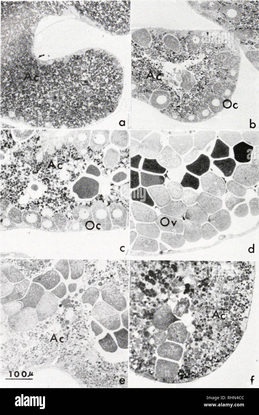 . The Biological bulletin. Biology; Zoology; Biology; Marine Biology. 578 R. MASUDA AND J. C. DAN. f FIGURE 1. Thick sections at various stages of Epon-embedded ovaries of Anthocidaris crassispina, stained with toluidine blue: a, stage I, January—accessory cells including numerous )bules occupy the ovariole; b, stage II, March—many growing oocytes are attached to the i wall; c, stage III, June—fully grown oocytes complete meiosis ; d, stage IV, August—. Please note that these images are extracted from scanned page images that may have been digitally enhanced for readability - coloration and ap Stock Photohttps://www.alamy.com/image-license-details/?v=1https://www.alamy.com/the-biological-bulletin-biology-zoology-biology-marine-biology-578-r-masuda-and-j-c-dan-f-figure-1-thick-sections-at-various-stages-of-epon-embedded-ovaries-of-anthocidaris-crassispina-stained-with-toluidine-blue-a-stage-i-januaryaccessory-cells-including-numerous-bules-occupy-the-ovariole-b-stage-ii-marchmany-growing-oocytes-are-attached-to-the-i-wall-c-stage-iii-junefully-grown-oocytes-complete-meiosis-d-stage-iv-august-please-note-that-these-images-are-extracted-from-scanned-page-images-that-may-have-been-digitally-enhanced-for-readability-coloration-and-ap-image234648412.html
RMRHN4CC–. The Biological bulletin. Biology; Zoology; Biology; Marine Biology. 578 R. MASUDA AND J. C. DAN. f FIGURE 1. Thick sections at various stages of Epon-embedded ovaries of Anthocidaris crassispina, stained with toluidine blue: a, stage I, January—accessory cells including numerous )bules occupy the ovariole; b, stage II, March—many growing oocytes are attached to the i wall; c, stage III, June—fully grown oocytes complete meiosis ; d, stage IV, August—. Please note that these images are extracted from scanned page images that may have been digitally enhanced for readability - coloration and ap
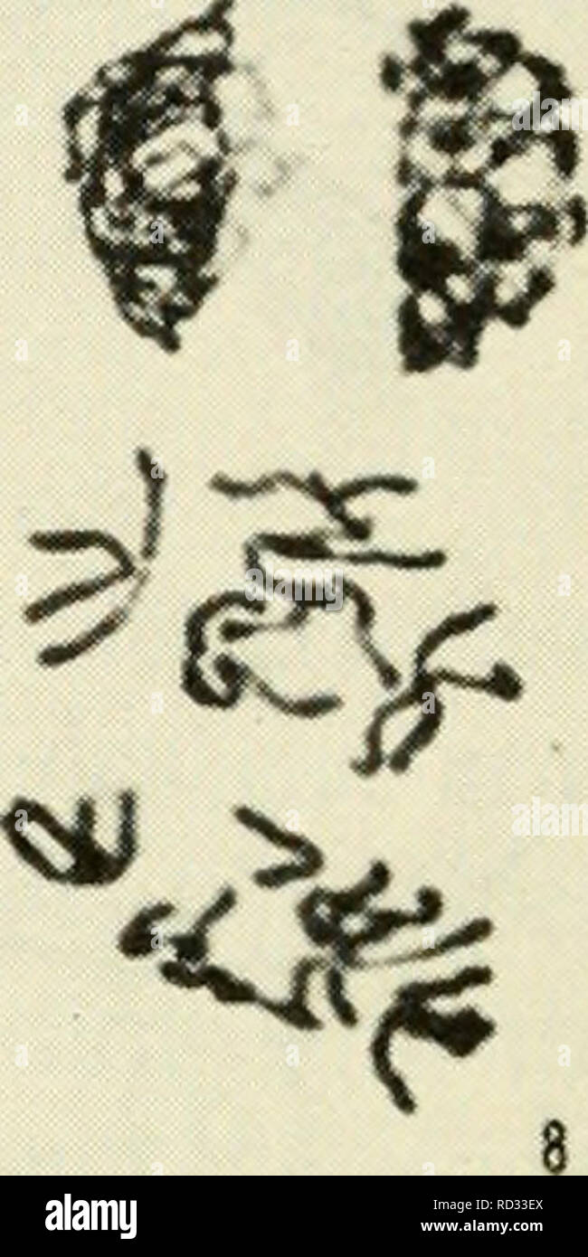 . Cytology. Cytology. '^f I / ^7 if'/ ,' '' ^ ^ A â ^ '^^h' 'A-. -t ^ ^ 11 ^ Figure 5-7. Stages of Meiosis in Microsporocytes of Podophyllum: (1) zygotene; (2) pachytene; (3) diplotene; (4) diakinesis; (5) metaphase I; (6) anaphase I; (7) telophase I; (8) prophase II; (9) metaphase II; (10) anaphase II; (11) early telophase II; (12) late telophase II. (Courtesy of Miss N. Gabriele MLihling, Montana State University.) 128 / CHAPTER 5. Please note that these images are extracted from scanned page images that may have been digitally enhanced for readability - coloration and appearance of these Stock Photohttps://www.alamy.com/image-license-details/?v=1https://www.alamy.com/cytology-cytology-f-i-7-if-a-h-a-t-11-figure-5-7-stages-of-meiosis-in-microsporocytes-of-podophyllum-1-zygotene-2-pachytene-3-diplotene-4-diakinesis-5-metaphase-i-6-anaphase-i-7-telophase-i-8-prophase-ii-9-metaphase-ii-10-anaphase-ii-11-early-telophase-ii-12-late-telophase-ii-courtesy-of-miss-n-gabriele-mlihling-montana-state-university-128-chapter-5-please-note-that-these-images-are-extracted-from-scanned-page-images-that-may-have-been-digitally-enhanced-for-readability-coloration-and-appearance-of-these-image231793938.html
RMRD33EX–. Cytology. Cytology. '^f I / ^7 if'/ ,' '' ^ ^ A â ^ '^^h' 'A-. -t ^ ^ 11 ^ Figure 5-7. Stages of Meiosis in Microsporocytes of Podophyllum: (1) zygotene; (2) pachytene; (3) diplotene; (4) diakinesis; (5) metaphase I; (6) anaphase I; (7) telophase I; (8) prophase II; (9) metaphase II; (10) anaphase II; (11) early telophase II; (12) late telophase II. (Courtesy of Miss N. Gabriele MLihling, Montana State University.) 128 / CHAPTER 5. Please note that these images are extracted from scanned page images that may have been digitally enhanced for readability - coloration and appearance of these
 . The Biological bulletin. Biology; Zoology; Biology; Marine Biology. 3. FIGURE 1. Anaphase II with adjacent polar body I, from the ostium of an oviduct ligated as close as possible to the anterior end. X 790. FIGURE 2. Section adjacent to that shown in Figure 1. < 790. FIGURE 3. Completed meiosis in an egg from the coelom of an animal with oviducts ligated at the ostia. Three chromosomes of the haploid set of eleven can be seen at the lower left. Portions of polar bodies I and II are visible outside the egg. • 970. FIGURE 4. Two interphase nuclei in an egg from the coelom kept for 40 hours Stock Photohttps://www.alamy.com/image-license-details/?v=1https://www.alamy.com/the-biological-bulletin-biology-zoology-biology-marine-biology-3-figure-1-anaphase-ii-with-adjacent-polar-body-i-from-the-ostium-of-an-oviduct-ligated-as-close-as-possible-to-the-anterior-end-x-790-figure-2-section-adjacent-to-that-shown-in-figure-1-lt-790-figure-3-completed-meiosis-in-an-egg-from-the-coelom-of-an-animal-with-oviducts-ligated-at-the-ostia-three-chromosomes-of-the-haploid-set-of-eleven-can-be-seen-at-the-lower-left-portions-of-polar-bodies-i-and-ii-are-visible-outside-the-egg-970-figure-4-two-interphase-nuclei-in-an-egg-from-the-coelom-kept-for-40-hours-image234665845.html
RMRHNXK1–. The Biological bulletin. Biology; Zoology; Biology; Marine Biology. 3. FIGURE 1. Anaphase II with adjacent polar body I, from the ostium of an oviduct ligated as close as possible to the anterior end. X 790. FIGURE 2. Section adjacent to that shown in Figure 1. < 790. FIGURE 3. Completed meiosis in an egg from the coelom of an animal with oviducts ligated at the ostia. Three chromosomes of the haploid set of eleven can be seen at the lower left. Portions of polar bodies I and II are visible outside the egg. • 970. FIGURE 4. Two interphase nuclei in an egg from the coelom kept for 40 hours
 . Cytology, with special reference to the metazoan nucleus. Cells. ii MEIOSIS IN ASCARIS 51 By the later stage shown in Fig. 20, C, synizesis has completely dis- appeared, and all the chromatin is in the form of a long, doubly split thread (only one split is visible in the plane of the figure). As there are really two bivalent chromosomes present, these must be joined tempor-. P Fig. 20. Meiosis in Ascaris megalocephala bivalens. (A-M, after Brauer, A .m.A., 1893 ; N-P, after O. Hertwig, A .m.A., 1890.) A, B, synizesis and syndesis ; C, D, E, formation of the definitive bivalents. In E the lef Stock Photohttps://www.alamy.com/image-license-details/?v=1https://www.alamy.com/cytology-with-special-reference-to-the-metazoan-nucleus-cells-ii-meiosis-in-ascaris-51-by-the-later-stage-shown-in-fig-20-c-synizesis-has-completely-dis-appeared-and-all-the-chromatin-is-in-the-form-of-a-long-doubly-split-thread-only-one-split-is-visible-in-the-plane-of-the-figure-as-there-are-really-two-bivalent-chromosomes-present-these-must-be-joined-tempor-p-fig-20-meiosis-in-ascaris-megalocephala-bivalens-a-m-after-brauer-a-ma-1893-n-p-after-o-hertwig-a-ma-1890-a-b-synizesis-and-syndesis-c-d-e-formation-of-the-definitive-bivalents-in-e-the-lef-image231804466.html
RMRD3GXX–. Cytology, with special reference to the metazoan nucleus. Cells. ii MEIOSIS IN ASCARIS 51 By the later stage shown in Fig. 20, C, synizesis has completely dis- appeared, and all the chromatin is in the form of a long, doubly split thread (only one split is visible in the plane of the figure). As there are really two bivalent chromosomes present, these must be joined tempor-. P Fig. 20. Meiosis in Ascaris megalocephala bivalens. (A-M, after Brauer, A .m.A., 1893 ; N-P, after O. Hertwig, A .m.A., 1890.) A, B, synizesis and syndesis ; C, D, E, formation of the definitive bivalents. In E the lef
 . The Biological bulletin. Biology; Zoology; Biology; Marine Biology. 5X0 R. MASUDA AND J. C. DAN ^ , ij**-j*!Kft^, « • • • 'v%a£.v0. f FIGURE 2. Thick sections of Epon-embedded ovariole of Hemicentrotus pulcherrimus: a, stage I, November—numerous accessory cells occupy the lumen and a few small oocytes are present along the wall; b, stage II, December—fully grown oocytes are seen along the wall; c, 'ate of stage III, January—fully grown oocytes complete meiosis, mature eggs occupy the. Please note that these images are extracted from scanned page images that may have been digitally enhanced f Stock Photohttps://www.alamy.com/image-license-details/?v=1https://www.alamy.com/the-biological-bulletin-biology-zoology-biology-marine-biology-5x0-r-masuda-and-j-c-dan-ij-j!kft-vav0-f-figure-2-thick-sections-of-epon-embedded-ovariole-of-hemicentrotus-pulcherrimus-a-stage-i-novembernumerous-accessory-cells-occupy-the-lumen-and-a-few-small-oocytes-are-present-along-the-wall-b-stage-ii-decemberfully-grown-oocytes-are-seen-along-the-wall-c-ate-of-stage-iii-januaryfully-grown-oocytes-complete-meiosis-mature-eggs-occupy-the-please-note-that-these-images-are-extracted-from-scanned-page-images-that-may-have-been-digitally-enhanced-f-image234648392.html
RMRHN4BM–. The Biological bulletin. Biology; Zoology; Biology; Marine Biology. 5X0 R. MASUDA AND J. C. DAN ^ , ij**-j*!Kft^, « • • • 'v%a£.v0. f FIGURE 2. Thick sections of Epon-embedded ovariole of Hemicentrotus pulcherrimus: a, stage I, November—numerous accessory cells occupy the lumen and a few small oocytes are present along the wall; b, stage II, December—fully grown oocytes are seen along the wall; c, 'ate of stage III, January—fully grown oocytes complete meiosis, mature eggs occupy the. Please note that these images are extracted from scanned page images that may have been digitally enhanced f
 . Cytological studies of five interspecific hybrids of Crepis leontodontoides. Karyokinesis; Crepis. c d Fig. 13. Meiosis in F, C. leontodontoides-marschalli. a, I—M, with four bivalents; b, I-A, showing' three large marscluilli chromo- somes ; c, II-A, after a 6-3 distribution at I-A; d, II-T, five large and two small nuclei. Mr A Ac 7£ Fig. 14. Somatic metaphases. a, of C aurea; b, of F! (7. leontodontoides-aurea.. Please note that these images are extracted from scanned page images that may have been digitally enhanced for readability - coloration and appearance of these illustrations may Stock Photohttps://www.alamy.com/image-license-details/?v=1https://www.alamy.com/cytological-studies-of-five-interspecific-hybrids-of-crepis-leontodontoides-karyokinesis-crepis-c-d-fig-13-meiosis-in-f-c-leontodontoides-marschalli-a-im-with-four-bivalents-b-i-a-showing-three-large-marscluilli-chromo-somes-c-ii-a-after-a-6-3-distribution-at-i-a-d-ii-t-five-large-and-two-small-nuclei-mr-a-ac-7-fig-14-somatic-metaphases-a-of-c-aurea-b-of-f!-7-leontodontoides-aurea-please-note-that-these-images-are-extracted-from-scanned-page-images-that-may-have-been-digitally-enhanced-for-readability-coloration-and-appearance-of-these-illustrations-may-image231778532.html
RMRD2BTM–. Cytological studies of five interspecific hybrids of Crepis leontodontoides. Karyokinesis; Crepis. c d Fig. 13. Meiosis in F, C. leontodontoides-marschalli. a, I—M, with four bivalents; b, I-A, showing' three large marscluilli chromo- somes ; c, II-A, after a 6-3 distribution at I-A; d, II-T, five large and two small nuclei. Mr A Ac 7£ Fig. 14. Somatic metaphases. a, of C aurea; b, of F! (7. leontodontoides-aurea.. Please note that these images are extracted from scanned page images that may have been digitally enhanced for readability - coloration and appearance of these illustrations may
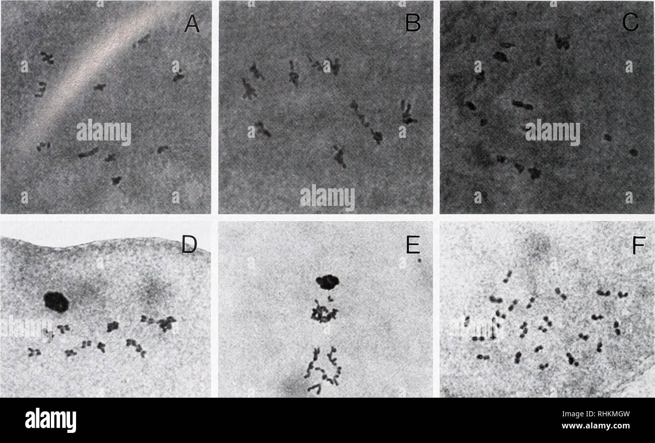 . The Biological bulletin. Biology; Zoology; Biology; Marine Biology. 312 X. GUO AND S. K. ALLEN, JR.. Figure 1. Synapsis and meiosis in eggs from triploid Pacific oysters: A—metaphase I eggs from diploids with 10 bivalents; B—complete synapsis in metaphase I eggs from triploids with 10 trivalents; C—incomplete synapsis in metaphase I eggs from triploids with a mixture of uni-, bi-, and trivalents; D—metaphase 11 in eggs from triploids; E—anaphase II in eggs from triploids; and F—metaphase of the first mitotic division of an embryo from a tnploid (female) x diploid cross. The orcein staining p Stock Photohttps://www.alamy.com/image-license-details/?v=1https://www.alamy.com/the-biological-bulletin-biology-zoology-biology-marine-biology-312-x-guo-and-s-k-allen-jr-figure-1-synapsis-and-meiosis-in-eggs-from-triploid-pacific-oysters-ametaphase-i-eggs-from-diploids-with-10-bivalents-bcomplete-synapsis-in-metaphase-i-eggs-from-triploids-with-10-trivalents-cincomplete-synapsis-in-metaphase-i-eggs-from-triploids-with-a-mixture-of-uni-bi-and-trivalents-dmetaphase-11-in-eggs-from-triploids-eanaphase-ii-in-eggs-from-triploids-and-fmetaphase-of-the-first-mitotic-division-of-an-embryo-from-a-tnploid-female-x-diploid-cross-the-orcein-staining-p-image234617177.html
RMRHKMGW–. The Biological bulletin. Biology; Zoology; Biology; Marine Biology. 312 X. GUO AND S. K. ALLEN, JR.. Figure 1. Synapsis and meiosis in eggs from triploid Pacific oysters: A—metaphase I eggs from diploids with 10 bivalents; B—complete synapsis in metaphase I eggs from triploids with 10 trivalents; C—incomplete synapsis in metaphase I eggs from triploids with a mixture of uni-, bi-, and trivalents; D—metaphase 11 in eggs from triploids; E—anaphase II in eggs from triploids; and F—metaphase of the first mitotic division of an embryo from a tnploid (female) x diploid cross. The orcein staining p
![. Bulletin of the Southern California Academy of Sciences. Science; Natural history; Natural history. Bi:i.i,uri., !Su. Cai.ii. At akkmv ok SVikiNCK.s Vol. 50. Part 2, 1951 broad ;m(l (ipni ami the petals sdiiicw hat rcHcxcd, rccallin;^ tin- Ihidlrya (plalr 'J i. This plani sii^,m'sts lliat the luliriil is at least partially t'eiliK'. Dr. I 111 has studied tniir cnlK-ctidns ot" the Inhrid troin Torrey Pines ( '///", .^(h(l. MdJ, Ml)7A) and one from' Del Mar {^M7). Just as in each ot' the parental species, the ha])loid ihidiuosonie number is 17. lie reports that meiosis, microspore Stock Photo . Bulletin of the Southern California Academy of Sciences. Science; Natural history; Natural history. Bi:i.i,uri., !Su. Cai.ii. At akkmv ok SVikiNCK.s Vol. 50. Part 2, 1951 broad ;m(l (ipni ami the petals sdiiicw hat rcHcxcd, rccallin;^ tin- Ihidlrya (plalr 'J i. This plani sii^,m'sts lliat the luliriil is at least partially t'eiliK'. Dr. I 111 has studied tniir cnlK-ctidns ot" the Inhrid troin Torrey Pines ( '///", .^(h(l. MdJ, Ml)7A) and one from' Del Mar {^M7). Just as in each ot' the parental species, the ha])loid ihidiuosonie number is 17. lie reports that meiosis, microspore Stock Photo](https://c8.alamy.com/comp/RGCKTW/bulletin-of-the-southern-california-academy-of-sciences-science-natural-history-natural-history-biiiuri-!su-caiii-at-akkmv-ok-svikincks-vol-50-part-2-1951-broad-ml-ipni-ami-the-petals-sdiiicw-hat-rchcxcd-rccallin-tin-ihidlrya-plalr-j-i-this-plani-siimsts-lliat-the-luliriil-is-at-least-partially-teilik-dr-i-111-has-studied-tniir-cnlk-ctidns-otquot-the-inhrid-troin-torrey-pines-quot-hl-mdj-ml7a-and-one-from-del-mar-m7-just-as-in-each-ot-the-parental-species-the-ha-loid-ihidiuosonie-number-is-17-lie-reports-that-meiosis-microspore-RGCKTW.jpg) . Bulletin of the Southern California Academy of Sciences. Science; Natural history; Natural history. Bi:i.i,uri., !Su. Cai.ii. At akkmv ok SVikiNCK.s Vol. 50. Part 2, 1951 broad ;m(l (ipni ami the petals sdiiicw hat rcHcxcd, rccallin;^ tin- Ihidlrya (plalr 'J i. This plani sii^,m'sts lliat the luliriil is at least partially t'eiliK'. Dr. I 111 has studied tniir cnlK-ctidns ot" the Inhrid troin Torrey Pines ( '///", .^(h(l. MdJ, Ml)7A) and one from' Del Mar {^M7). Just as in each ot' the parental species, the ha])loid ihidiuosonie number is 17. lie reports that meiosis, microspore Stock Photohttps://www.alamy.com/image-license-details/?v=1https://www.alamy.com/bulletin-of-the-southern-california-academy-of-sciences-science-natural-history-natural-history-biiiuri-!su-caiii-at-akkmv-ok-svikincks-vol-50-part-2-1951-broad-ml-ipni-ami-the-petals-sdiiicw-hat-rchcxcd-rccallin-tin-ihidlrya-plalr-j-i-this-plani-siimsts-lliat-the-luliriil-is-at-least-partially-teilik-dr-i-111-has-studied-tniir-cnlk-ctidns-otquot-the-inhrid-troin-torrey-pines-quot-hl-mdj-ml7a-and-one-from-del-mar-m7-just-as-in-each-ot-the-parental-species-the-ha-loid-ihidiuosonie-number-is-17-lie-reports-that-meiosis-microspore-image233848297.html
. Bulletin of the Southern California Academy of Sciences. Science; Natural history; Natural history. Bi:i.i,uri., !Su. Cai.ii. At akkmv ok SVikiNCK.s Vol. 50. Part 2, 1951 broad ;m(l (ipni ami the petals sdiiicw hat rcHcxcd, rccallin;^ tin- Ihidlrya (plalr 'J i. This plani sii^,m'sts lliat the luliriil is at least partially t'eiliK'. Dr. I 111 has studied tniir cnlK-ctidns ot" the Inhrid troin Torrey Pines ( '///", .^(h(l. MdJ, Ml)7A) and one from' Del Mar {^M7). Just as in each ot' the parental species, the ha])loid ihidiuosonie number is 17. lie reports that meiosis, microspore Stock Photohttps://www.alamy.com/image-license-details/?v=1https://www.alamy.com/bulletin-of-the-southern-california-academy-of-sciences-science-natural-history-natural-history-biiiuri-!su-caiii-at-akkmv-ok-svikincks-vol-50-part-2-1951-broad-ml-ipni-ami-the-petals-sdiiicw-hat-rchcxcd-rccallin-tin-ihidlrya-plalr-j-i-this-plani-siimsts-lliat-the-luliriil-is-at-least-partially-teilik-dr-i-111-has-studied-tniir-cnlk-ctidns-otquot-the-inhrid-troin-torrey-pines-quot-hl-mdj-ml7a-and-one-from-del-mar-m7-just-as-in-each-ot-the-parental-species-the-ha-loid-ihidiuosonie-number-is-17-lie-reports-that-meiosis-microspore-image233848297.htmlRMRGCKTW–. Bulletin of the Southern California Academy of Sciences. Science; Natural history; Natural history. Bi:i.i,uri., !Su. Cai.ii. At akkmv ok SVikiNCK.s Vol. 50. Part 2, 1951 broad ;m(l (ipni ami the petals sdiiicw hat rcHcxcd, rccallin;^ tin- Ihidlrya (plalr 'J i. This plani sii^,m'sts lliat the luliriil is at least partially t'eiliK'. Dr. I 111 has studied tniir cnlK-ctidns ot" the Inhrid troin Torrey Pines ( '///", .^(h(l. MdJ, Ml)7A) and one from' Del Mar {^M7). Just as in each ot' the parental species, the ha])loid ihidiuosonie number is 17. lie reports that meiosis, microspore
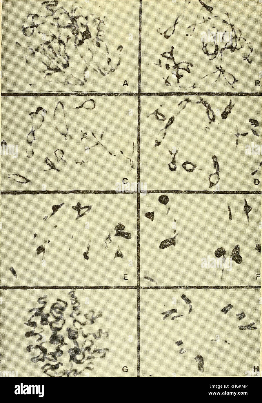 . Boletin de la Sociedad de BiologÃ-a de Concepción. Sociedad de BiologÃ-a de Concepción; Biology; Biology. xâ^ Fig. 1.âEstadios de la meiosis en células gonádicas del macho de Aucacris eumera Hebard (Orthoptera, Acrididae, Omexechinae). A.â Paquiteno. B.â Diplo- teno medio. Câ Diploteno final. D.â Diacinesis. E-F.â Metafase I. G.âProfase II. H.âMetafase II. â 290- -f. Please note that these images are extracted from scanned page images that may have been digitally enhanced for readability - coloration and appearance of these illustrations may not perfectly resemble the original work.. Soc Stock Photohttps://www.alamy.com/image-license-details/?v=1https://www.alamy.com/boletin-de-la-sociedad-de-biolog-a-de-concepcin-sociedad-de-biolog-a-de-concepcin-biology-biology-x-fig-1estadios-de-la-meiosis-en-clulas-gondicas-del-macho-de-aucacris-eumera-hebard-orthoptera-acrididae-omexechinae-a-paquiteno-b-diplo-teno-medio-c-diploteno-final-d-diacinesis-e-f-metafase-i-gprofase-ii-hmetafase-ii-290-f-please-note-that-these-images-are-extracted-from-scanned-page-images-that-may-have-been-digitally-enhanced-for-readability-coloration-and-appearance-of-these-illustrations-may-not-perfectly-resemble-the-original-work-soc-image234550646.html
RMRHGKMP–. Boletin de la Sociedad de BiologÃ-a de Concepción. Sociedad de BiologÃ-a de Concepción; Biology; Biology. xâ^ Fig. 1.âEstadios de la meiosis en células gonádicas del macho de Aucacris eumera Hebard (Orthoptera, Acrididae, Omexechinae). A.â Paquiteno. B.â Diplo- teno medio. Câ Diploteno final. D.â Diacinesis. E-F.â Metafase I. G.âProfase II. H.âMetafase II. â 290- -f. Please note that these images are extracted from scanned page images that may have been digitally enhanced for readability - coloration and appearance of these illustrations may not perfectly resemble the original work.. Soc
 . The British fern gazette. Ferns. R1T.FERNGAZ. 10(2) 1969 PLATF I â '-.- * * I 4 -if J * ⢠,* * i« ** *⢠â > v, -4 B ^â¢^. * ⢠-«*? â¢A. **«* y T^ i * â¢: AMi t + +. PLATE II: A: Meiosis in A. adiantum-nigrum ex Kynancs Cove, diakinesis with 72 bivalents. B, C, D, E&F: Meiosis in Asplenium adiantum-nigrum Phyllitis scolopendrium. B, C & D: GV 100:1A. E&F: JDL 1846. B & F: 108 univalents. D: 4 bivalents & 100 univalents. C & E: 6 bivalents & 96 univalents. Magnification / 1000, Leitz N.A. 1.4 2 mm apochromat with X10 eyepiece. to face page 64. Please not Stock Photohttps://www.alamy.com/image-license-details/?v=1https://www.alamy.com/the-british-fern-gazette-ferns-r1tferngaz-102-1969-platf-i-i-4-if-j-i-gt-v-4-b-a-y-t-i-ami-t-plate-ii-a-meiosis-in-a-adiantum-nigrum-ex-kynancs-cove-diakinesis-with-72-bivalents-b-c-d-eampf-meiosis-in-asplenium-adiantum-nigrum-phyllitis-scolopendrium-b-c-amp-d-gv-1001a-eampf-jdl-1846-b-amp-f-108-univalents-d-4-bivalents-amp-100-univalents-c-amp-e-6-bivalents-amp-96-univalents-magnification-1000-leitz-na-14-2-mm-apochromat-with-x10-eyepiece-to-face-page-64-please-not-image234254039.html
RMRH35BK–. The British fern gazette. Ferns. R1T.FERNGAZ. 10(2) 1969 PLATF I â '-.- * * I 4 -if J * ⢠,* * i« ** *⢠â > v, -4 B ^â¢^. * ⢠-«*? â¢A. **«* y T^ i * â¢: AMi t + +. PLATE II: A: Meiosis in A. adiantum-nigrum ex Kynancs Cove, diakinesis with 72 bivalents. B, C, D, E&F: Meiosis in Asplenium adiantum-nigrum Phyllitis scolopendrium. B, C & D: GV 100:1A. E&F: JDL 1846. B & F: 108 univalents. D: 4 bivalents & 100 univalents. C & E: 6 bivalents & 96 univalents. Magnification / 1000, Leitz N.A. 1.4 2 mm apochromat with X10 eyepiece. to face page 64. Please not