Sternal angle Black & White Stock Photos
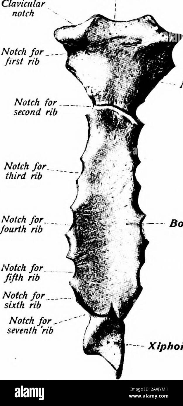
 A manual of anatomy . de. The superior margin is attachedor fused to the manubrium forming an angle at the junction that isusually apDreciable to the touch. This is the sternal angle {angiilussterni). The lateral portions of this margin complete the facet forthe second costal cartilage. The inferior margin is convex and its 36 OSTEOLOGY middle part is connected to the xyphoid process. Lateral to thisthe sixth and seventh costal cartilages are attached. Each lateralmargin is thick and irregular presenting facets at the extremitiesof the ridges that accommodate the third, fourth and fifth costal Stock Photohttps://www.alamy.com/image-license-details/?v=1https://www.alamy.com/a-manual-of-anatomy-de-the-superior-margin-is-attachedor-fused-to-the-manubrium-forming-an-angle-at-the-junction-that-isusually-apdreciable-to-the-touch-this-is-the-sternal-angle-angiilussterni-the-lateral-portions-of-this-margin-complete-the-facet-forthe-second-costal-cartilage-the-inferior-margin-is-convex-and-its-36-osteology-middle-part-is-connected-to-the-xyphoid-process-lateral-to-thisthe-sixth-and-seventh-costal-cartilages-are-attached-each-lateralmargin-is-thick-and-irregular-presenting-facets-at-the-extremitiesof-the-ridges-that-accommodate-the-third-fourth-and-fifth-costal-image343394929.html
RM2AXJYMH–A manual of anatomy . de. The superior margin is attachedor fused to the manubrium forming an angle at the junction that isusually apDreciable to the touch. This is the sternal angle {angiilussterni). The lateral portions of this margin complete the facet forthe second costal cartilage. The inferior margin is convex and its 36 OSTEOLOGY middle part is connected to the xyphoid process. Lateral to thisthe sixth and seventh costal cartilages are attached. Each lateralmargin is thick and irregular presenting facets at the extremitiesof the ridges that accommodate the third, fourth and fifth costal
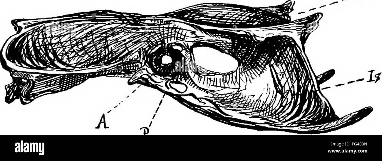
 . A text-book of agricultural zoology. Zoology, Economic. THE SKELETON AND ANATOMY OF BIKDS. 341 except the first and last pair. There is a large hreast-bone or sternum (14). In flying birds this sternum has a deep sternal ridge or keel, to which are attached the powerful muscles which move the wings. The pectoral arch (fig. 177) consists of a pair of scapulae (Sc), clavicles (/), and coracoid bones (Co). The scapula is an elongated simple bone; the coracoids are distinct and very strong, and articulate with the upper angle of the sternum ; the clavicles form the V-shaped bone popularly called Stock Photohttps://www.alamy.com/image-license-details/?v=1https://www.alamy.com/a-text-book-of-agricultural-zoology-zoology-economic-the-skeleton-and-anatomy-of-bikds-341-except-the-first-and-last-pair-there-is-a-large-hreast-bone-or-sternum-14-in-flying-birds-this-sternum-has-a-deep-sternal-ridge-or-keel-to-which-are-attached-the-powerful-muscles-which-move-the-wings-the-pectoral-arch-fig-177-consists-of-a-pair-of-scapulae-sc-clavicles-and-coracoid-bones-co-the-scapula-is-an-elongated-simple-bone-the-coracoids-are-distinct-and-very-strong-and-articulate-with-the-upper-angle-of-the-sternum-the-clavicles-form-the-v-shaped-bone-popularly-called-image216446825.html
RMPG403N–. A text-book of agricultural zoology. Zoology, Economic. THE SKELETON AND ANATOMY OF BIKDS. 341 except the first and last pair. There is a large hreast-bone or sternum (14). In flying birds this sternum has a deep sternal ridge or keel, to which are attached the powerful muscles which move the wings. The pectoral arch (fig. 177) consists of a pair of scapulae (Sc), clavicles (/), and coracoid bones (Co). The scapula is an elongated simple bone; the coracoids are distinct and very strong, and articulate with the upper angle of the sternum ; the clavicles form the V-shaped bone popularly called
 A manual of anatomy . al ligament. The lateral sternal lines are two lines at the right and left bordersof the sternum. The parasternal lines are two vertical lines midway between themidsternal and midclavicular lines. Some place these lines midwaybetween the lateral sternal and the midclavicular lines. The midaxillary lines (two) are drawn from the apex of the axilla(armpit) with the arms extended at a right angle to the body. The scapular lines (two) are drawn vertically through the inferiorangle of each scapula. The midvertebral line (one) is drawn along the spinous processesof the thoracic Stock Photohttps://www.alamy.com/image-license-details/?v=1https://www.alamy.com/a-manual-of-anatomy-al-ligament-the-lateral-sternal-lines-are-two-lines-at-the-right-and-left-bordersof-the-sternum-the-parasternal-lines-are-two-vertical-lines-midway-between-themidsternal-and-midclavicular-lines-some-place-these-lines-midwaybetween-the-lateral-sternal-and-the-midclavicular-lines-the-midaxillary-lines-two-are-drawn-from-the-apex-of-the-axillaarmpit-with-the-arms-extended-at-a-right-angle-to-the-body-the-scapular-lines-two-are-drawn-vertically-through-the-inferiorangle-of-each-scapula-the-midvertebral-line-one-is-drawn-along-the-spinous-processesof-the-thoracic-image343356855.html
RM2AXH74R–A manual of anatomy . al ligament. The lateral sternal lines are two lines at the right and left bordersof the sternum. The parasternal lines are two vertical lines midway between themidsternal and midclavicular lines. Some place these lines midwaybetween the lateral sternal and the midclavicular lines. The midaxillary lines (two) are drawn from the apex of the axilla(armpit) with the arms extended at a right angle to the body. The scapular lines (two) are drawn vertically through the inferiorangle of each scapula. The midvertebral line (one) is drawn along the spinous processesof the thoracic
![. Monographs of North American rodentia [microform]. Rodentia; Paleontology; Rongeurs; Paléontologie. " j'%1 r 578 MONOORAPlia OF NORTH AMERICAN KODENTIA. and knobbed at the sternal extremity, where the cross-section would be decidedly triangular. The scapula is about 1.75 inches long by 0.90 broad at the widest part, and presents numerous well-marked features. The general contour is that of an inequilateral triangle with the postero-superior corner rounded off, and the anterior angle pro(?uced into a neck. The lower border, which is much the longest, is nearly straight; the jwsterior c Stock Photo . Monographs of North American rodentia [microform]. Rodentia; Paleontology; Rongeurs; Paléontologie. " j'%1 r 578 MONOORAPlia OF NORTH AMERICAN KODENTIA. and knobbed at the sternal extremity, where the cross-section would be decidedly triangular. The scapula is about 1.75 inches long by 0.90 broad at the widest part, and presents numerous well-marked features. The general contour is that of an inequilateral triangle with the postero-superior corner rounded off, and the anterior angle pro(?uced into a neck. The lower border, which is much the longest, is nearly straight; the jwsterior c Stock Photo](https://c8.alamy.com/comp/RJ64TN/monographs-of-north-american-rodentia-microform-rodentia-paleontology-rongeurs-palontologie-quot-j1-r-578-monooraplia-of-north-american-kodentia-and-knobbed-at-the-sternal-extremity-where-the-cross-section-would-be-decidedly-triangular-the-scapula-is-about-175-inches-long-by-090-broad-at-the-widest-part-and-presents-numerous-well-marked-features-the-general-contour-is-that-of-an-inequilateral-triangle-with-the-postero-superior-corner-rounded-off-and-the-anterior-angle-prouced-into-a-neck-the-lower-border-which-is-much-the-longest-is-nearly-straight-the-jwsterior-c-RJ64TN.jpg) . Monographs of North American rodentia [microform]. Rodentia; Paleontology; Rongeurs; Paléontologie. " j'%1 r 578 MONOORAPlia OF NORTH AMERICAN KODENTIA. and knobbed at the sternal extremity, where the cross-section would be decidedly triangular. The scapula is about 1.75 inches long by 0.90 broad at the widest part, and presents numerous well-marked features. The general contour is that of an inequilateral triangle with the postero-superior corner rounded off, and the anterior angle pro(?uced into a neck. The lower border, which is much the longest, is nearly straight; the jwsterior c Stock Photohttps://www.alamy.com/image-license-details/?v=1https://www.alamy.com/monographs-of-north-american-rodentia-microform-rodentia-paleontology-rongeurs-palontologie-quot-j1-r-578-monooraplia-of-north-american-kodentia-and-knobbed-at-the-sternal-extremity-where-the-cross-section-would-be-decidedly-triangular-the-scapula-is-about-175-inches-long-by-090-broad-at-the-widest-part-and-presents-numerous-well-marked-features-the-general-contour-is-that-of-an-inequilateral-triangle-with-the-postero-superior-corner-rounded-off-and-the-anterior-angle-prouced-into-a-neck-the-lower-border-which-is-much-the-longest-is-nearly-straight-the-jwsterior-c-image234934133.html
. Monographs of North American rodentia [microform]. Rodentia; Paleontology; Rongeurs; Paléontologie. " j'%1 r 578 MONOORAPlia OF NORTH AMERICAN KODENTIA. and knobbed at the sternal extremity, where the cross-section would be decidedly triangular. The scapula is about 1.75 inches long by 0.90 broad at the widest part, and presents numerous well-marked features. The general contour is that of an inequilateral triangle with the postero-superior corner rounded off, and the anterior angle pro(?uced into a neck. The lower border, which is much the longest, is nearly straight; the jwsterior c Stock Photohttps://www.alamy.com/image-license-details/?v=1https://www.alamy.com/monographs-of-north-american-rodentia-microform-rodentia-paleontology-rongeurs-palontologie-quot-j1-r-578-monooraplia-of-north-american-kodentia-and-knobbed-at-the-sternal-extremity-where-the-cross-section-would-be-decidedly-triangular-the-scapula-is-about-175-inches-long-by-090-broad-at-the-widest-part-and-presents-numerous-well-marked-features-the-general-contour-is-that-of-an-inequilateral-triangle-with-the-postero-superior-corner-rounded-off-and-the-anterior-angle-prouced-into-a-neck-the-lower-border-which-is-much-the-longest-is-nearly-straight-the-jwsterior-c-image234934133.htmlRMRJ64TN–. Monographs of North American rodentia [microform]. Rodentia; Paleontology; Rongeurs; Paléontologie. " j'%1 r 578 MONOORAPlia OF NORTH AMERICAN KODENTIA. and knobbed at the sternal extremity, where the cross-section would be decidedly triangular. The scapula is about 1.75 inches long by 0.90 broad at the widest part, and presents numerous well-marked features. The general contour is that of an inequilateral triangle with the postero-superior corner rounded off, and the anterior angle pro(?uced into a neck. The lower border, which is much the longest, is nearly straight; the jwsterior c
 . The anatomy of the domestic animals . Veterinary anatomy. THE SKELETON OF THE HORSE Ventral border Costal"' cartilages"--.^ Cariniform cartilage Ribs costarum). Except in the case of the first, the cartilage does not continue the direction of the rib, but forms with the latter an angle which is open in front, and increases from second to last. More or less extensive ossification is to be regarded as a normal occurrence, especially in the cartilages of the sternal ribs. The Sternum The sternum of the horse is shaped somewhat like a canoe; it is compressed laterally, except in its p Stock Photohttps://www.alamy.com/image-license-details/?v=1https://www.alamy.com/the-anatomy-of-the-domestic-animals-veterinary-anatomy-the-skeleton-of-the-horse-ventral-border-costalquot-cartilagesquot-cariniform-cartilage-ribs-costarum-except-in-the-case-of-the-first-the-cartilage-does-not-continue-the-direction-of-the-rib-but-forms-with-the-latter-an-angle-which-is-open-in-front-and-increases-from-second-to-last-more-or-less-extensive-ossification-is-to-be-regarded-as-a-normal-occurrence-especially-in-the-cartilages-of-the-sternal-ribs-the-sternum-the-sternum-of-the-horse-is-shaped-somewhat-like-a-canoe-it-is-compressed-laterally-except-in-its-p-image232327785.html
RMRDYCCW–. The anatomy of the domestic animals . Veterinary anatomy. THE SKELETON OF THE HORSE Ventral border Costal"' cartilages"--.^ Cariniform cartilage Ribs costarum). Except in the case of the first, the cartilage does not continue the direction of the rib, but forms with the latter an angle which is open in front, and increases from second to last. More or less extensive ossification is to be regarded as a normal occurrence, especially in the cartilages of the sternal ribs. The Sternum The sternum of the horse is shaped somewhat like a canoe; it is compressed laterally, except in its p
 . A text-book of agricultural zoology. Zoology, Economic. THE SKELETON AND ANATOMY OF BIKDS. 341 except the first and last pair. There is a large hreast-bone or sternum (14). In flying birds this sternum has a deep sternal ridge or keel, to which are attached the powerful muscles which move the wings. The pectoral arch (fig. 177) consists of a pair of scapulae (Sc), clavicles (/), and coracoid bones (Co). The scapula is an elongated simple bone; the coracoids are distinct and very strong, and articulate with the upper angle of the sternum ; the clavicles form the V-shaped bone popularly called Stock Photohttps://www.alamy.com/image-license-details/?v=1https://www.alamy.com/a-text-book-of-agricultural-zoology-zoology-economic-the-skeleton-and-anatomy-of-bikds-341-except-the-first-and-last-pair-there-is-a-large-hreast-bone-or-sternum-14-in-flying-birds-this-sternum-has-a-deep-sternal-ridge-or-keel-to-which-are-attached-the-powerful-muscles-which-move-the-wings-the-pectoral-arch-fig-177-consists-of-a-pair-of-scapulae-sc-clavicles-and-coracoid-bones-co-the-scapula-is-an-elongated-simple-bone-the-coracoids-are-distinct-and-very-strong-and-articulate-with-the-upper-angle-of-the-sternum-the-clavicles-form-the-v-shaped-bone-popularly-called-image232132967.html
RMRDJFY3–. A text-book of agricultural zoology. Zoology, Economic. THE SKELETON AND ANATOMY OF BIKDS. 341 except the first and last pair. There is a large hreast-bone or sternum (14). In flying birds this sternum has a deep sternal ridge or keel, to which are attached the powerful muscles which move the wings. The pectoral arch (fig. 177) consists of a pair of scapulae (Sc), clavicles (/), and coracoid bones (Co). The scapula is an elongated simple bone; the coracoids are distinct and very strong, and articulate with the upper angle of the sternum ; the clavicles form the V-shaped bone popularly called
 . The anatomy of the domestic animals . Veterinary anatomy. Sternal extremity Fig. 22.—Left Eighth Rib of Horse; ERAL View. Lat- Costo- chondral junction Sternal extremity of costal cartilage Fig. 23.—Right Eighth Rib and Costal Cartilage of Horse; Medial View. impression where the brachial vein curves around it; above this there is commonly a small tubercle (Tuberculum scaleni) which indicates the lower limit of the in- sertion of the scalenus muscle. The costal groove is absent. The head is large and has two facets of unequal extent, which meet at an acute angle in front; the smaller one fac Stock Photohttps://www.alamy.com/image-license-details/?v=1https://www.alamy.com/the-anatomy-of-the-domestic-animals-veterinary-anatomy-sternal-extremity-fig-22left-eighth-rib-of-horse-eral-view-lat-costo-chondral-junction-sternal-extremity-of-costal-cartilage-fig-23right-eighth-rib-and-costal-cartilage-of-horse-medial-view-impression-where-the-brachial-vein-curves-around-it-above-this-there-is-commonly-a-small-tubercle-tuberculum-scaleni-which-indicates-the-lower-limit-of-the-in-sertion-of-the-scalenus-muscle-the-costal-groove-is-absent-the-head-is-large-and-has-two-facets-of-unequal-extent-which-meet-at-an-acute-angle-in-front-the-smaller-one-fac-image232327796.html
RMRDYCD8–. The anatomy of the domestic animals . Veterinary anatomy. Sternal extremity Fig. 22.—Left Eighth Rib of Horse; ERAL View. Lat- Costo- chondral junction Sternal extremity of costal cartilage Fig. 23.—Right Eighth Rib and Costal Cartilage of Horse; Medial View. impression where the brachial vein curves around it; above this there is commonly a small tubercle (Tuberculum scaleni) which indicates the lower limit of the in- sertion of the scalenus muscle. The costal groove is absent. The head is large and has two facets of unequal extent, which meet at an acute angle in front; the smaller one fac