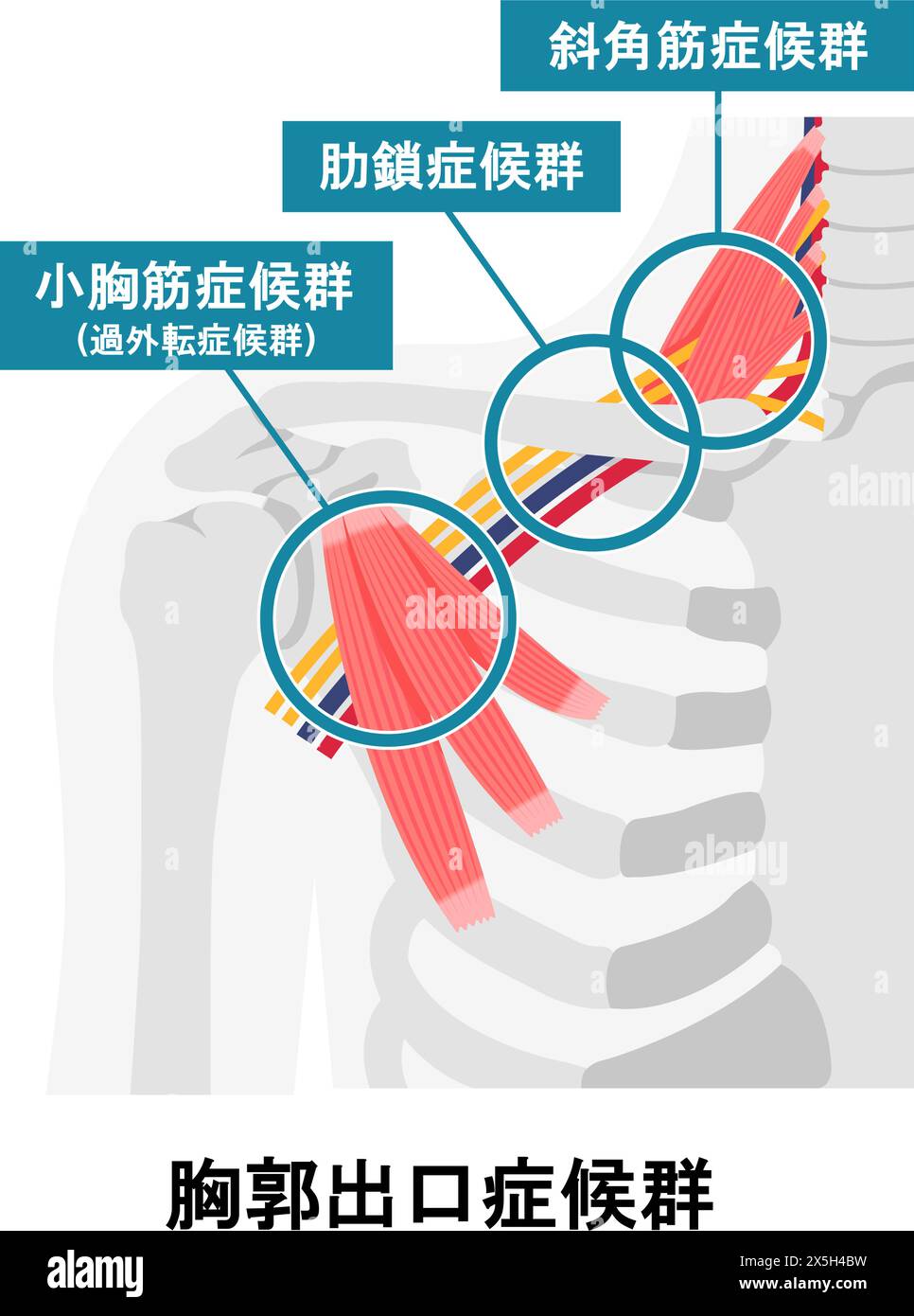
Thoracic nerve Cut Out Stock Images
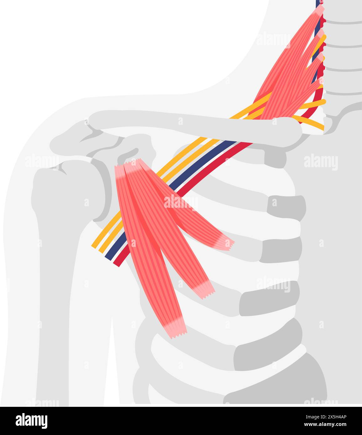 Vector illustration of where thoracic outlet syndrome occurs Stock Vectorhttps://www.alamy.com/image-license-details/?v=1https://www.alamy.com/vector-illustration-of-where-thoracic-outlet-syndrome-occurs-image605812782.html
RF2X5H4AP–Vector illustration of where thoracic outlet syndrome occurs
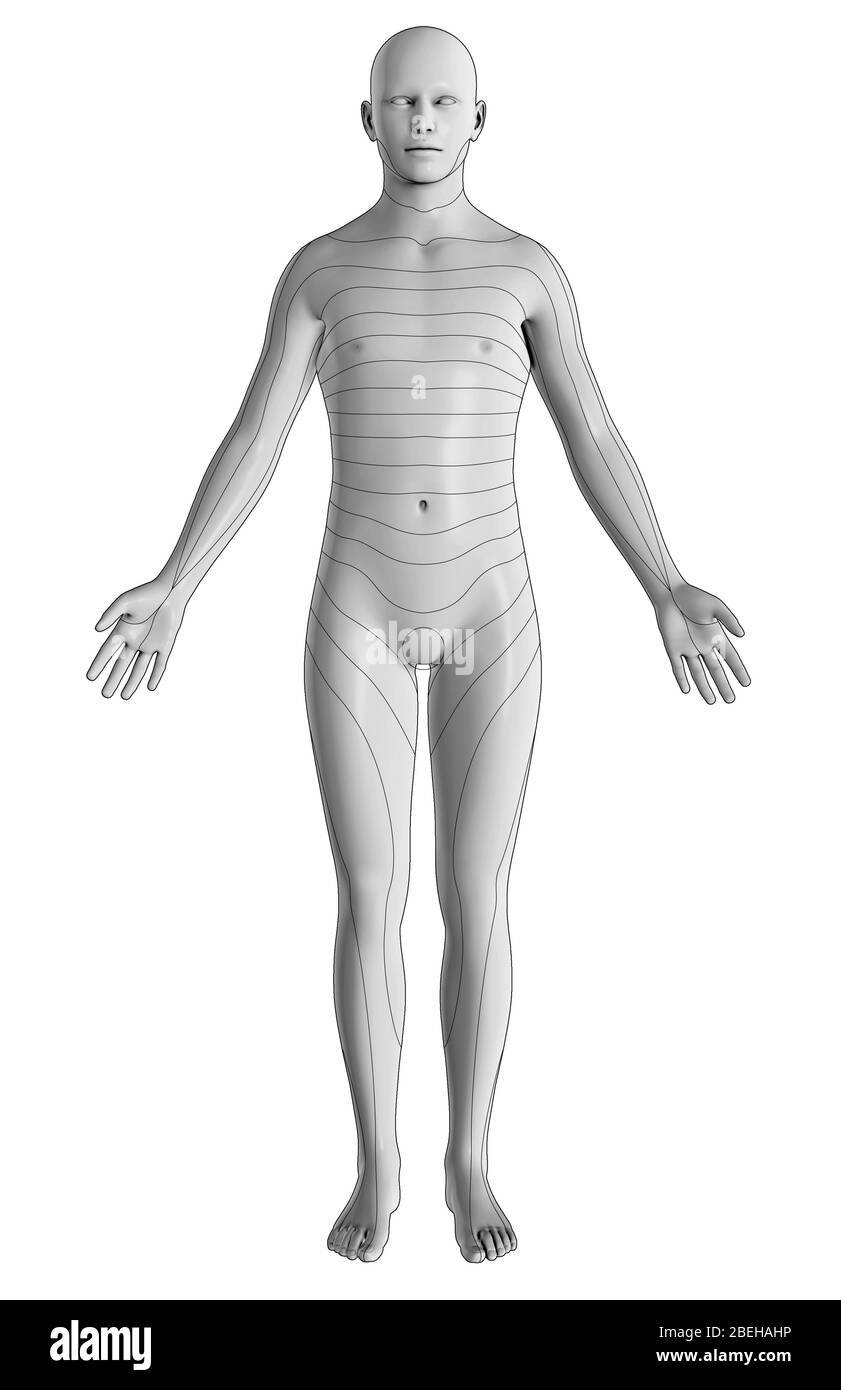 An illustration of the dermatomes of the body from an anterior view. Dermatomes are regions of skin supplied by specific spinal nerves, which relay sensory information to the brain. This includes eight cervical nerves (excludes C1), twelve thoracic nerves, five lumbar nerves, and five sacral nerves. Stock Photohttps://www.alamy.com/image-license-details/?v=1https://www.alamy.com/an-illustration-of-the-dermatomes-of-the-body-from-an-anterior-view-dermatomes-are-regions-of-skin-supplied-by-specific-spinal-nerves-which-relay-sensory-information-to-the-brain-this-includes-eight-cervical-nerves-excludes-c1-twelve-thoracic-nerves-five-lumbar-nerves-and-five-sacral-nerves-image353194066.html
RM2BEHAHP–An illustration of the dermatomes of the body from an anterior view. Dermatomes are regions of skin supplied by specific spinal nerves, which relay sensory information to the brain. This includes eight cervical nerves (excludes C1), twelve thoracic nerves, five lumbar nerves, and five sacral nerves.
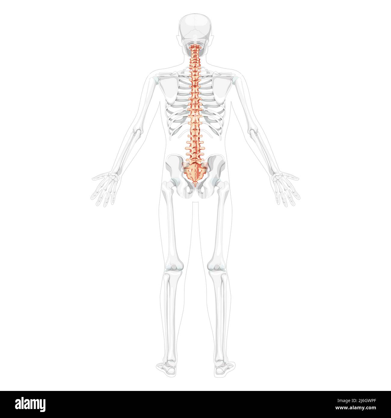 Human vertebral column back posterior view with partly transparent skeleton position, spinal cord, thoracic lumbar spine, sacrum and coccyx. Vector flat, realistic isolated illustration anatomy Stock Vectorhttps://www.alamy.com/image-license-details/?v=1https://www.alamy.com/human-vertebral-column-back-posterior-view-with-partly-transparent-skeleton-position-spinal-cord-thoracic-lumbar-spine-sacrum-and-coccyx-vector-flat-realistic-isolated-illustration-anatomy-image468739335.html
RF2J6GWPF–Human vertebral column back posterior view with partly transparent skeleton position, spinal cord, thoracic lumbar spine, sacrum and coccyx. Vector flat, realistic isolated illustration anatomy
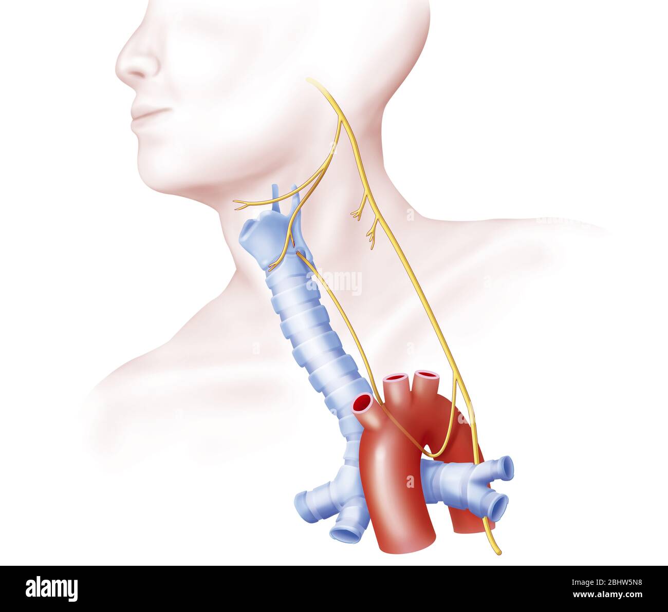 Upper and lower laryngeal nerves from the Wave nerg. Represented here on the left side, the laryngeal nerves come from the vagus nerve (10th pair of c Stock Photohttps://www.alamy.com/image-license-details/?v=1https://www.alamy.com/upper-and-lower-laryngeal-nerves-from-the-wave-nerg-represented-here-on-the-left-side-the-laryngeal-nerves-come-from-the-vagus-nerve-10th-pair-of-c-image355209828.html
RM2BHW5N8–Upper and lower laryngeal nerves from the Wave nerg. Represented here on the left side, the laryngeal nerves come from the vagus nerve (10th pair of c
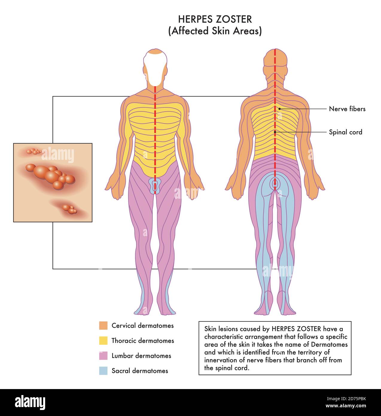 Medical diagram of affected skin areas of Herpes Zoster with annotations. Stock Vectorhttps://www.alamy.com/image-license-details/?v=1https://www.alamy.com/medical-diagram-of-affected-skin-areas-of-herpes-zoster-with-annotations-image383058023.html
RF2D75PBK–Medical diagram of affected skin areas of Herpes Zoster with annotations.
 The nerve supply of the upper body Stock Photohttps://www.alamy.com/image-license-details/?v=1https://www.alamy.com/stock-photo-the-nerve-supply-of-the-upper-body-13171690.html
RFACJM3R–The nerve supply of the upper body
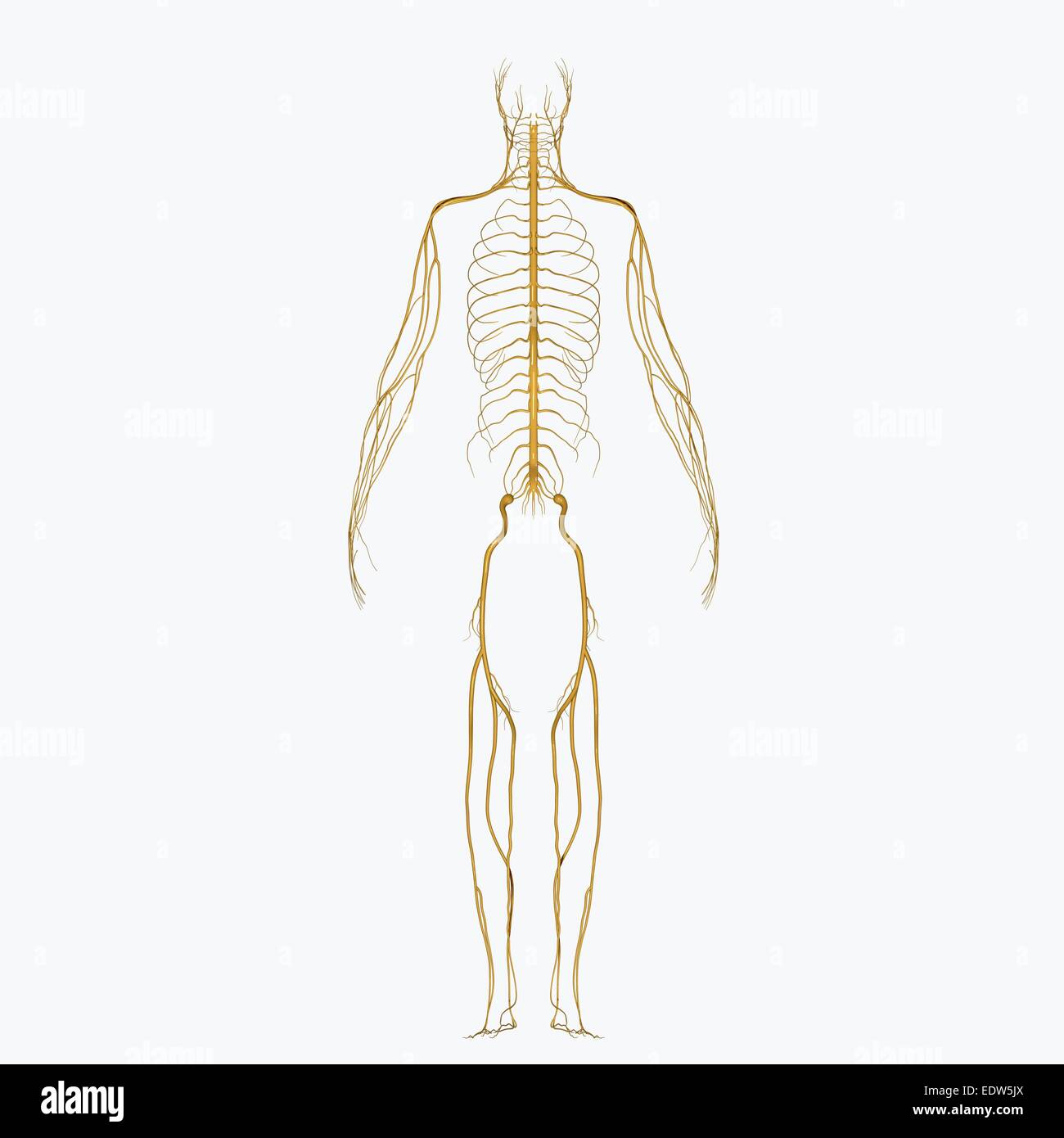 Nerves Stock Photohttps://www.alamy.com/image-license-details/?v=1https://www.alamy.com/stock-photo-nerves-77385250.html
RMEDW5JX–Nerves
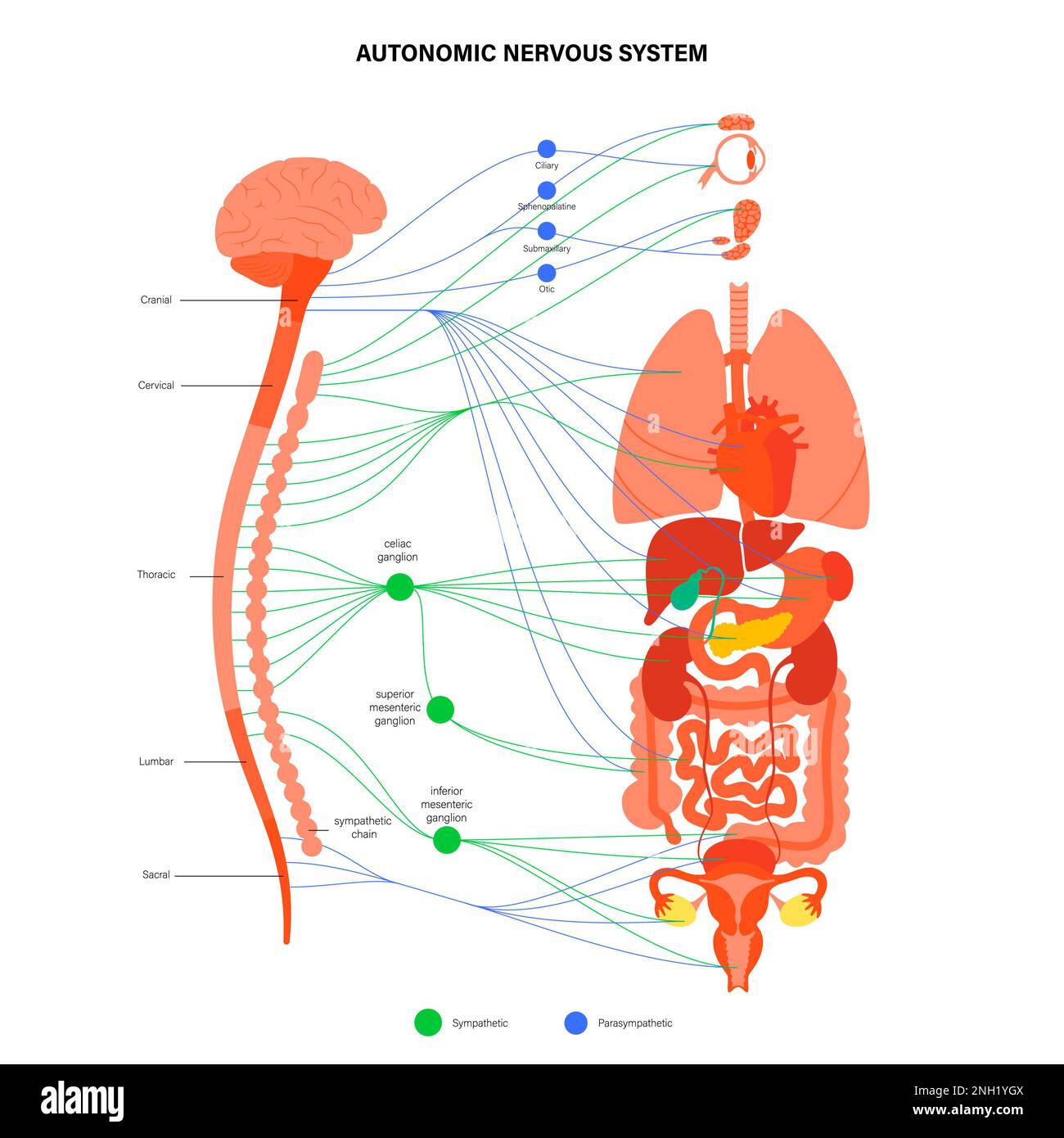
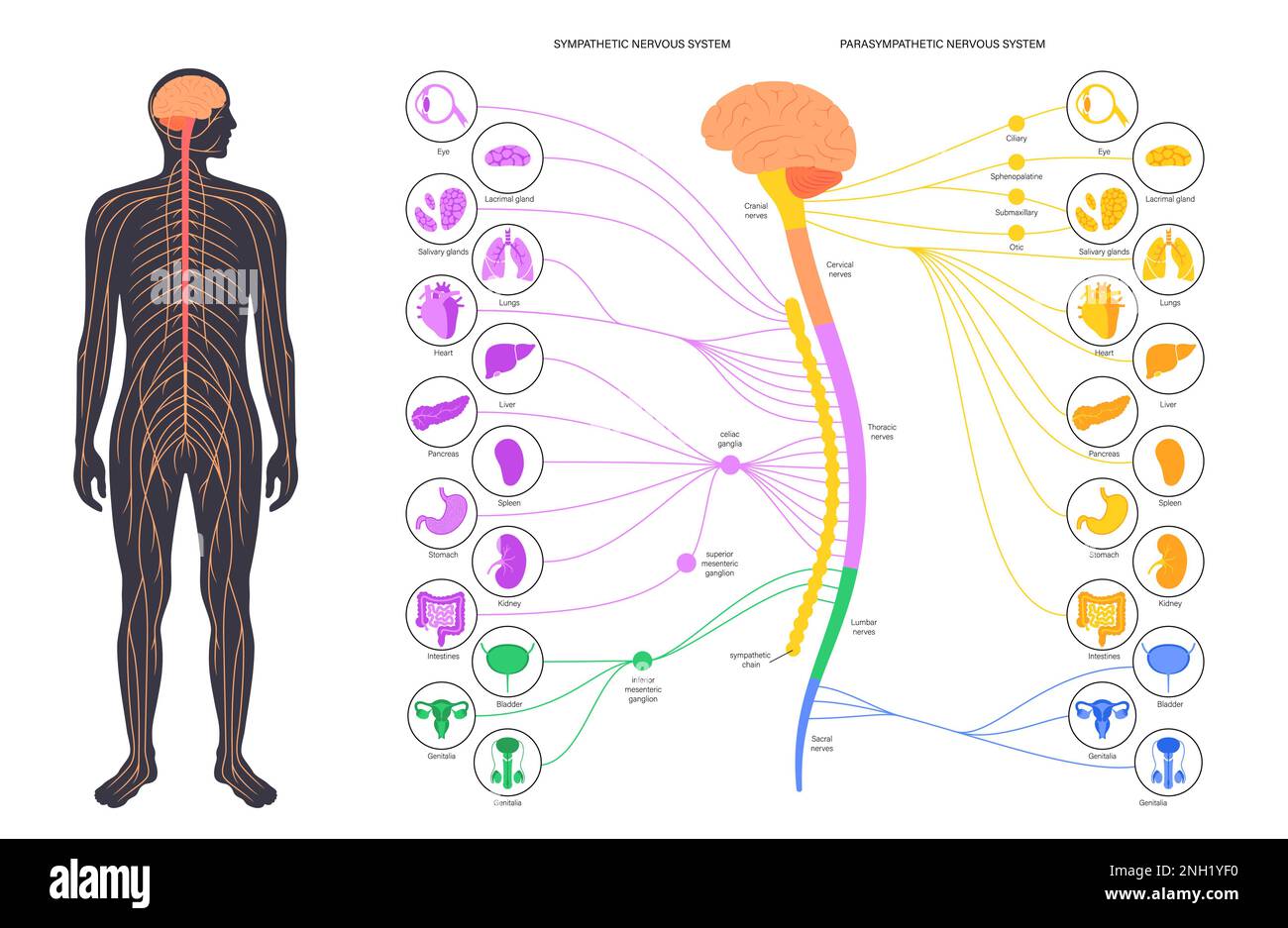 Autonomic nervous system, illustration Stock Photohttps://www.alamy.com/image-license-details/?v=1https://www.alamy.com/autonomic-nervous-system-illustration-image526803732.html
RF2NH1YF0–Autonomic nervous system, illustration
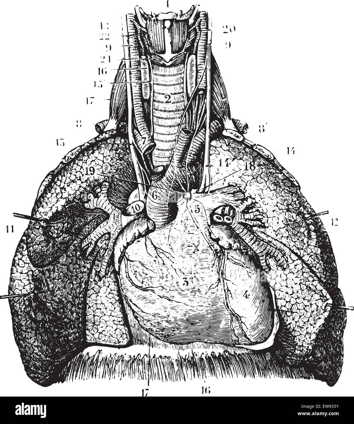 Main reports of the lungs. (Thoracic organs seen by their front face), vintage engraved illustration. Usual Medicine Dictionary Stock Vectorhttps://www.alamy.com/image-license-details/?v=1https://www.alamy.com/stock-photo-main-reports-of-the-lungs-thoracic-organs-seen-by-their-front-face-84407959.html
RFEW935Y–Main reports of the lungs. (Thoracic organs seen by their front face), vintage engraved illustration. Usual Medicine Dictionary
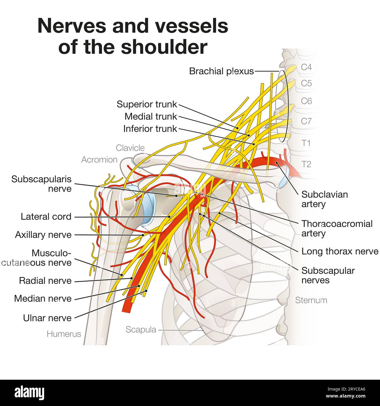 The shoulder region houses a complex network of nerves and vessels, including the brachial plexus, arteries, and veins, essential for limb innervation Stock Photohttps://www.alamy.com/image-license-details/?v=1https://www.alamy.com/the-shoulder-region-houses-a-complex-network-of-nerves-and-vessels-including-the-brachial-plexus-arteries-and-veins-essential-for-limb-innervation-image567602174.html
RF2RYCEA6–The shoulder region houses a complex network of nerves and vessels, including the brachial plexus, arteries, and veins, essential for limb innervation
 Dorsal vertebra Stock Photohttps://www.alamy.com/image-license-details/?v=1https://www.alamy.com/dorsal-vertebra-image407105441.html
RM2EJ9741–Dorsal vertebra
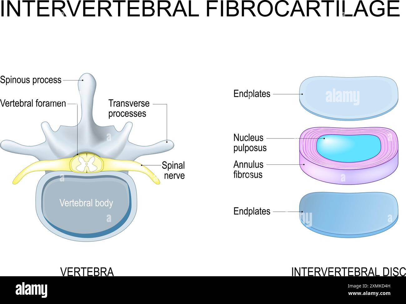 Intervertebral disc structure from Nucleus pulposus and Annulus fibrosus to Endplates. Vertebra anatomy. Spinal Column. Vector illustration Stock Vectorhttps://www.alamy.com/image-license-details/?v=1https://www.alamy.com/intervertebral-disc-structure-from-nucleus-pulposus-and-annulus-fibrosus-to-endplates-vertebra-anatomy-spinal-column-vector-illustration-image615083409.html
RF2XMKD4H–Intervertebral disc structure from Nucleus pulposus and Annulus fibrosus to Endplates. Vertebra anatomy. Spinal Column. Vector illustration
RF2HJEWA4–Spinal Simple vector icon. Illustration symbol design template for web mobile UI element.
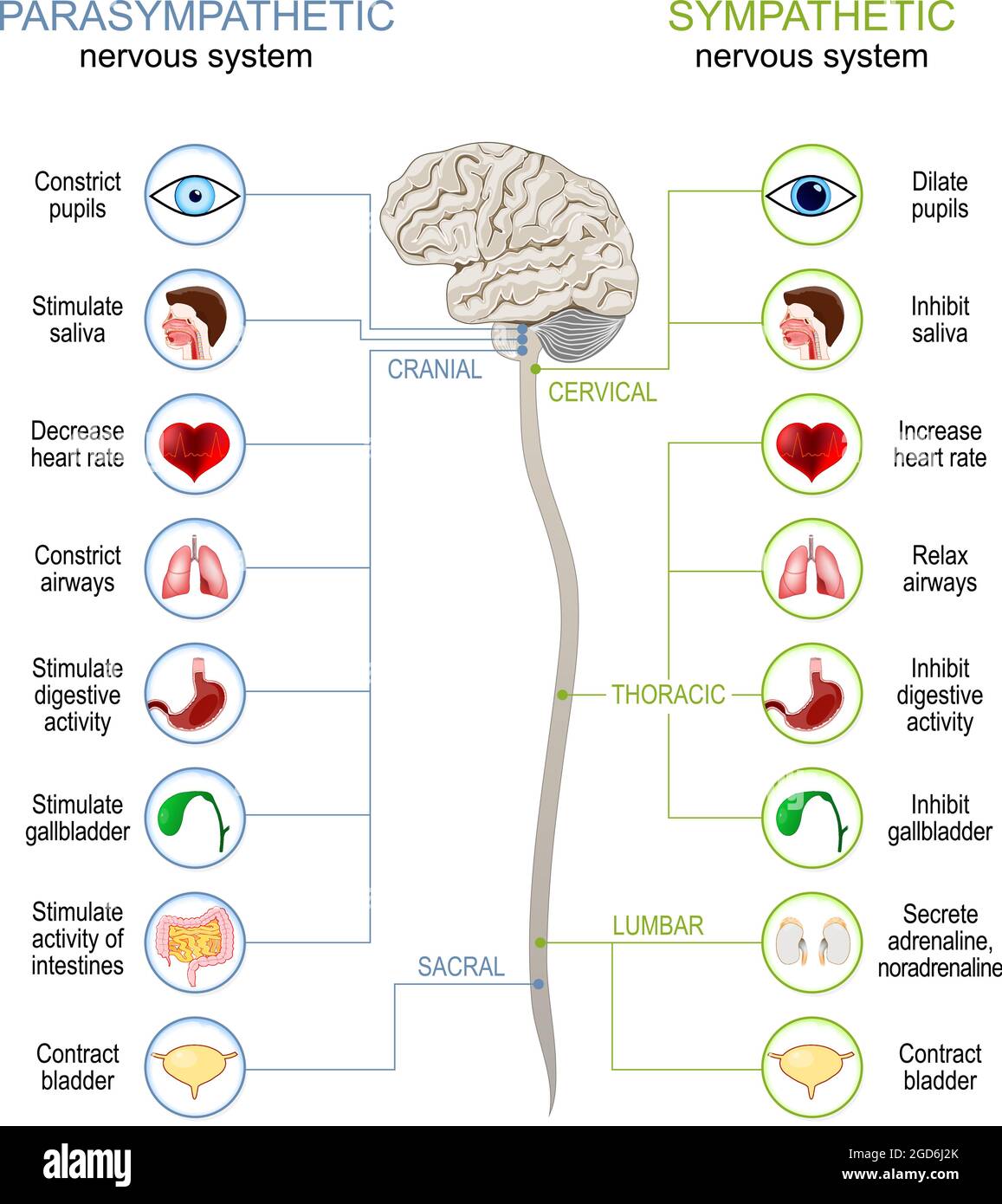 Sympathetic And Parasympathetic Nervous System. Difference. diagram with connected inner organs, brain and spinal cord. vector illustration Stock Vectorhttps://www.alamy.com/image-license-details/?v=1https://www.alamy.com/sympathetic-and-parasympathetic-nervous-system-difference-diagram-with-connected-inner-organs-brain-and-spinal-cord-vector-illustration-image438395627.html
RF2GD6J2K–Sympathetic And Parasympathetic Nervous System. Difference. diagram with connected inner organs, brain and spinal cord. vector illustration
 Spine diagram showing back pain Stock Vectorhttps://www.alamy.com/image-license-details/?v=1https://www.alamy.com/spine-diagram-showing-back-pain-image444651841.html
RF2GRBHXW–Spine diagram showing back pain
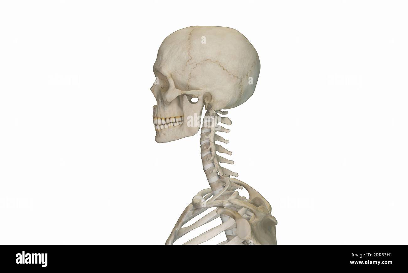 Side view of cervical section of spine Stock Photohttps://www.alamy.com/image-license-details/?v=1https://www.alamy.com/side-view-of-cervical-section-of-spine-image564937549.html
RF2RR33H1–Side view of cervical section of spine
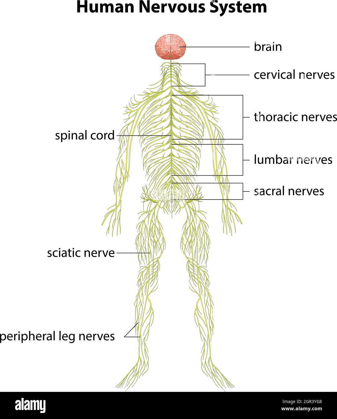 Human nervous system Stock Vectorhttps://www.alamy.com/image-license-details/?v=1https://www.alamy.com/human-nervous-system-image444483768.html
RF2GR3YG8–Human nervous system
 Spinal cord and pelvis Stock Photohttps://www.alamy.com/image-license-details/?v=1https://www.alamy.com/stock-photo-spinal-cord-and-pelvis-77385817.html
RFEDW6B5–Spinal cord and pelvis
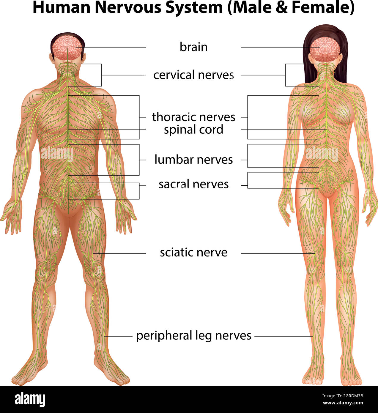 Human nervous system Stock Vectorhttps://www.alamy.com/image-license-details/?v=1https://www.alamy.com/human-nervous-system-image444697439.html
RF2GRDM3B–Human nervous system
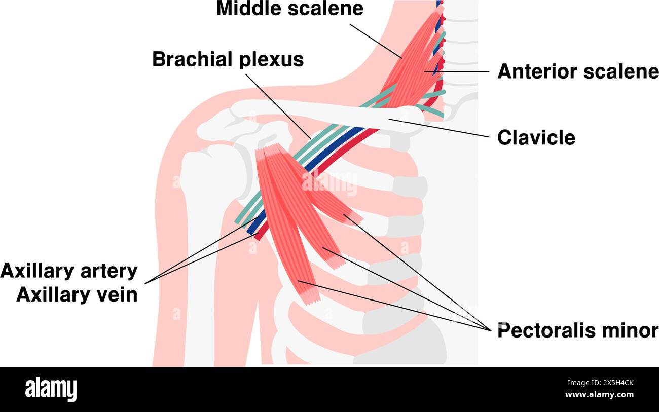 Vector illustration of where thoracic outlet syndrome occurs Stock Vectorhttps://www.alamy.com/image-license-details/?v=1https://www.alamy.com/vector-illustration-of-where-thoracic-outlet-syndrome-occurs-image605812835.html
RF2X5H4CK–Vector illustration of where thoracic outlet syndrome occurs
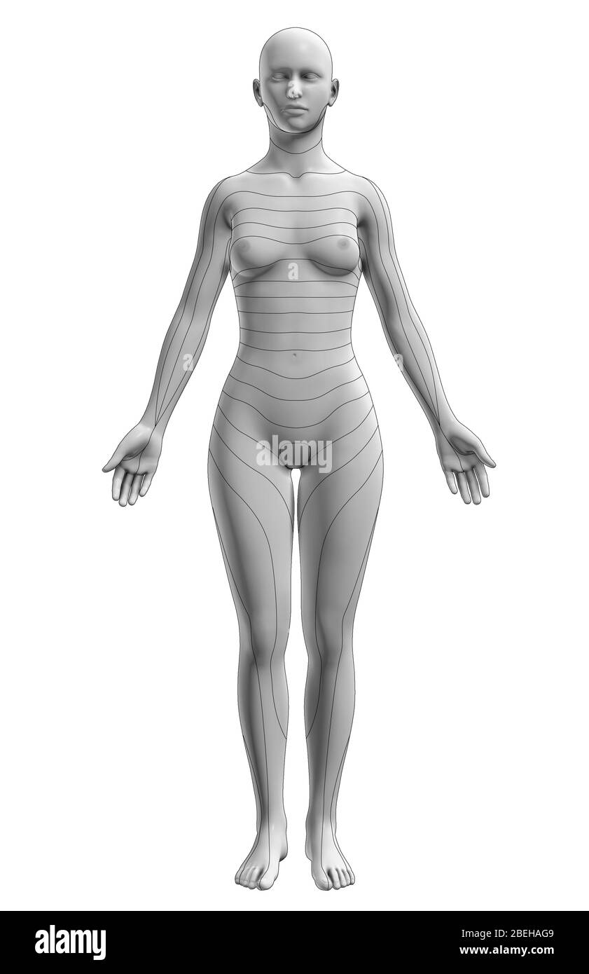 Dermatomes, Anterior View Stock Photohttps://www.alamy.com/image-license-details/?v=1https://www.alamy.com/dermatomes-anterior-view-image353194025.html
RM2BEHAG9–Dermatomes, Anterior View
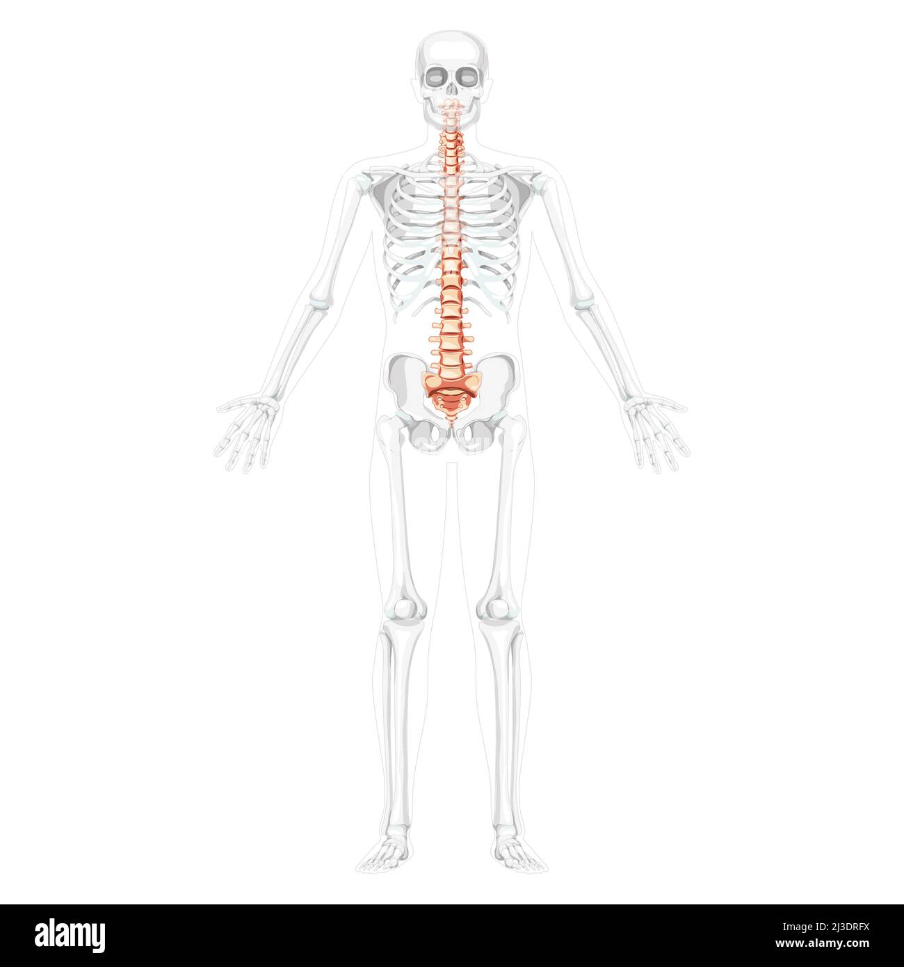 Human vertebral column front anterior view with partly transparent skeleton position, thoracic lumbar spine, sacrum and coccyx. Vector flat natural colors, realistic isolated illustration anatomy Stock Vectorhttps://www.alamy.com/image-license-details/?v=1https://www.alamy.com/human-vertebral-column-front-anterior-view-with-partly-transparent-skeleton-position-thoracic-lumbar-spine-sacrum-and-coccyx-vector-flat-natural-colors-realistic-isolated-illustration-anatomy-image466827758.html
RF2J3DRFX–Human vertebral column front anterior view with partly transparent skeleton position, thoracic lumbar spine, sacrum and coccyx. Vector flat natural colors, realistic isolated illustration anatomy
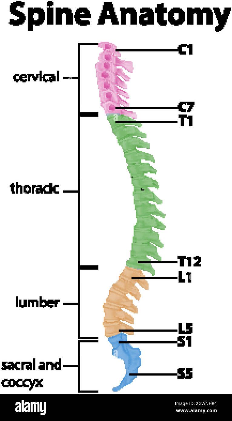 Anatomy of the spine or spinal curves infographic Stock Vectorhttps://www.alamy.com/image-license-details/?v=1https://www.alamy.com/anatomy-of-the-spine-or-spinal-curves-infographic-image446100568.html
RF2GWNHR4–Anatomy of the spine or spinal curves infographic
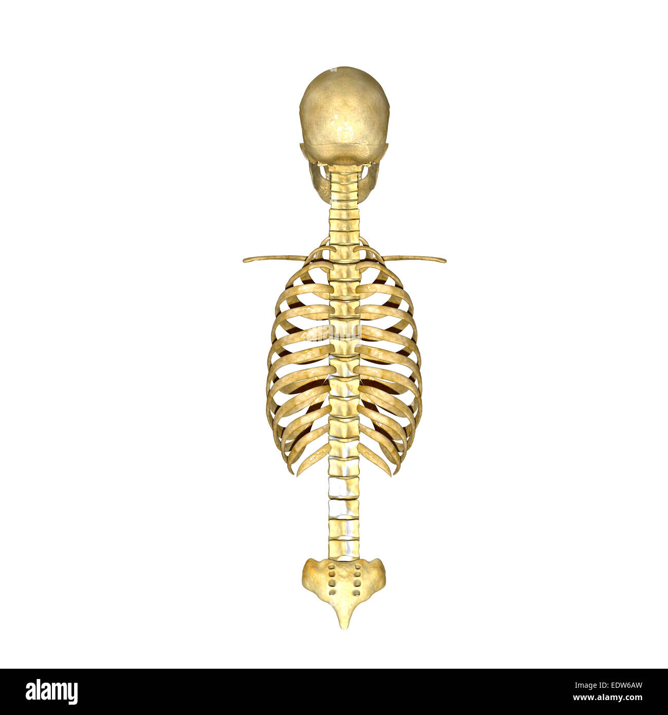 Rib cage spinal cord Stock Photohttps://www.alamy.com/image-license-details/?v=1https://www.alamy.com/stock-photo-rib-cage-spinal-cord-77385809.html
RFEDW6AW–Rib cage spinal cord
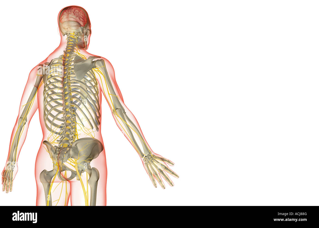 The nerve supply of the upper body Stock Photohttps://www.alamy.com/image-license-details/?v=1https://www.alamy.com/stock-photo-the-nerve-supply-of-the-upper-body-13167711.html
RFACJ88G–The nerve supply of the upper body
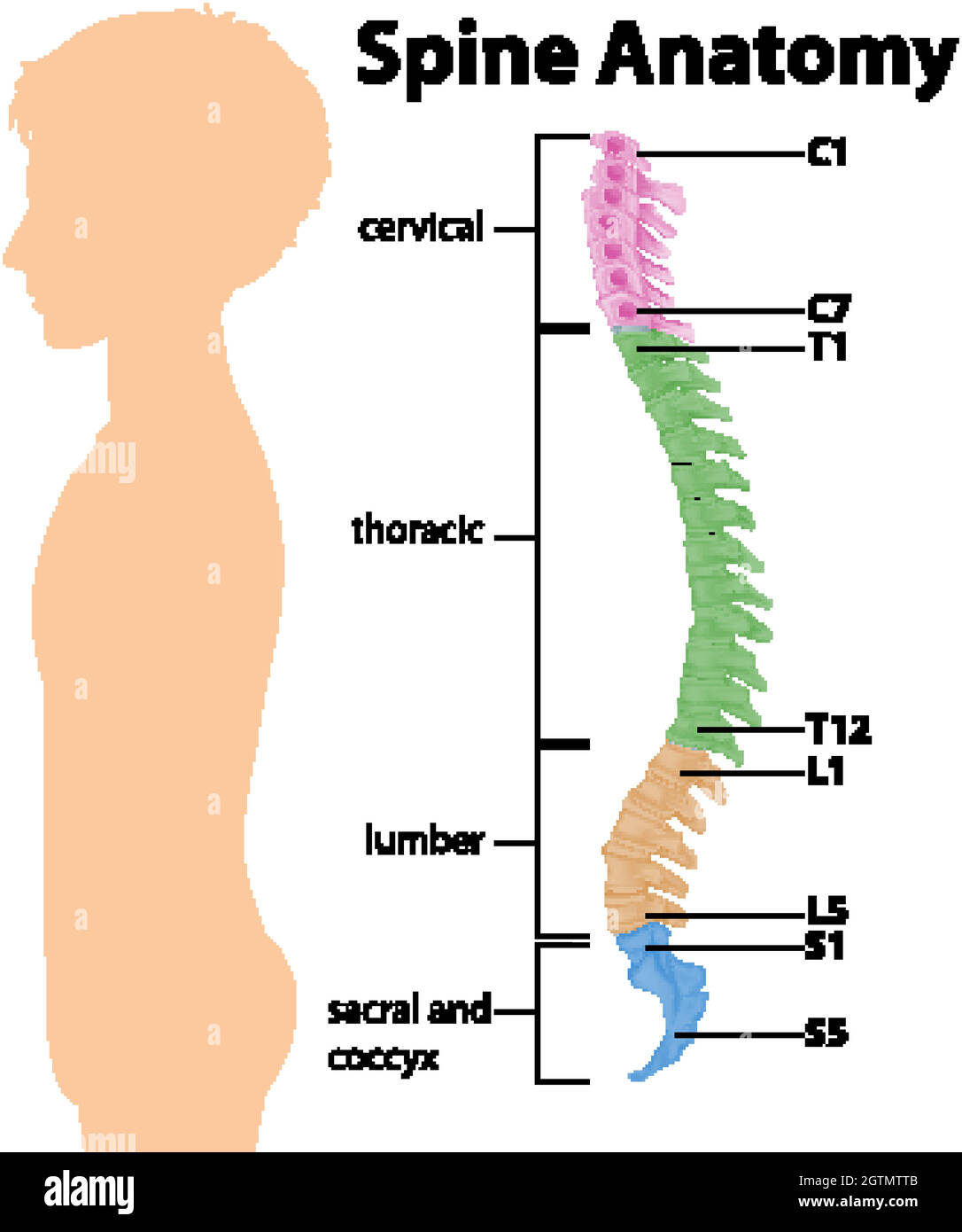 Anatomy of the spine or spinal curves infographic Stock Vectorhttps://www.alamy.com/image-license-details/?v=1https://www.alamy.com/anatomy-of-the-spine-or-spinal-curves-infographic-image445469483.html
RF2GTMTTB–Anatomy of the spine or spinal curves infographic
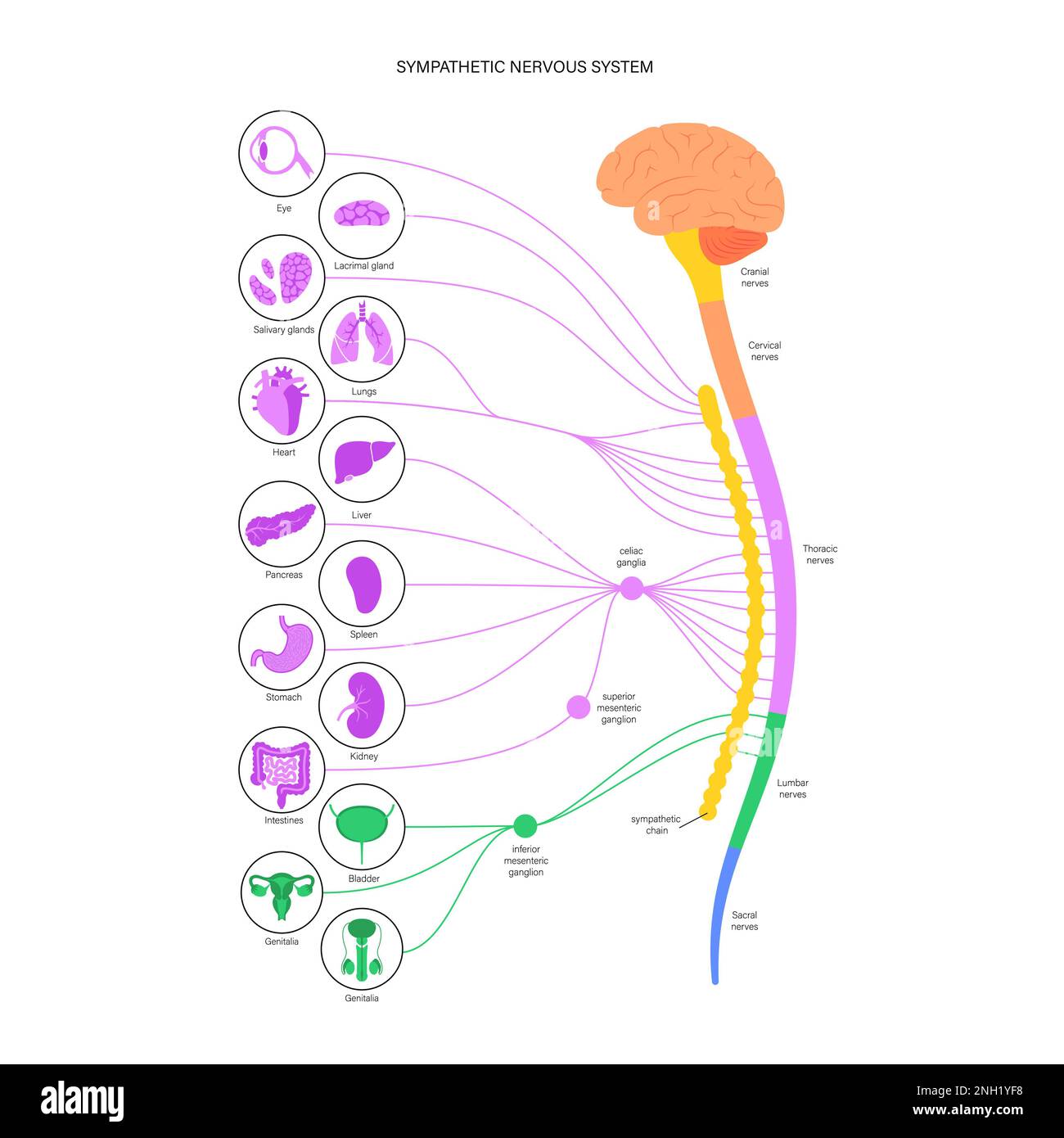 Sympathetic nervous system, illustration Stock Photohttps://www.alamy.com/image-license-details/?v=1https://www.alamy.com/sympathetic-nervous-system-illustration-image526803740.html
RF2NH1YF8–Sympathetic nervous system, illustration
 Main reports of the lungs. (Thoracic organs seen by their front face), vintage engraved illustration. Usual Medicine Dictionary Stock Vectorhttps://www.alamy.com/image-license-details/?v=1https://www.alamy.com/stock-photo-main-reports-of-the-lungs-thoracic-organs-seen-by-their-front-face-84419982.html
RFEW9JFA–Main reports of the lungs. (Thoracic organs seen by their front face), vintage engraved illustration. Usual Medicine Dictionary
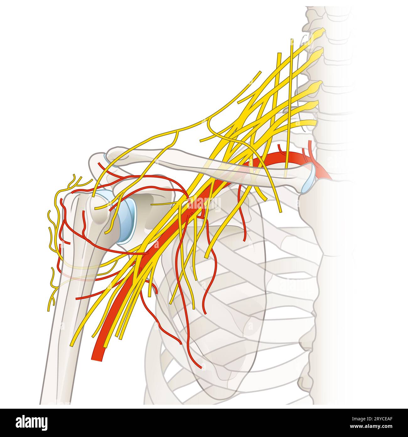 The shoulder region houses a complex network of nerves and vessels, including the brachial plexus, arteries, and veins, essential for limb innervation Stock Photohttps://www.alamy.com/image-license-details/?v=1https://www.alamy.com/the-shoulder-region-houses-a-complex-network-of-nerves-and-vessels-including-the-brachial-plexus-arteries-and-veins-essential-for-limb-innervation-image567602183.html
RF2RYCEAF–The shoulder region houses a complex network of nerves and vessels, including the brachial plexus, arteries, and veins, essential for limb innervation
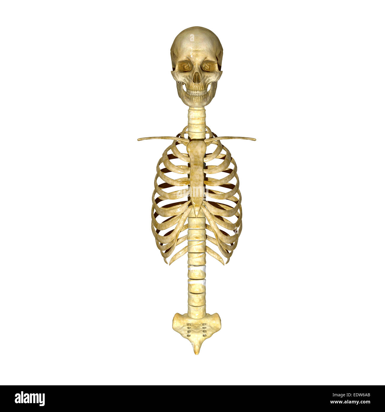 Rib cage, skull and spinal cord Stock Photohttps://www.alamy.com/image-license-details/?v=1https://www.alamy.com/stock-photo-rib-cage-skull-and-spinal-cord-77385795.html
RFEDW6AB–Rib cage, skull and spinal cord
 asian woman Stock Photohttps://www.alamy.com/image-license-details/?v=1https://www.alamy.com/asian-woman-image343651766.html
RM2AY2K9A–asian woman
 The development of the human body; a manual of human embryology . 8.—Diagram showing the CutaneousDistribution of the Spinal Nerves.—{Head.) St 164 THE DEVELOPMENT OF THE HUMAN BODY. more or less distinct zones, and being therefore segmented(Pig. 78). But a considerable commingling of adjacentdermatomes has also occurred. Thus, while the distributionof the cutaneous branches of the fourth thoracic nerve, asdetermined experimentally in the monkey (Macacus), is dis-tinctly zonal or segmental, the nipple lying practically in themiddle line of the zone, the upper half of its area is also suppliedo Stock Photohttps://www.alamy.com/image-license-details/?v=1https://www.alamy.com/the-development-of-the-human-body-a-manual-of-human-embryology-8diagram-showing-the-cutaneousdistribution-of-the-spinal-nerveshead-st-164-the-development-of-the-human-body-more-or-less-distinct-zones-and-being-therefore-segmentedpig-78-but-a-considerable-commingling-of-adjacentdermatomes-has-also-occurred-thus-while-the-distributionof-the-cutaneous-branches-of-the-fourth-thoracic-nerve-asdetermined-experimentally-in-the-monkey-macacus-is-dis-tinctly-zonal-or-segmental-the-nipple-lying-practically-in-themiddle-line-of-the-zone-the-upper-half-of-its-area-is-also-suppliedo-image342706527.html
RM2AWFHJR–The development of the human body; a manual of human embryology . 8.—Diagram showing the CutaneousDistribution of the Spinal Nerves.—{Head.) St 164 THE DEVELOPMENT OF THE HUMAN BODY. more or less distinct zones, and being therefore segmented(Pig. 78). But a considerable commingling of adjacentdermatomes has also occurred. Thus, while the distributionof the cutaneous branches of the fourth thoracic nerve, asdetermined experimentally in the monkey (Macacus), is dis-tinctly zonal or segmental, the nipple lying practically in themiddle line of the zone, the upper half of its area is also suppliedo
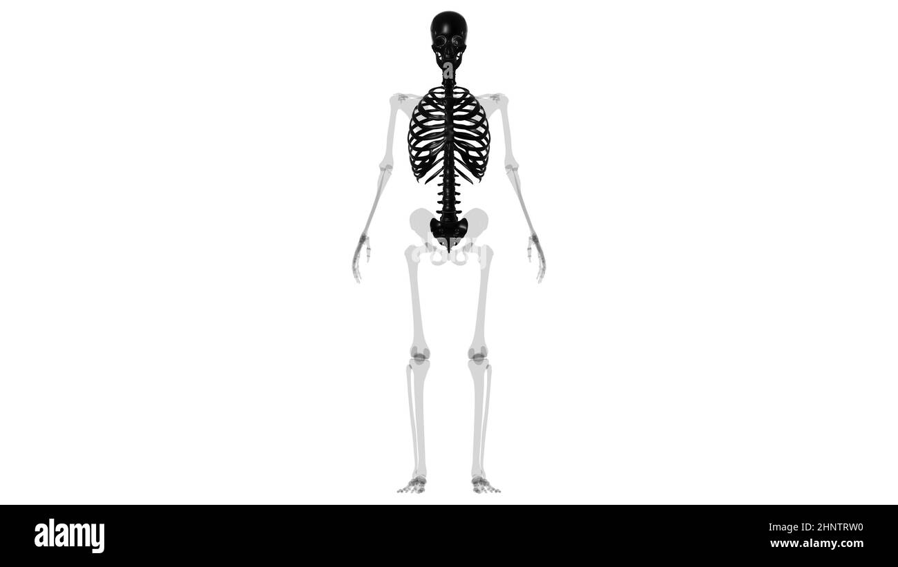 Human Skeleton Axial Skeleton Anatomy 3D Illustration Stock Photohttps://www.alamy.com/image-license-details/?v=1https://www.alamy.com/human-skeleton-axial-skeleton-anatomy-3d-illustration-image460922924.html
RF2HNTRW0–Human Skeleton Axial Skeleton Anatomy 3D Illustration
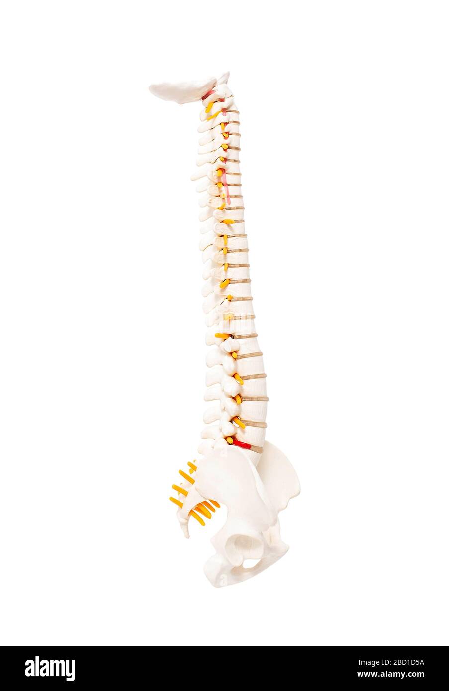 Mock up human spine on a white background. The concept of segments and divisions of the spine, the structure and anatomy of the bone marrow, nerves an Stock Photohttps://www.alamy.com/image-license-details/?v=1https://www.alamy.com/mock-up-human-spine-on-a-white-background-the-concept-of-segments-and-divisions-of-the-spine-the-structure-and-anatomy-of-the-bone-marrow-nerves-an-image352230182.html
RF2BD1D5A–Mock up human spine on a white background. The concept of segments and divisions of the spine, the structure and anatomy of the bone marrow, nerves an
 Nervous system nerve body system anatomical internal organ graphic illustration Stock Vectorhttps://www.alamy.com/image-license-details/?v=1https://www.alamy.com/nervous-system-nerve-body-system-anatomical-internal-organ-graphic-illustration-image482395821.html
RF2K0R0P5–Nervous system nerve body system anatomical internal organ graphic illustration
 Third thoracic vertebrae (T3): The thoracic spinal nerve 3 passes through underneath T3 3d illustration Stock Photohttps://www.alamy.com/image-license-details/?v=1https://www.alamy.com/third-thoracic-vertebrae-t3-the-thoracic-spinal-nerve-3-passes-through-underneath-t3-3d-illustration-image596578093.html
RF2WJGDCD–Third thoracic vertebrae (T3): The thoracic spinal nerve 3 passes through underneath T3 3d illustration
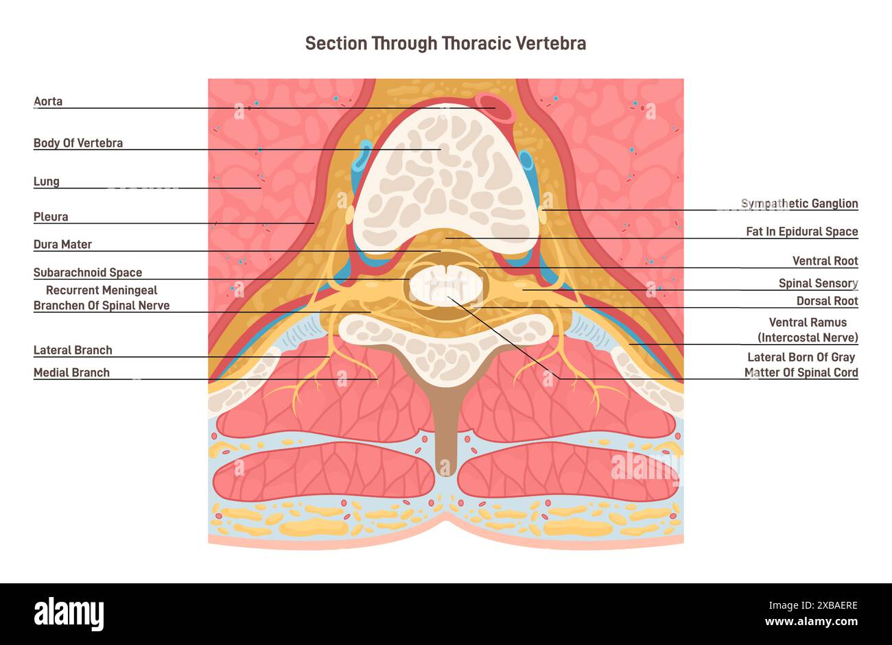 Cross section through thoracic vertebra. Spinal cord anatomy. Middle segment of the vertebral column with nerves, blood vessels, muscle and fat tissue. Human anatomy banner. Flat vector illustration Stock Vectorhttps://www.alamy.com/image-license-details/?v=1https://www.alamy.com/cross-section-through-thoracic-vertebra-spinal-cord-anatomy-middle-segment-of-the-vertebral-column-with-nerves-blood-vessels-muscle-and-fat-tissue-human-anatomy-banner-flat-vector-illustration-image609355250.html
RF2XBAERE–Cross section through thoracic vertebra. Spinal cord anatomy. Middle segment of the vertebral column with nerves, blood vessels, muscle and fat tissue. Human anatomy banner. Flat vector illustration
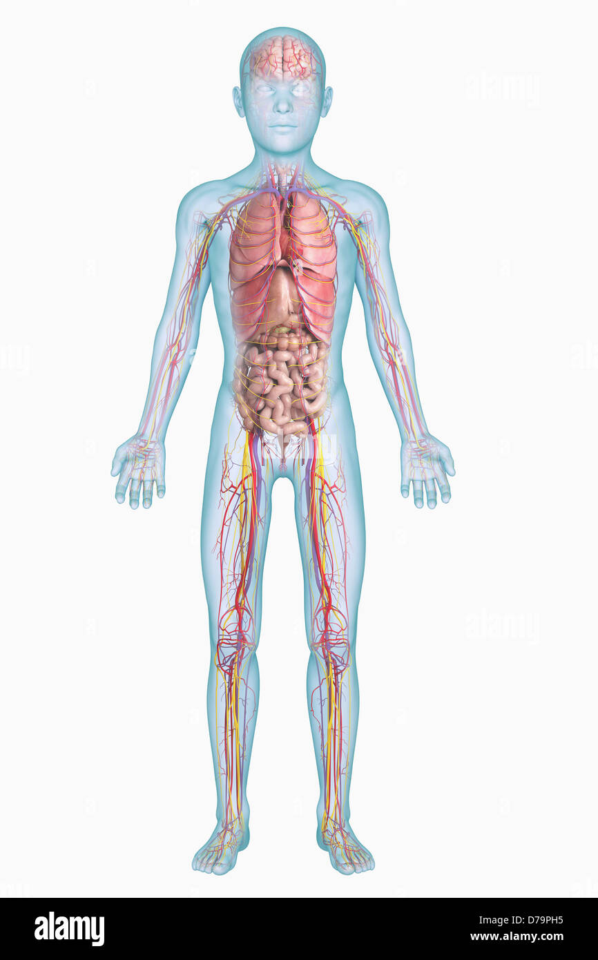 Internal Anatomy Pre-Adolescent) Stock Photohttps://www.alamy.com/image-license-details/?v=1https://www.alamy.com/stock-photo-internal-anatomy-pre-adolescent-56148993.html
RMD79PH5–Internal Anatomy Pre-Adolescent)
 Vector illustration of where thoracic outlet syndrome occurs Stock Vectorhttps://www.alamy.com/image-license-details/?v=1https://www.alamy.com/vector-illustration-of-where-thoracic-outlet-syndrome-occurs-image605812910.html
RF2X5H4FA–Vector illustration of where thoracic outlet syndrome occurs
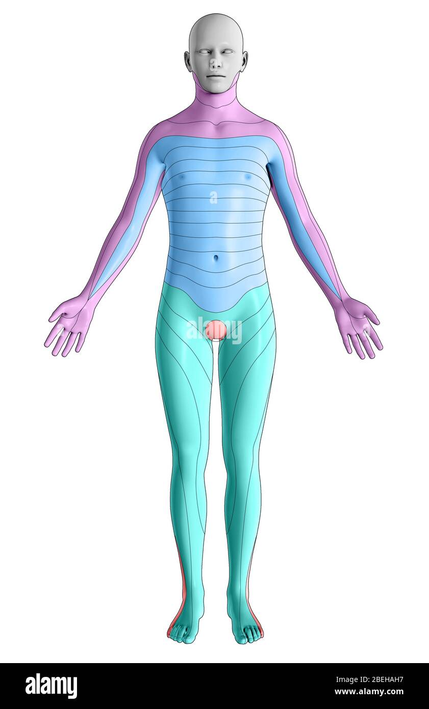 Dermatomes, Anterior View Stock Photohttps://www.alamy.com/image-license-details/?v=1https://www.alamy.com/dermatomes-anterior-view-image353194051.html
RM2BEHAH7–Dermatomes, Anterior View
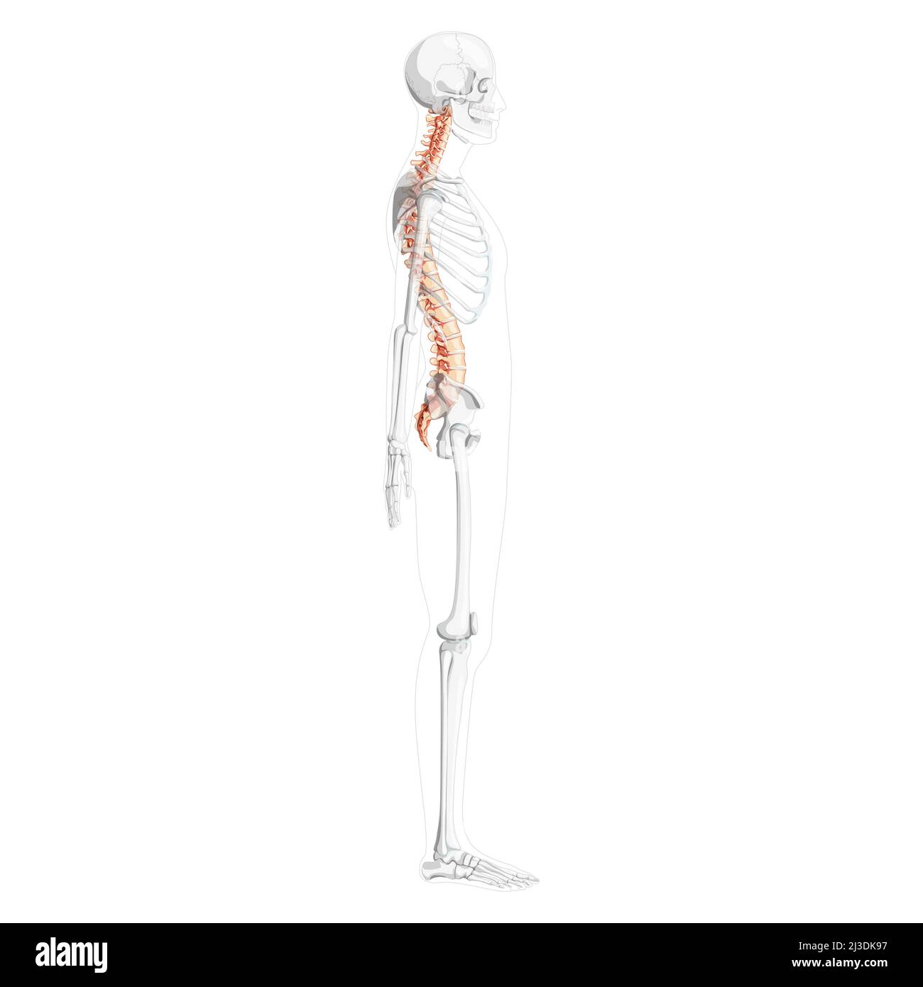 Human vertebral column side lateral view with partly transparent skeleton position, spinal cord, thoracic lumbar spine, sacrum. Vector flat natural colors, realistic isolated illustration anatomy Stock Vectorhttps://www.alamy.com/image-license-details/?v=1https://www.alamy.com/human-vertebral-column-side-lateral-view-with-partly-transparent-skeleton-position-spinal-cord-thoracic-lumbar-spine-sacrum-vector-flat-natural-colors-realistic-isolated-illustration-anatomy-image466824435.html
RF2J3DK97–Human vertebral column side lateral view with partly transparent skeleton position, spinal cord, thoracic lumbar spine, sacrum. Vector flat natural colors, realistic isolated illustration anatomy
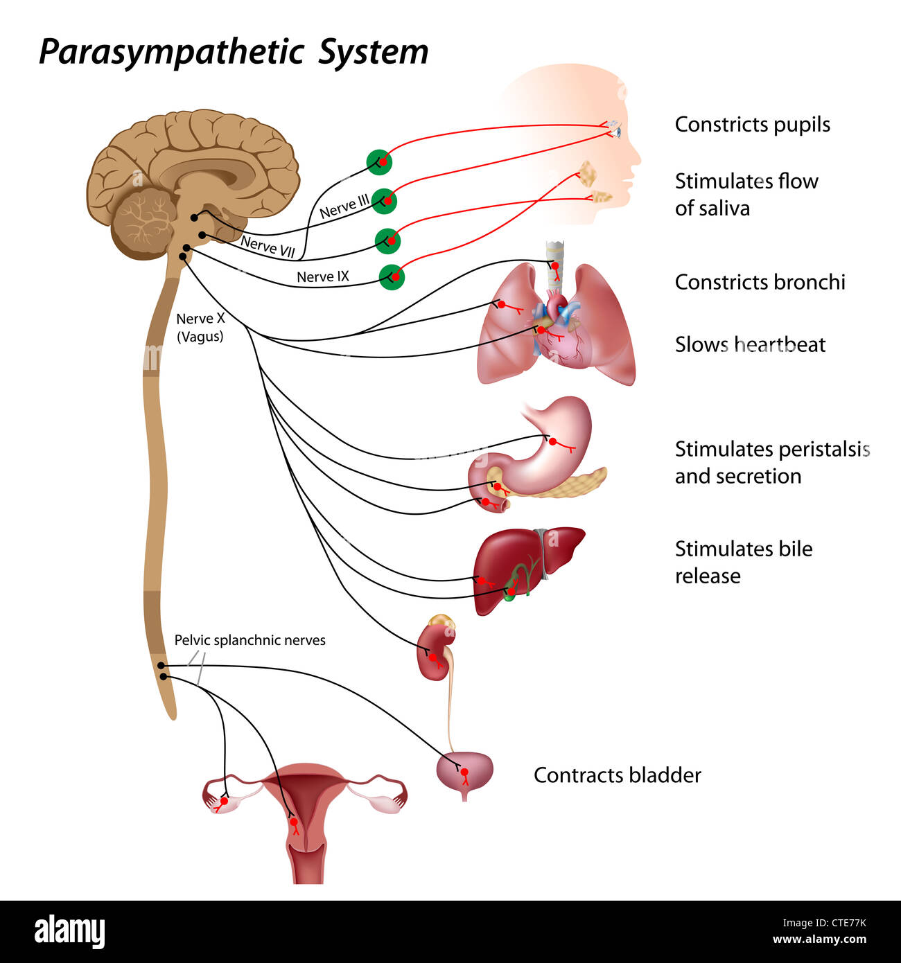 Parasympathetic pathway of the ANS Stock Photohttps://www.alamy.com/image-license-details/?v=1https://www.alamy.com/stock-photo-parasympathetic-pathway-of-the-ans-49485511.html
RFCTE77K–Parasympathetic pathway of the ANS
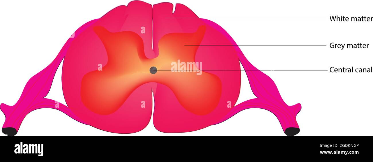 Anatomy of the Spinal Cord, The spinal cord, detailed structure of spinal cord, Anatomy of spinal cord, Labeled anatomy of spinal cord Stock Vectorhttps://www.alamy.com/image-license-details/?v=1https://www.alamy.com/anatomy-of-the-spinal-cord-the-spinal-cord-detailed-structure-of-spinal-cord-anatomy-of-spinal-cord-labeled-anatomy-of-spinal-cord-image438683750.html
RF2GDKNGP–Anatomy of the Spinal Cord, The spinal cord, detailed structure of spinal cord, Anatomy of spinal cord, Labeled anatomy of spinal cord
 The nerve supply of the upper body Stock Photohttps://www.alamy.com/image-license-details/?v=1https://www.alamy.com/stock-photo-the-nerve-supply-of-the-upper-body-13167849.html
RFACJ8KP–The nerve supply of the upper body
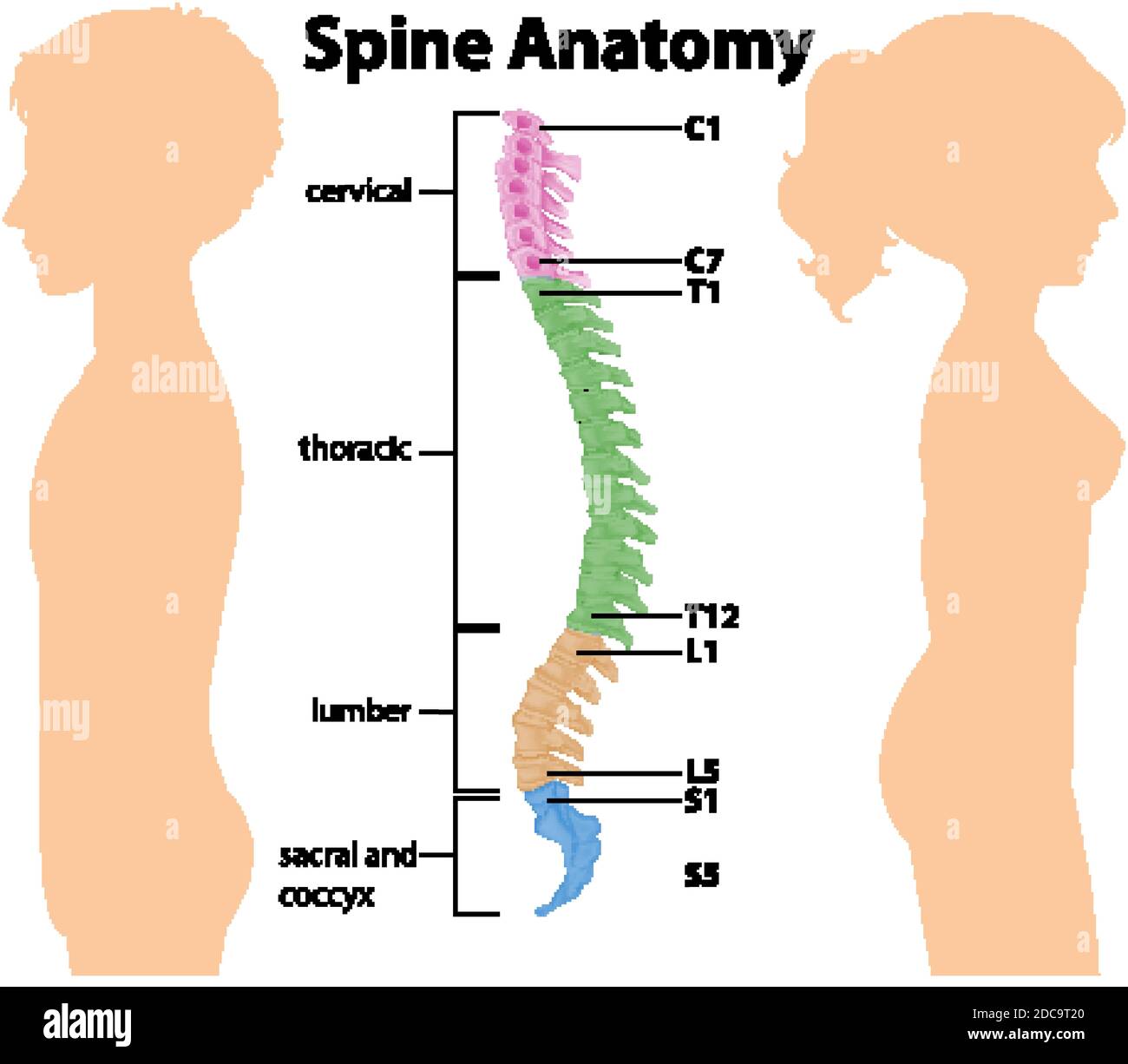 Anatomy of the spine or spinal curves infographic illustration Stock Vectorhttps://www.alamy.com/image-license-details/?v=1https://www.alamy.com/anatomy-of-the-spine-or-spinal-curves-infographic-illustration-image386220408.html
RF2DC9T20–Anatomy of the spine or spinal curves infographic illustration
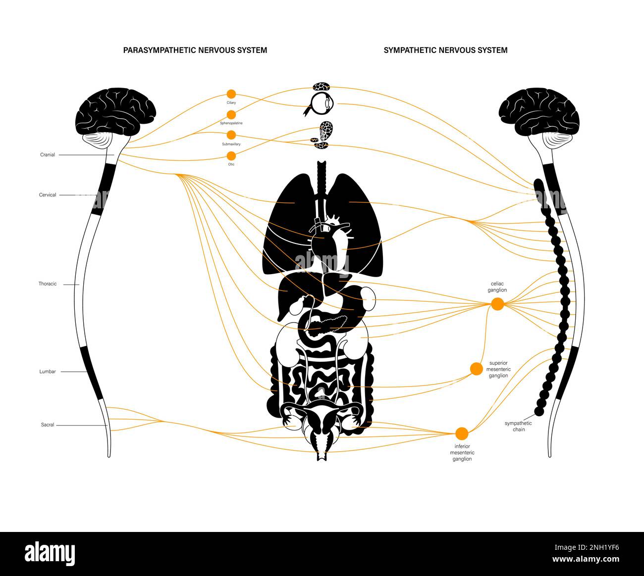 Autonomic nervous system, illustration Stock Photohttps://www.alamy.com/image-license-details/?v=1https://www.alamy.com/autonomic-nervous-system-illustration-image526803738.html
RF2NH1YF6–Autonomic nervous system, illustration
RF2ATDAX9–Spinal diagram icon, outline style
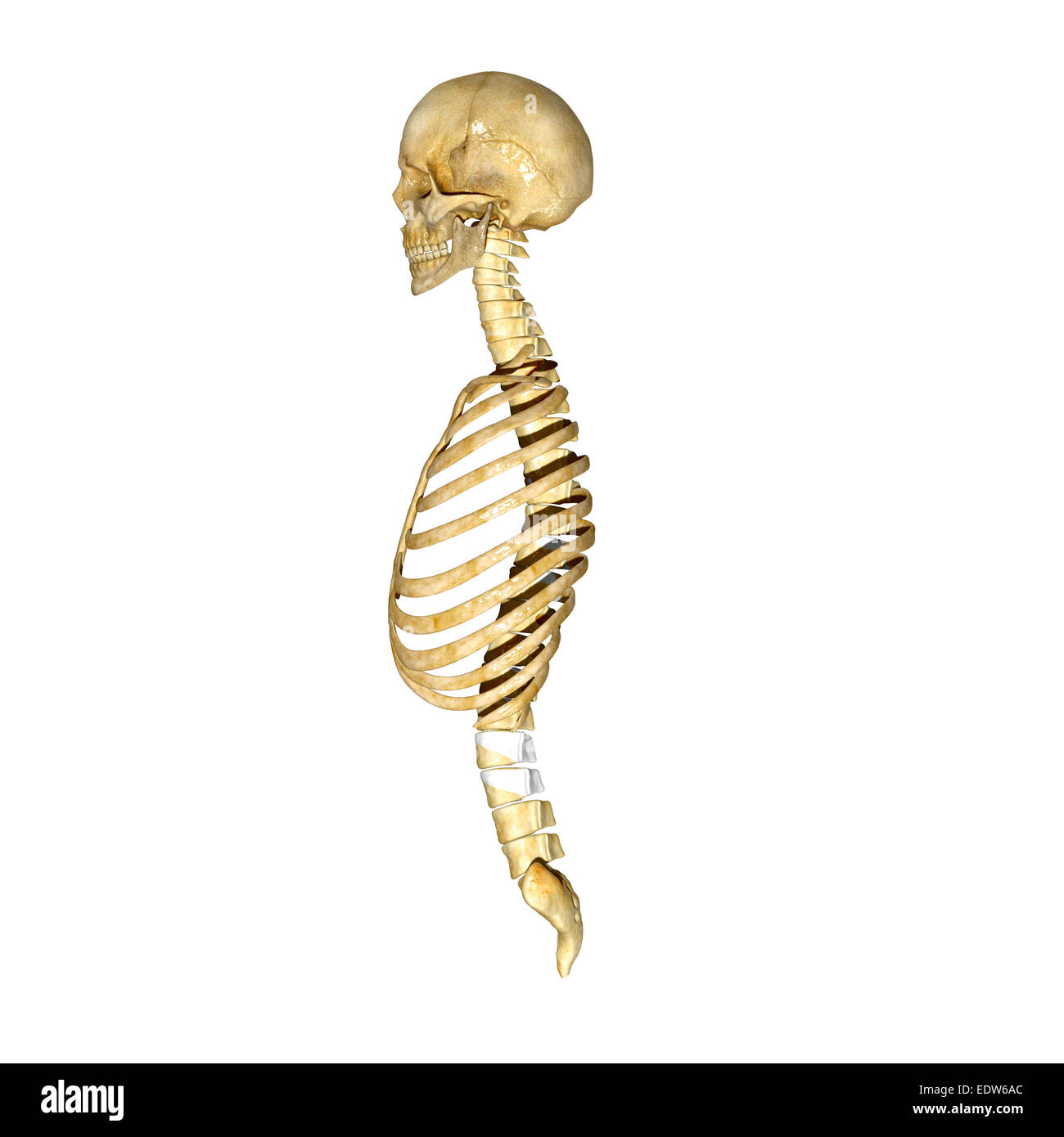 Rib cage, skull and spinal cord Stock Photohttps://www.alamy.com/image-license-details/?v=1https://www.alamy.com/stock-photo-rib-cage-skull-and-spinal-cord-77385796.html
RFEDW6AC–Rib cage, skull and spinal cord
 asian anatomy Stock Photohttps://www.alamy.com/image-license-details/?v=1https://www.alamy.com/asian-anatomy-image343651663.html
RM2AY2K5K–asian anatomy
 Skull Spinal cord Stock Photohttps://www.alamy.com/image-license-details/?v=1https://www.alamy.com/stock-photo-skull-spinal-cord-77385811.html
RFEDW6AY–Skull Spinal cord
 A manual of anatomy . y and laterally separating two grooves; the ventral, deeper, oneis for the subclavian vein, while the dorsal one is for the subclavian THE COSTAL CARTILAGES 39 artery and ventral division of the first thoracic nerve {sulcus sub-clavicB). Dorsal to the latter groove the surface is roughed for theattachment of the m. scalenius medius. The inferior surface issmooth and covered with pleura. The lateral margin is thickenedand rough dorsally for attachment of the first digitation of the m.serratus anterior. In the second rib there is no twist and the surfaces are turned sothat Stock Photohttps://www.alamy.com/image-license-details/?v=1https://www.alamy.com/a-manual-of-anatomy-y-and-laterally-separating-two-grooves-the-ventral-deeper-oneis-for-the-subclavian-vein-while-the-dorsal-one-is-for-the-subclavian-the-costal-cartilages-39-artery-and-ventral-division-of-the-first-thoracic-nerve-sulcus-sub-clavicb-dorsal-to-the-latter-groove-the-surface-is-roughed-for-theattachment-of-the-m-scalenius-medius-the-inferior-surface-issmooth-and-covered-with-pleura-the-lateral-margin-is-thickenedand-rough-dorsally-for-attachment-of-the-first-digitation-of-the-mserratus-anterior-in-the-second-rib-there-is-no-twist-and-the-surfaces-are-turned-sothat-image343394292.html
RM2AXJXWT–A manual of anatomy . y and laterally separating two grooves; the ventral, deeper, oneis for the subclavian vein, while the dorsal one is for the subclavian THE COSTAL CARTILAGES 39 artery and ventral division of the first thoracic nerve {sulcus sub-clavicB). Dorsal to the latter groove the surface is roughed for theattachment of the m. scalenius medius. The inferior surface issmooth and covered with pleura. The lateral margin is thickenedand rough dorsally for attachment of the first digitation of the m.serratus anterior. In the second rib there is no twist and the surfaces are turned sothat
 Third thoracic vertebrae (T3): The thoracic spinal nerve 3 passes through underneath T3 3d illustration Stock Photohttps://www.alamy.com/image-license-details/?v=1https://www.alamy.com/third-thoracic-vertebrae-t3-the-thoracic-spinal-nerve-3-passes-through-underneath-t3-3d-illustration-image596578082.html
RF2WJGDC2–Third thoracic vertebrae (T3): The thoracic spinal nerve 3 passes through underneath T3 3d illustration
 Vector illustration of where thoracic outlet syndrome occurs Stock Vectorhttps://www.alamy.com/image-license-details/?v=1https://www.alamy.com/vector-illustration-of-where-thoracic-outlet-syndrome-occurs-image605812837.html
RF2X5H4CN–Vector illustration of where thoracic outlet syndrome occurs
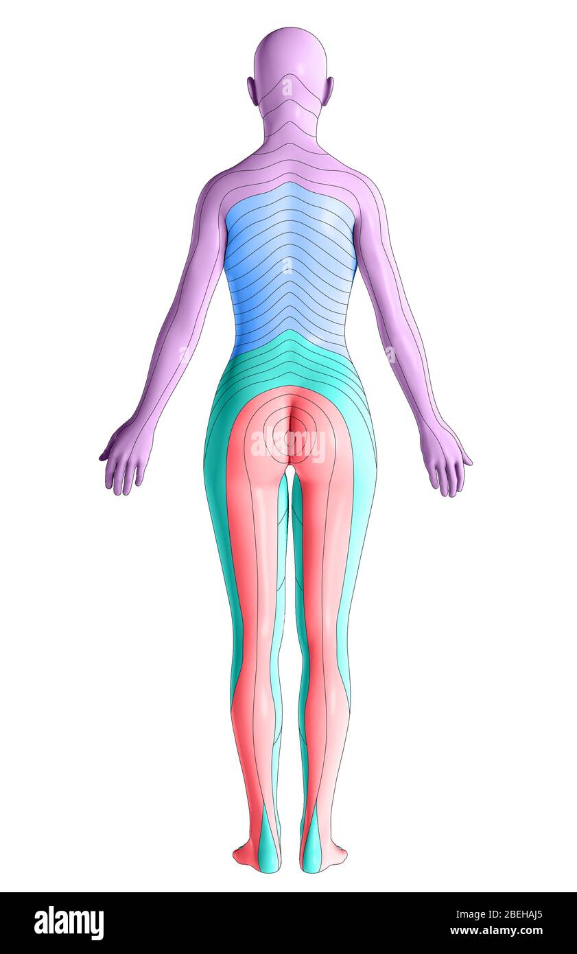 Dermatomes, Posterior View Stock Photohttps://www.alamy.com/image-license-details/?v=1https://www.alamy.com/dermatomes-posterior-view-image353194077.html
RM2BEHAJ5–Dermatomes, Posterior View
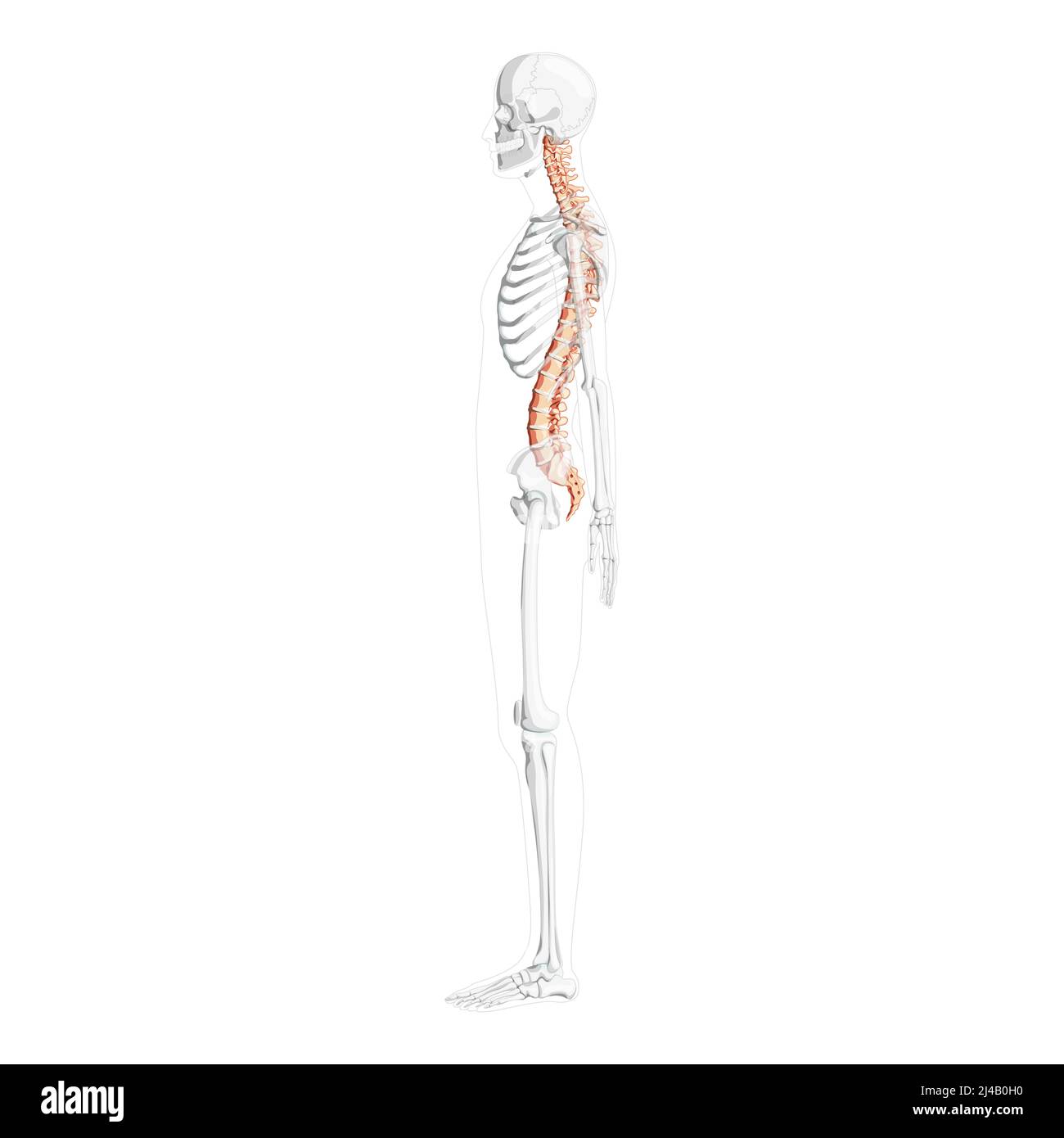 Human vertebral column lateral side view with partly transparent skeleton position, spinal cord, thoracic lumbar spine, coccyx. Vector flat natural colors, realistic isolated illustration anatomy Stock Vectorhttps://www.alamy.com/image-license-details/?v=1https://www.alamy.com/human-vertebral-column-lateral-side-view-with-partly-transparent-skeleton-position-spinal-cord-thoracic-lumbar-spine-coccyx-vector-flat-natural-colors-realistic-isolated-illustration-anatomy-image467380508.html
RF2J4B0H0–Human vertebral column lateral side view with partly transparent skeleton position, spinal cord, thoracic lumbar spine, coccyx. Vector flat natural colors, realistic isolated illustration anatomy
 Dermatome Stock Photohttps://www.alamy.com/image-license-details/?v=1https://www.alamy.com/stock-photo-dermatome-49485586.html
RFCTE7AA–Dermatome
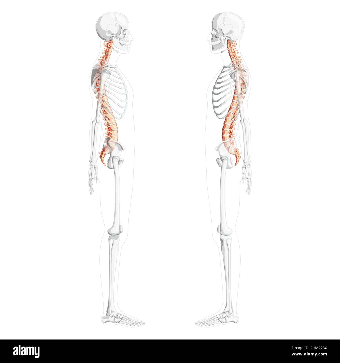 Human vertebral column side view with partly transparent skeleton position, spinal cord, thoracic lumbar spine, sacrum and coccyx. Vector flat natural colors, realistic isolated illustration anatomy Stock Vectorhttps://www.alamy.com/image-license-details/?v=1https://www.alamy.com/human-vertebral-column-side-view-with-partly-transparent-skeleton-position-spinal-cord-thoracic-lumbar-spine-sacrum-and-coccyx-vector-flat-natural-colors-realistic-isolated-illustration-anatomy-image459808270.html
RF2HM223X–Human vertebral column side view with partly transparent skeleton position, spinal cord, thoracic lumbar spine, sacrum and coccyx. Vector flat natural colors, realistic isolated illustration anatomy
 The nerves of the upper body Stock Photohttps://www.alamy.com/image-license-details/?v=1https://www.alamy.com/stock-photo-the-nerves-of-the-upper-body-13171216.html
RFACJJMH–The nerves of the upper body
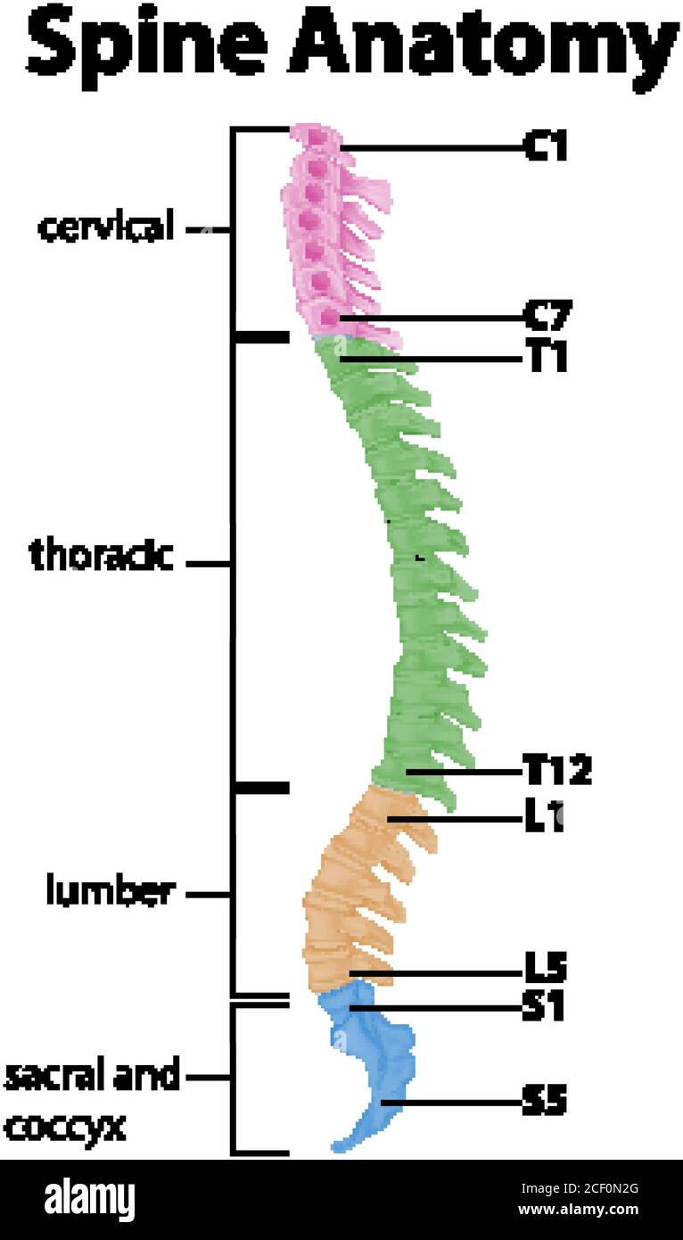 Anatomy of the spine or spinal curves infographic illustration Stock Vectorhttps://www.alamy.com/image-license-details/?v=1https://www.alamy.com/anatomy-of-the-spine-or-spinal-curves-infographic-illustration-image370654104.html
RF2CF0N2G–Anatomy of the spine or spinal curves infographic illustration
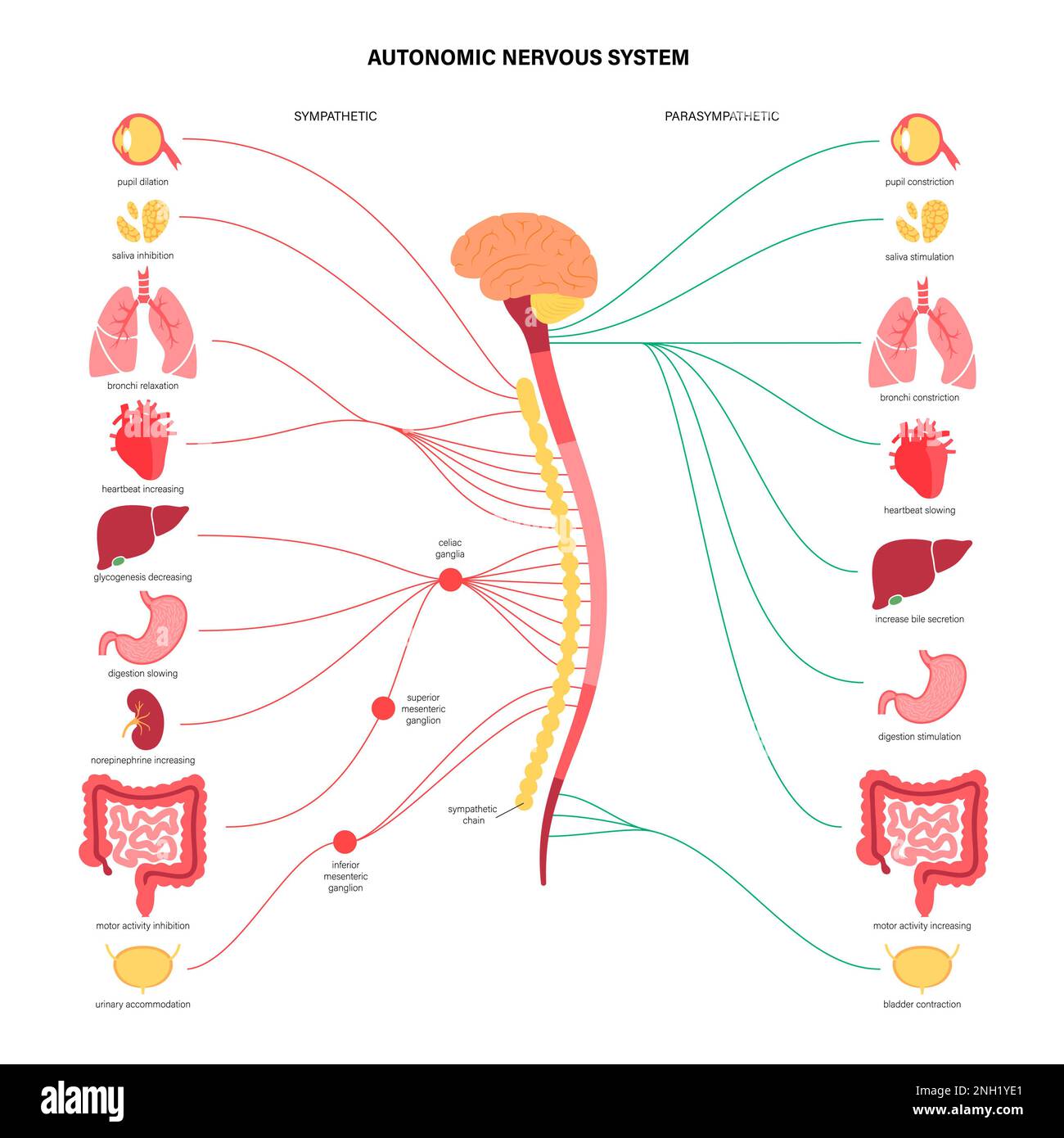 Autonomic nervous system, illustration Stock Photohttps://www.alamy.com/image-license-details/?v=1https://www.alamy.com/autonomic-nervous-system-illustration-image526803705.html
RF2NH1YE1–Autonomic nervous system, illustration
RF2H2C1E2–Spinal diagram icon. Outline spinal diagram vector icon color flat isolated
 african-american anatomy Stock Photohttps://www.alamy.com/image-license-details/?v=1https://www.alamy.com/african-american-anatomy-image343651711.html
RM2AY2K7B–african-american anatomy
 The development of the human body; a manual of human embryology . Fir. 117.—External Surface of the Os Innominatum showing theAttachment of Muscles and the Zones Supplied by the VariousNerves. 12, Twelfth thoracic nerve; / to V, lumbar nerves; 1 and 2, sacralnerves.—(Bolk.) that of the outer portion, and, extending to the tips of thetoes, they are reflected upon the plantar surface and so 236 THE DEVELOPMENT OF THE HUMAN BODY. loop upward on the posterior surface of the leg towardtheir point of origin from the trunk. In a transverse section through any part of the limb,. Fig. 118.—Sections thr Stock Photohttps://www.alamy.com/image-license-details/?v=1https://www.alamy.com/the-development-of-the-human-body-a-manual-of-human-embryology-fir-117external-surface-of-the-os-innominatum-showing-theattachment-of-muscles-and-the-zones-supplied-by-the-variousnerves-12-twelfth-thoracic-nerve-to-v-lumbar-nerves-1-and-2-sacralnervesbolk-that-of-the-outer-portion-and-extending-to-the-tips-of-thetoes-they-are-reflected-upon-the-plantar-surface-and-so-236-the-development-of-the-human-body-loop-upward-on-the-posterior-surface-of-the-leg-towardtheir-point-of-origin-from-the-trunk-in-a-transverse-section-through-any-part-of-the-limb-fig-118sections-thr-image342686107.html
RM2AWEKHF–The development of the human body; a manual of human embryology . Fir. 117.—External Surface of the Os Innominatum showing theAttachment of Muscles and the Zones Supplied by the VariousNerves. 12, Twelfth thoracic nerve; / to V, lumbar nerves; 1 and 2, sacralnerves.—(Bolk.) that of the outer portion, and, extending to the tips of thetoes, they are reflected upon the plantar surface and so 236 THE DEVELOPMENT OF THE HUMAN BODY. loop upward on the posterior surface of the leg towardtheir point of origin from the trunk. In a transverse section through any part of the limb,. Fig. 118.—Sections thr
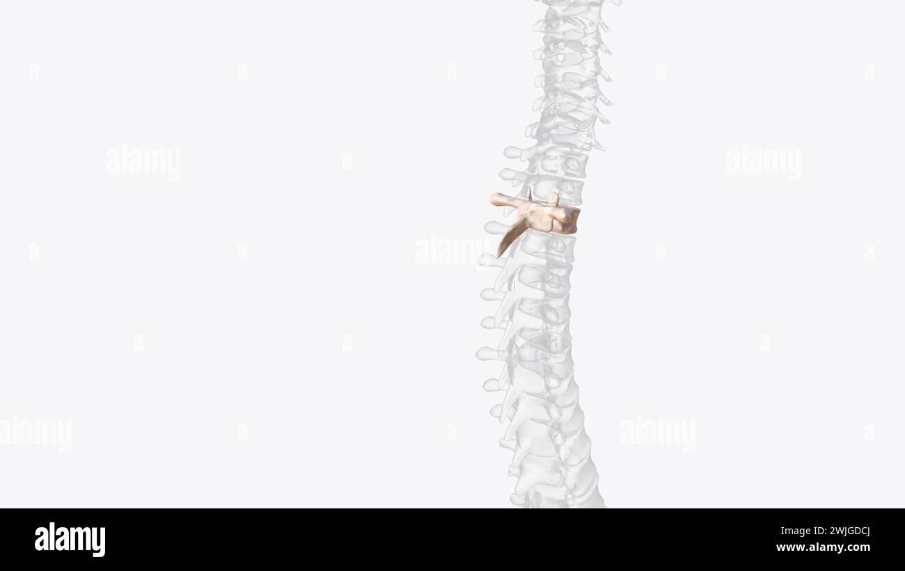 Third thoracic vertebrae (T3): The thoracic spinal nerve 3 passes through underneath T3 3d illustration Stock Photohttps://www.alamy.com/image-license-details/?v=1https://www.alamy.com/third-thoracic-vertebrae-t3-the-thoracic-spinal-nerve-3-passes-through-underneath-t3-3d-illustration-image596578098.html
RF2WJGDCJ–Third thoracic vertebrae (T3): The thoracic spinal nerve 3 passes through underneath T3 3d illustration
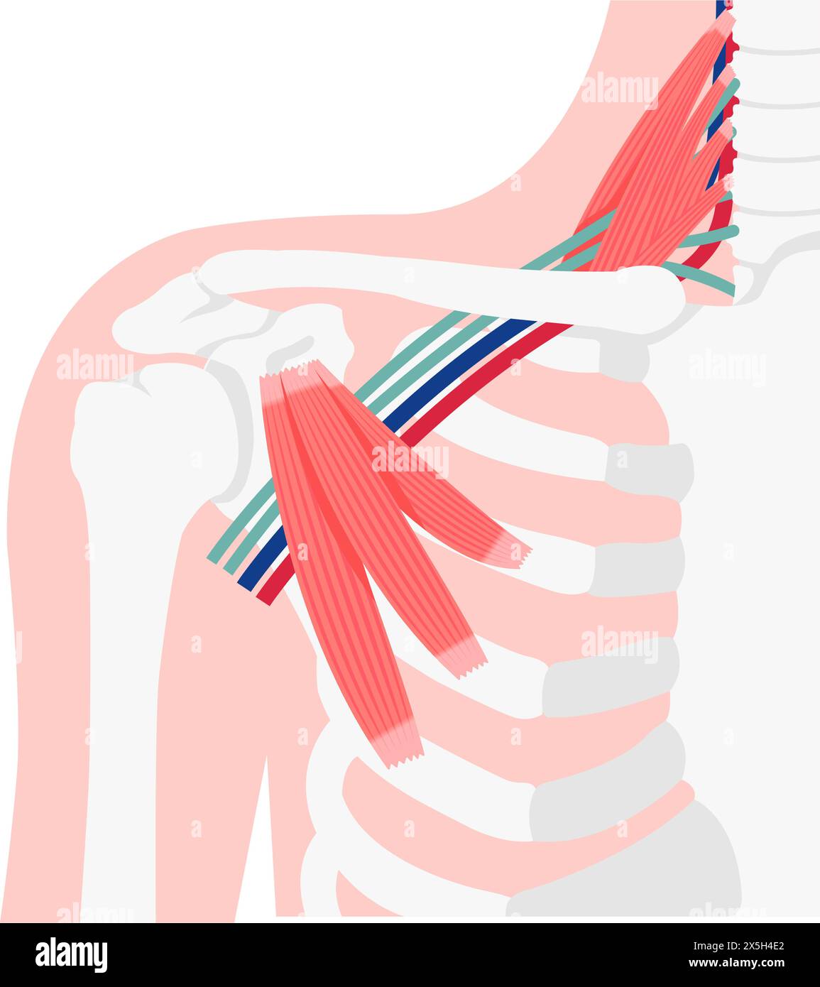 Vector illustration of where thoracic outlet syndrome occurs Stock Vectorhttps://www.alamy.com/image-license-details/?v=1https://www.alamy.com/vector-illustration-of-where-thoracic-outlet-syndrome-occurs-image605812874.html
RF2X5H4E2–Vector illustration of where thoracic outlet syndrome occurs
 Dermatomes, Posterior View Stock Photohttps://www.alamy.com/image-license-details/?v=1https://www.alamy.com/dermatomes-posterior-view-image353194034.html
RM2BEHAGJ–Dermatomes, Posterior View
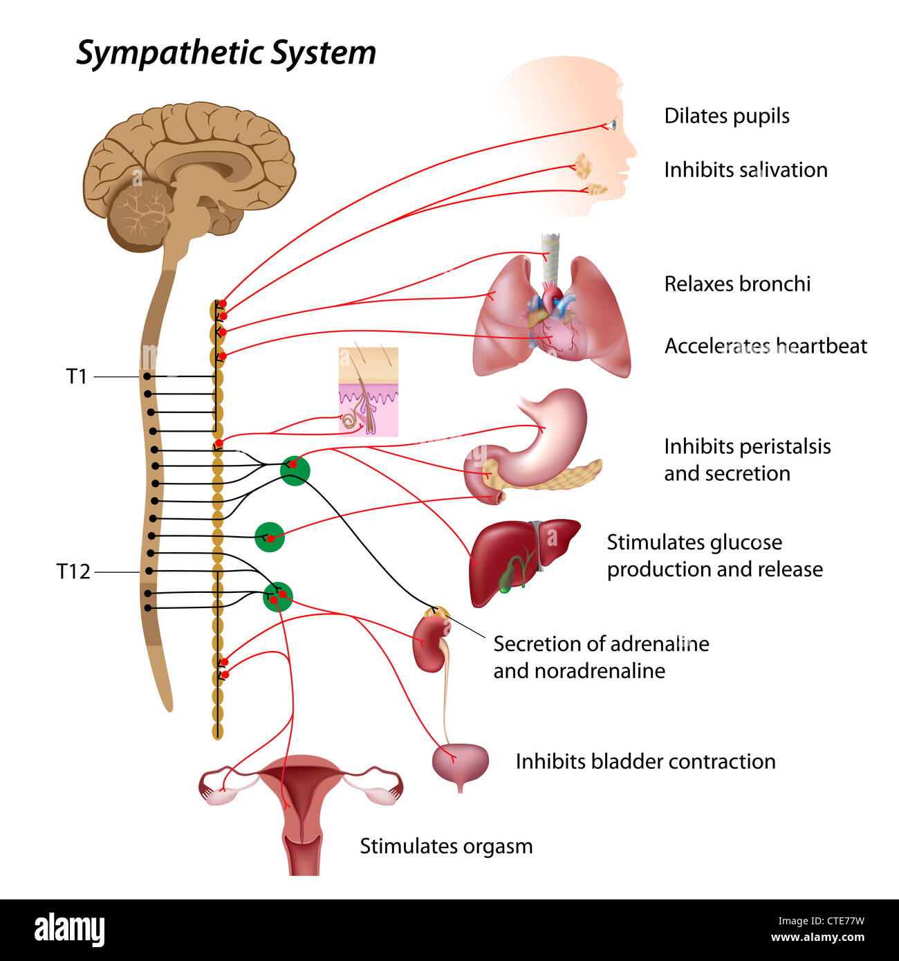 Sympathetic pathway of the ANS Stock Photohttps://www.alamy.com/image-license-details/?v=1https://www.alamy.com/stock-photo-sympathetic-pathway-of-the-ans-49485517.html
RFCTE77W–Sympathetic pathway of the ANS
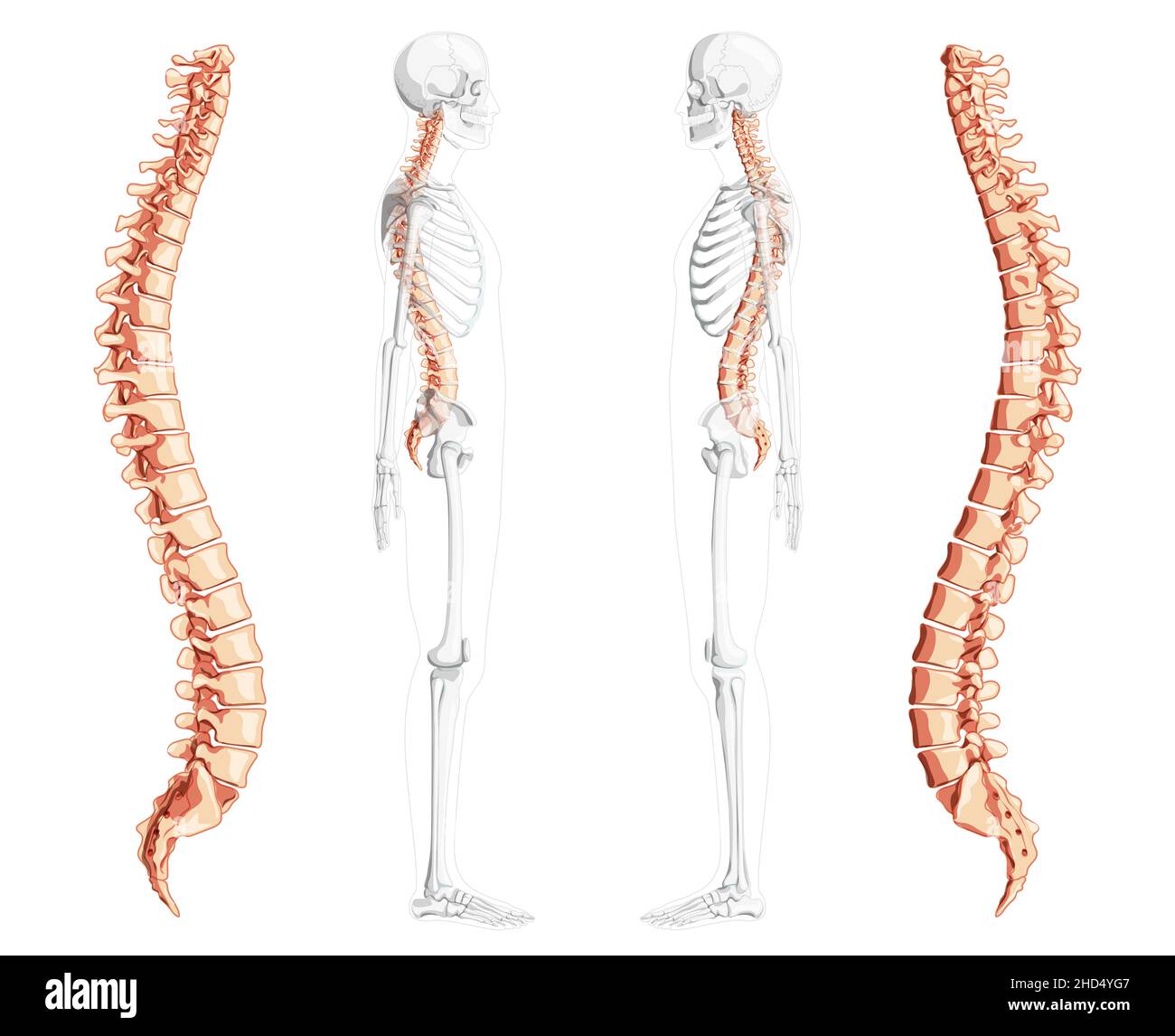 Human vertebral column side view with partly transparent skeleton position, spinal cord, thoracic lumbar spine, sacrum and coccyx. Vector flat natural colors, realistic isolated illustration anatomy Stock Vectorhttps://www.alamy.com/image-license-details/?v=1https://www.alamy.com/human-vertebral-column-side-view-with-partly-transparent-skeleton-position-spinal-cord-thoracic-lumbar-spine-sacrum-and-coccyx-vector-flat-natural-colors-realistic-isolated-illustration-anatomy-image455569527.html
RF2HD4YG7–Human vertebral column side view with partly transparent skeleton position, spinal cord, thoracic lumbar spine, sacrum and coccyx. Vector flat natural colors, realistic isolated illustration anatomy
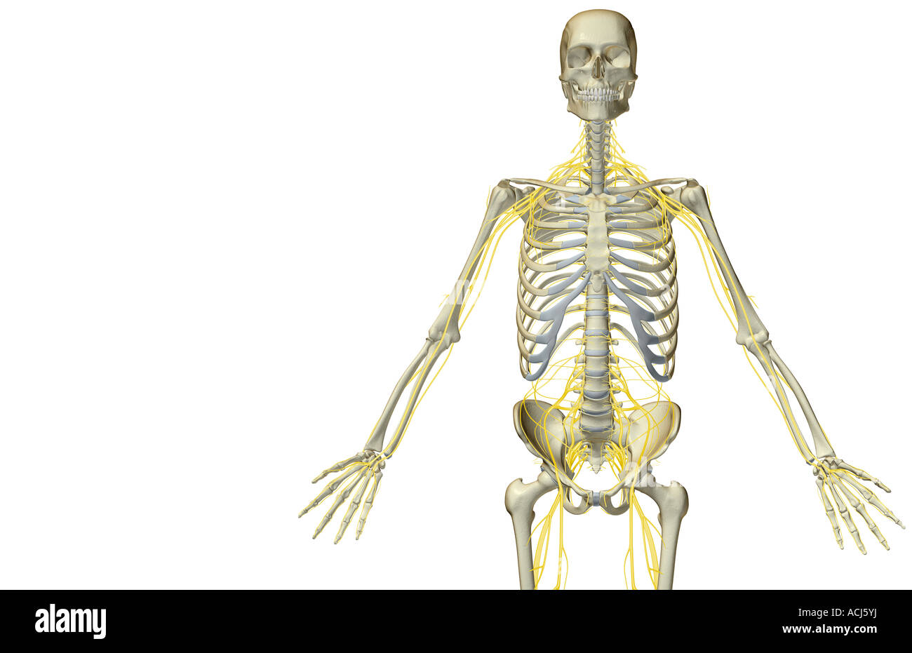 The nerves of the upper body Stock Photohttps://www.alamy.com/image-license-details/?v=1https://www.alamy.com/stock-photo-the-nerves-of-the-upper-body-13166933.html
RFACJ5YJ–The nerves of the upper body
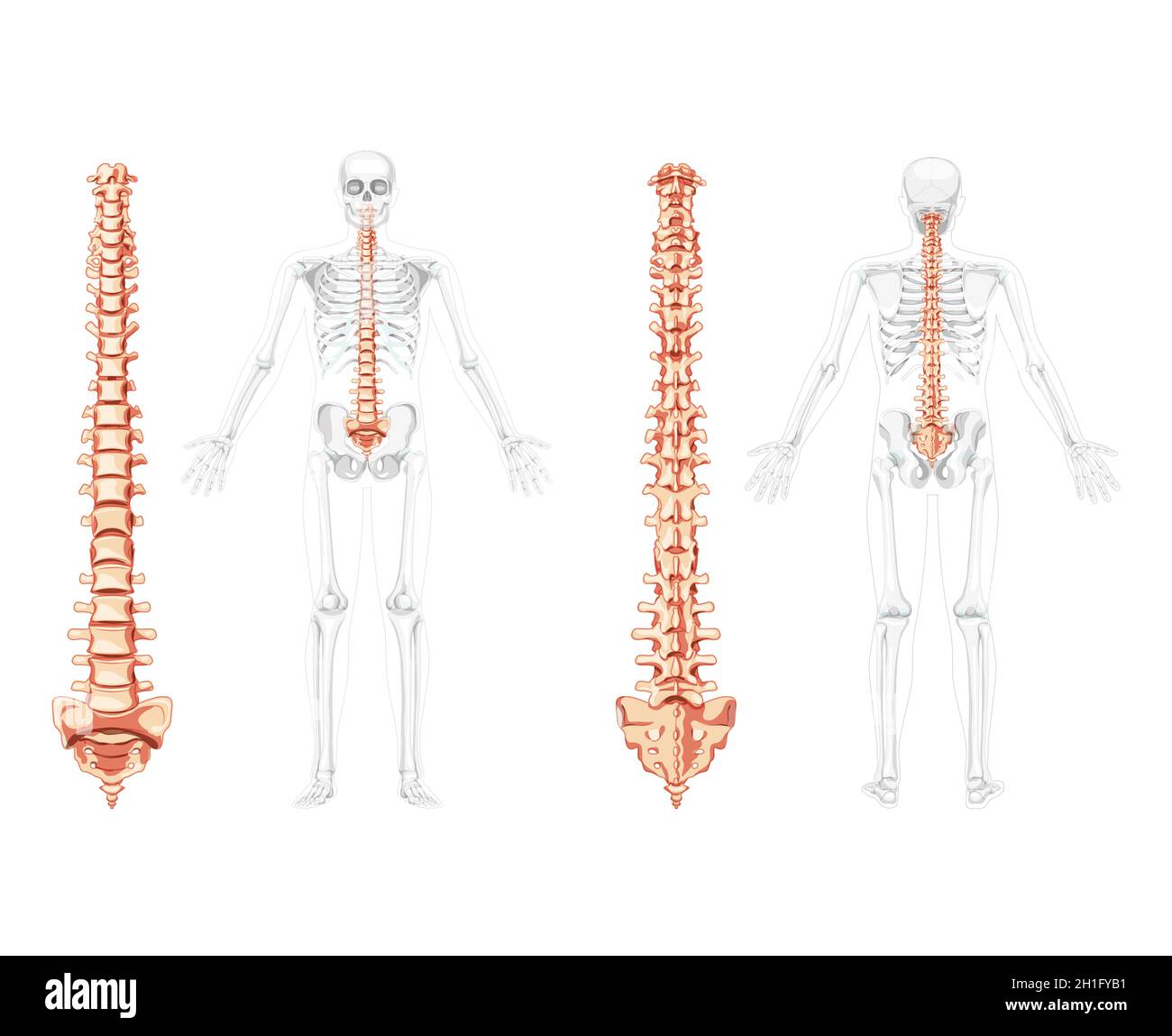 Human vertebral column in front, back with skeleton position, spinal cord, cervical, thoracic and lumbar spine, sacrum and coccyx. Vector flat natural colours, realistic isolated illustration anatomy Stock Vectorhttps://www.alamy.com/image-license-details/?v=1https://www.alamy.com/human-vertebral-column-in-front-back-with-skeleton-position-spinal-cord-cervical-thoracic-and-lumbar-spine-sacrum-and-coccyx-vector-flat-natural-colours-realistic-isolated-illustration-anatomy-image448434981.html
RF2H1FYB1–Human vertebral column in front, back with skeleton position, spinal cord, cervical, thoracic and lumbar spine, sacrum and coccyx. Vector flat natural colours, realistic isolated illustration anatomy
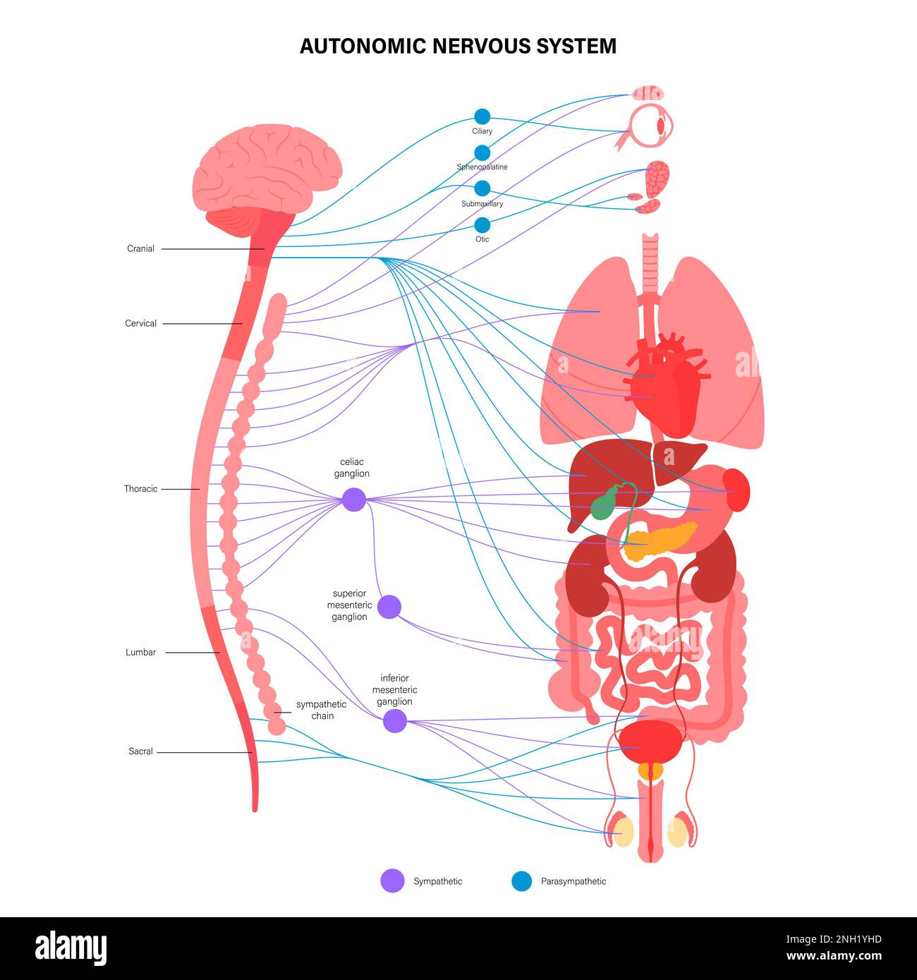 Autonomic nervous system, illustration Stock Photohttps://www.alamy.com/image-license-details/?v=1https://www.alamy.com/autonomic-nervous-system-illustration-image526803801.html
RF2NH1YHD–Autonomic nervous system, illustration
 Human vertebral column front back side view with partly transparent skeleton position, spinal cord, thoracic lumbar spine, sacrum. Vector flat natural colors, realistic isolated illustration anatomy Stock Vectorhttps://www.alamy.com/image-license-details/?v=1https://www.alamy.com/human-vertebral-column-front-back-side-view-with-partly-transparent-skeleton-position-spinal-cord-thoracic-lumbar-spine-sacrum-vector-flat-natural-colors-realistic-isolated-illustration-anatomy-image455764919.html
RF2HDDTPF–Human vertebral column front back side view with partly transparent skeleton position, spinal cord, thoracic lumbar spine, sacrum. Vector flat natural colors, realistic isolated illustration anatomy
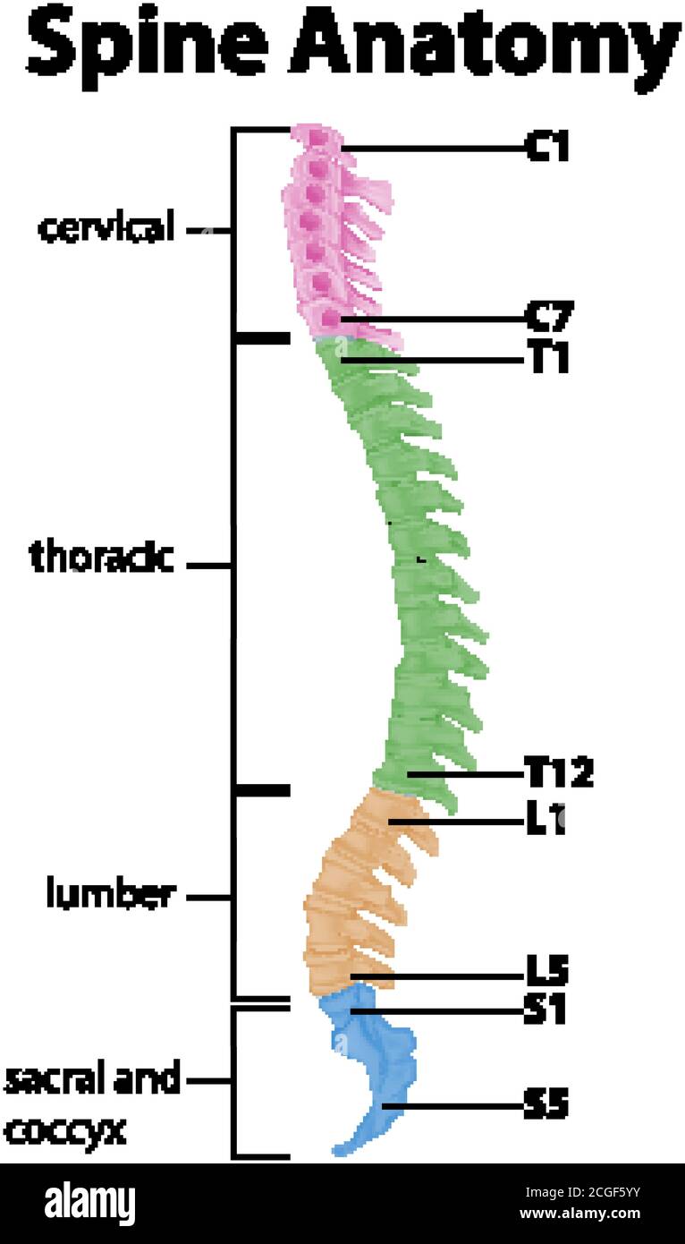 Anatomy of the spine or spinal curves infographic illustration Stock Vectorhttps://www.alamy.com/image-license-details/?v=1https://www.alamy.com/anatomy-of-the-spine-or-spinal-curves-infographic-illustration-image371586207.html
RF2CGF5YY–Anatomy of the spine or spinal curves infographic illustration
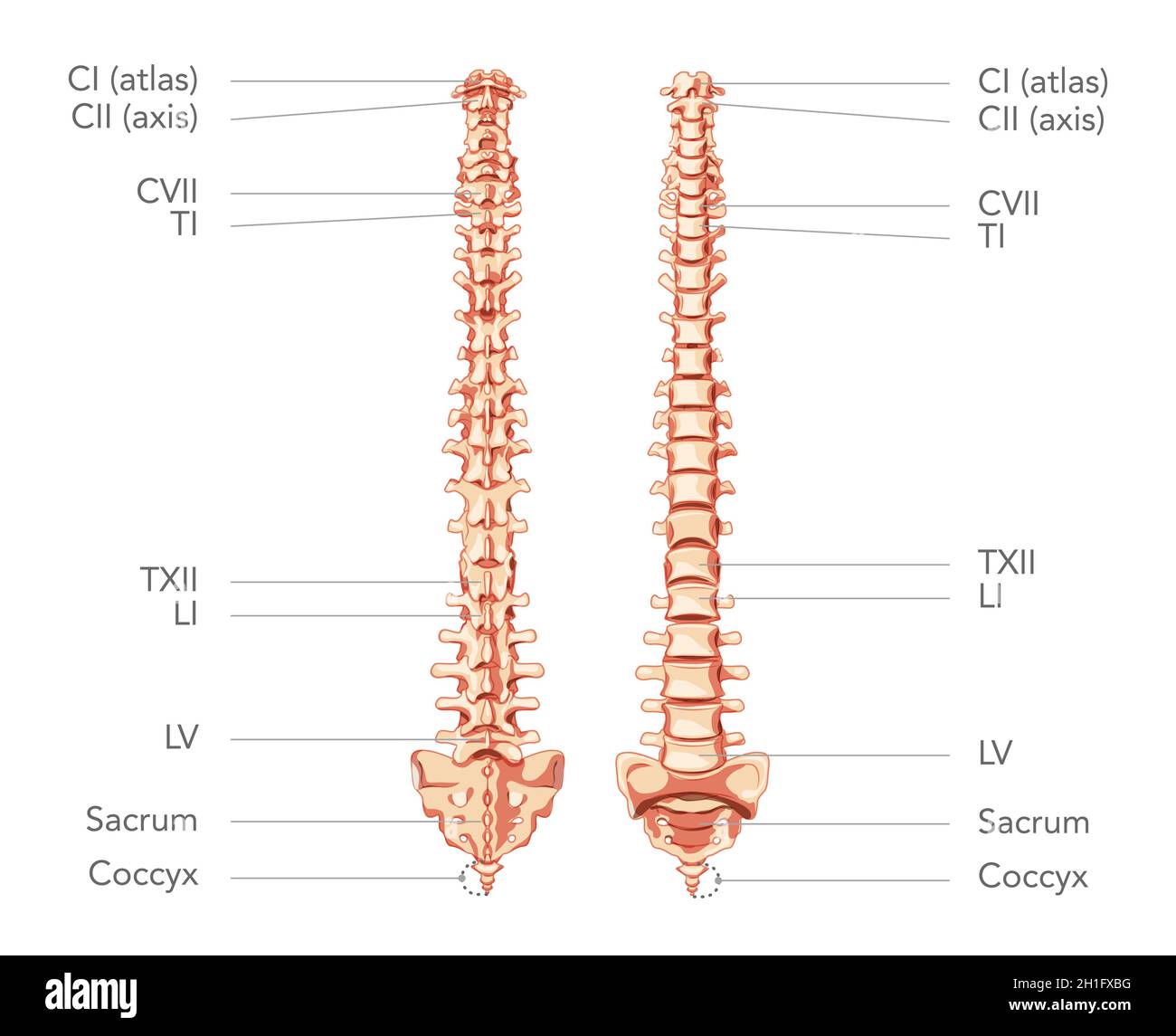 Human vertebral column in anterior posterior view, with spine parts labeled. Vector flat realistic concept illustration isolated on white background diagram for medical, educational or banner use Stock Vectorhttps://www.alamy.com/image-license-details/?v=1https://www.alamy.com/human-vertebral-column-in-anterior-posterior-view-with-spine-parts-labeled-vector-flat-realistic-concept-illustration-isolated-on-white-background-diagram-for-medical-educational-or-banner-use-image448434212.html
RF2H1FXBG–Human vertebral column in anterior posterior view, with spine parts labeled. Vector flat realistic concept illustration isolated on white background diagram for medical, educational or banner use
 african-american woman Stock Photohttps://www.alamy.com/image-license-details/?v=1https://www.alamy.com/african-american-woman-image343651784.html
RM2AY2KA0–african-american woman
 . Mammalian anatomy : with special reference to the cat . Mammals; Anatomy, Comparative; Cats. 208 ELEMENTS OF MAMMALIAN ANATOMY. branches of the sixth, seventh, and eighth cervical nerves and the first thoracic nerve form the brachial plexus. This may be displayed by removing the cephalo-humeral muscle and cutting through the pectoral muscles about two. Fig. 105. Ventral Aspect of the Brachial Plexus and Chief Nerves of the Arm. 6, 7, 8 and 1, Sixth, seventh, and eighth cervical and first dorsal nerves; at, ath, anterior thoracic nerves; a and b, to muscles of the forearm; c and d, to the joi Stock Photohttps://www.alamy.com/image-license-details/?v=1https://www.alamy.com/mammalian-anatomy-with-special-reference-to-the-cat-mammals-anatomy-comparative-cats-208-elements-of-mammalian-anatomy-branches-of-the-sixth-seventh-and-eighth-cervical-nerves-and-the-first-thoracic-nerve-form-the-brachial-plexus-this-may-be-displayed-by-removing-the-cephalo-humeral-muscle-and-cutting-through-the-pectoral-muscles-about-two-fig-105-ventral-aspect-of-the-brachial-plexus-and-chief-nerves-of-the-arm-6-7-8-and-1-sixth-seventh-and-eighth-cervical-and-first-dorsal-nerves-at-ath-anterior-thoracic-nerves-a-and-b-to-muscles-of-the-forearm-c-and-d-to-the-joi-image232141829.html
RMRDJY7H–. Mammalian anatomy : with special reference to the cat . Mammals; Anatomy, Comparative; Cats. 208 ELEMENTS OF MAMMALIAN ANATOMY. branches of the sixth, seventh, and eighth cervical nerves and the first thoracic nerve form the brachial plexus. This may be displayed by removing the cephalo-humeral muscle and cutting through the pectoral muscles about two. Fig. 105. Ventral Aspect of the Brachial Plexus and Chief Nerves of the Arm. 6, 7, 8 and 1, Sixth, seventh, and eighth cervical and first dorsal nerves; at, ath, anterior thoracic nerves; a and b, to muscles of the forearm; c and d, to the joi
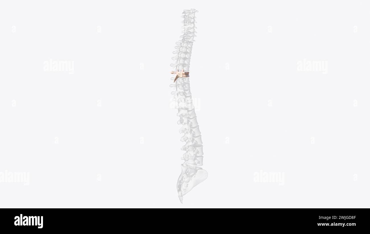 Sixth thoracic vertebra (T6) Edit 3d illustration The thoracic spinal nerve 6 (T6) passes out underneath it 3d illustration Stock Photohttps://www.alamy.com/image-license-details/?v=1https://www.alamy.com/sixth-thoracic-vertebra-t6-edit-3d-illustration-the-thoracic-spinal-nerve-6-t6-passes-out-underneath-it-3d-illustration-image596577983.html
RF2WJGD8F–Sixth thoracic vertebra (T6) Edit 3d illustration The thoracic spinal nerve 6 (T6) passes out underneath it 3d illustration
 Vector illustration of where thoracic outlet syndrome occurs Stock Vectorhttps://www.alamy.com/image-license-details/?v=1https://www.alamy.com/vector-illustration-of-where-thoracic-outlet-syndrome-occurs-image605812790.html
RF2X5H4B2–Vector illustration of where thoracic outlet syndrome occurs
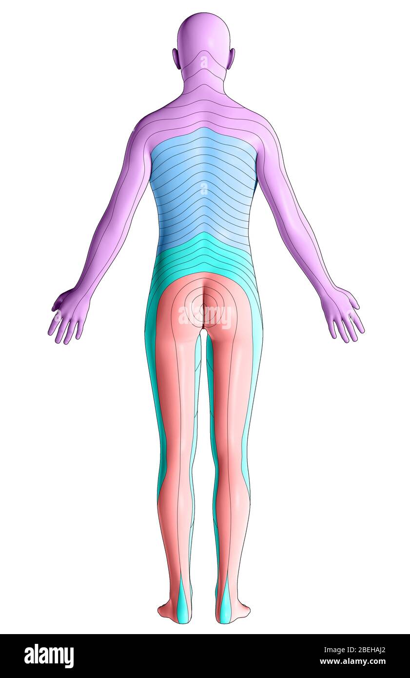 Dermatomes, Posterior View Stock Photohttps://www.alamy.com/image-license-details/?v=1https://www.alamy.com/dermatomes-posterior-view-image353194074.html
RM2BEHAJ2–Dermatomes, Posterior View
 The nerves of the upper body Stock Photohttps://www.alamy.com/image-license-details/?v=1https://www.alamy.com/stock-photo-the-nerves-of-the-upper-body-13172098.html
RFACJN9R–The nerves of the upper body
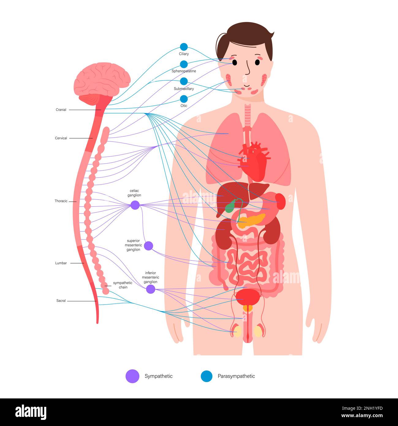 Autonomic nervous system, illustration Stock Photohttps://www.alamy.com/image-license-details/?v=1https://www.alamy.com/autonomic-nervous-system-illustration-image526803745.html
RF2NH1YFD–Autonomic nervous system, illustration
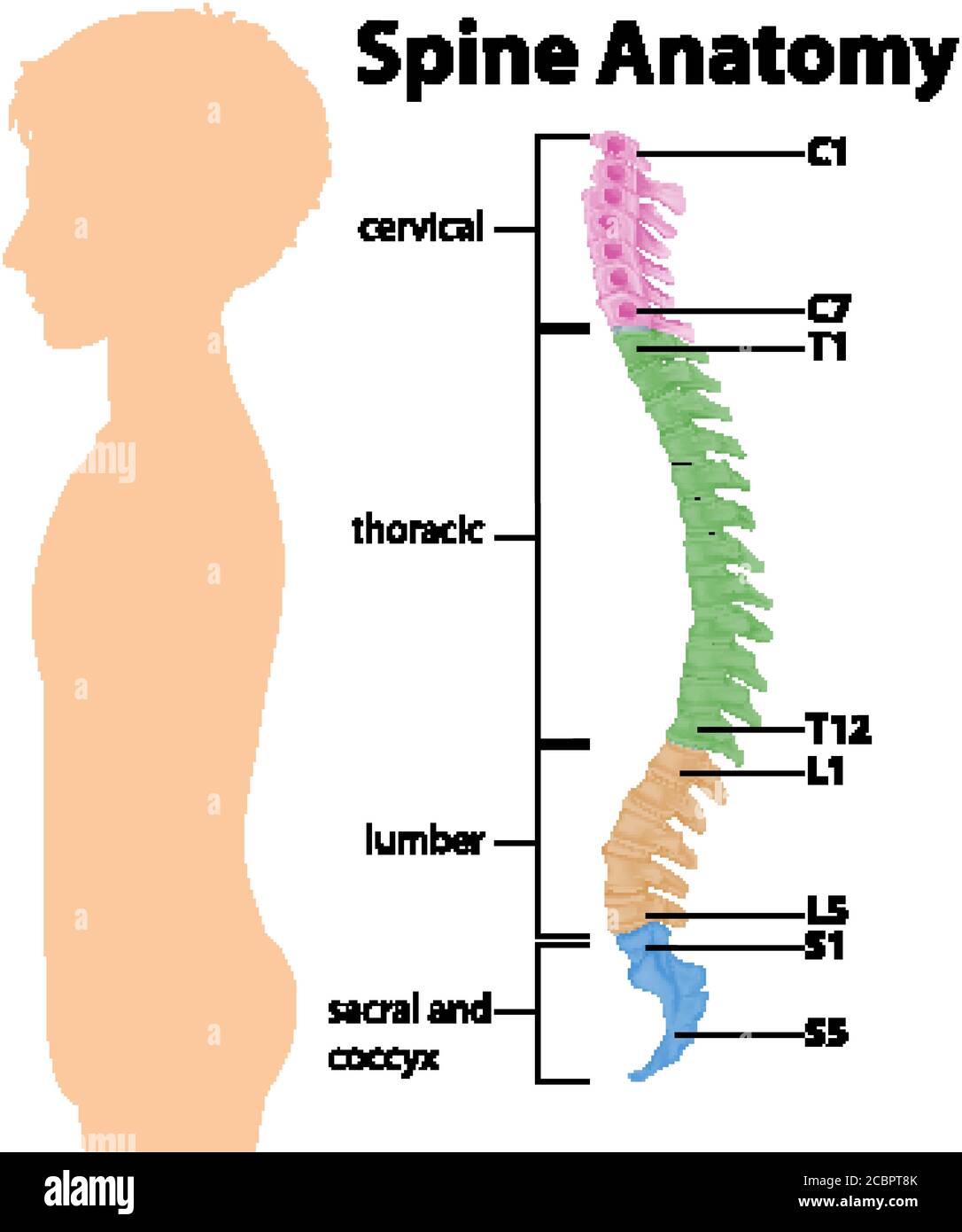 Anatomy of the spine or spinal curves infographic illustration Stock Vectorhttps://www.alamy.com/image-license-details/?v=1https://www.alamy.com/anatomy-of-the-spine-or-spinal-curves-infographic-illustration-image368680947.html
RF2CBPT8K–Anatomy of the spine or spinal curves infographic illustration
 . Anatomical technology as applied to the domestic cat; an introduction to human, veterinary, and comparative anatomy. Cats; Dissection; Mammals. TSE BRACHIAL PLEXUS. 381 771. levtchr ana- sea and. th-om(raideiis /ectal cph. ). Fig. 106.—Diagbam of the Eight Brachial Plexus, Ventral View ; x about 3. nerve of the M. liiUssimus dorsi or tbe long subscapular nerve (§ 1033). N. thr. ant. (ental. cd.), N. thoracicus anterior (entalis s. caudalis)—The anterior thoracic nerve, ental or caudal division. N. thr. post., N. thoracicus posterior—The posterior thoracic nerve or the external respiratory o Stock Photohttps://www.alamy.com/image-license-details/?v=1https://www.alamy.com/anatomical-technology-as-applied-to-the-domestic-cat-an-introduction-to-human-veterinary-and-comparative-anatomy-cats-dissection-mammals-tse-brachial-plexus-381-771-levtchr-ana-sea-and-th-omraideiis-ectal-cph-fig-106diagbam-of-the-eight-brachial-plexus-ventral-view-x-about-3-nerve-of-the-m-liiussimus-dorsi-or-tbe-long-subscapular-nerve-1033-n-thr-ant-ental-cd-n-thoracicus-anterior-entalis-s-caudalisthe-anterior-thoracic-nerve-ental-or-caudal-division-n-thr-post-n-thoracicus-posteriorthe-posterior-thoracic-nerve-or-the-external-respiratory-o-image232347880.html
RMRE0A2G–. Anatomical technology as applied to the domestic cat; an introduction to human, veterinary, and comparative anatomy. Cats; Dissection; Mammals. TSE BRACHIAL PLEXUS. 381 771. levtchr ana- sea and. th-om(raideiis /ectal cph. ). Fig. 106.—Diagbam of the Eight Brachial Plexus, Ventral View ; x about 3. nerve of the M. liiUssimus dorsi or tbe long subscapular nerve (§ 1033). N. thr. ant. (ental. cd.), N. thoracicus anterior (entalis s. caudalis)—The anterior thoracic nerve, ental or caudal division. N. thr. post., N. thoracicus posterior—The posterior thoracic nerve or the external respiratory o
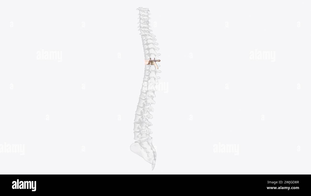 Sixth thoracic vertebra (T6) Edit 3d illustration The thoracic spinal nerve 6 (T6) passes out underneath it 3d illustration Stock Photohttps://www.alamy.com/image-license-details/?v=1https://www.alamy.com/sixth-thoracic-vertebra-t6-edit-3d-illustration-the-thoracic-spinal-nerve-6-t6-passes-out-underneath-it-3d-illustration-image596577991.html
RF2WJGD8R–Sixth thoracic vertebra (T6) Edit 3d illustration The thoracic spinal nerve 6 (T6) passes out underneath it 3d illustration
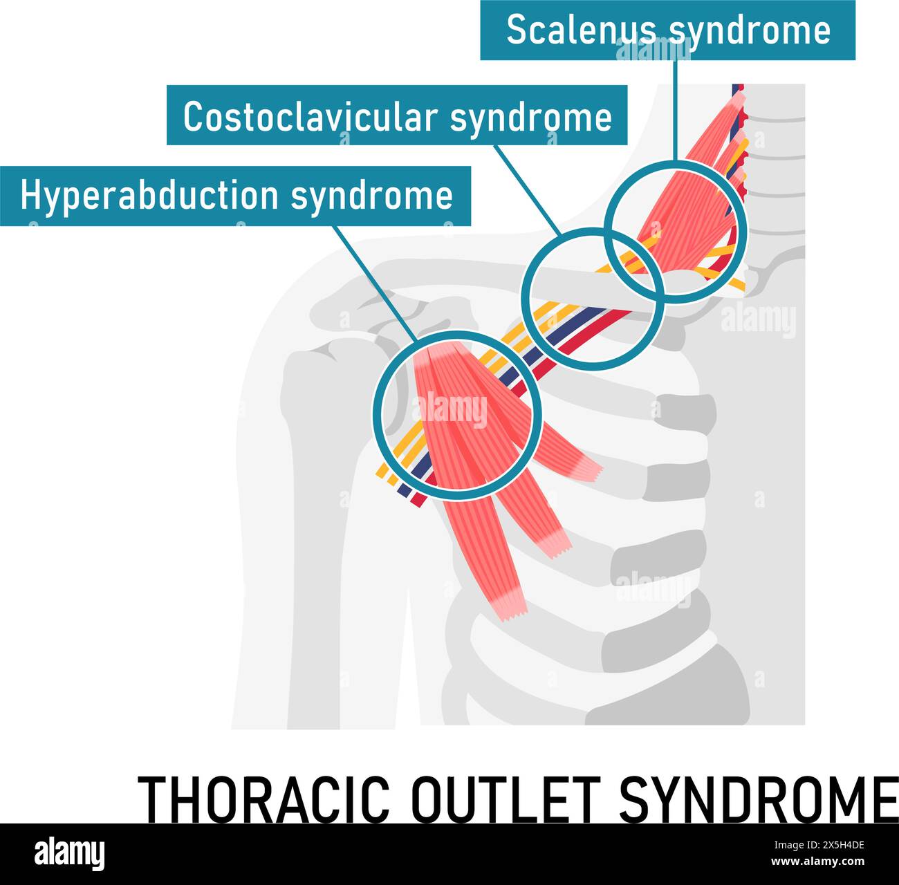 Vector illustration of where thoracic outlet syndrome occurs Stock Vectorhttps://www.alamy.com/image-license-details/?v=1https://www.alamy.com/vector-illustration-of-where-thoracic-outlet-syndrome-occurs-image605812858.html
RF2X5H4DE–Vector illustration of where thoracic outlet syndrome occurs
 Dermatomes, Posterior View Stock Photohttps://www.alamy.com/image-license-details/?v=1https://www.alamy.com/dermatomes-posterior-view-image353194037.html
RM2BEHAGN–Dermatomes, Posterior View
 Autonomic nervous system, illustration Stock Photohttps://www.alamy.com/image-license-details/?v=1https://www.alamy.com/autonomic-nervous-system-illustration-image526803786.html
RF2NH1YGX–Autonomic nervous system, illustration
 A manual of anatomy . below. {SohoUa and McMurrich.) supraspinous ligaments. The superior portion is inserted, as thesplenius capitis into the mastoid process and the superior nuchal line.The inferior portion, as the splenius cervicis, is inserted into dorsaltubercles of the first three or four cervical vertebrae. Actions .—Extension and lateral movement of the vertebral column.The splenius capitis assists in flexion, rotation and raising the head. 164 MYOLOGY Nerve Stipply.-nerves. -Dorsal rami of the cervical and superior thoracic FOURTH LAYER The m. sacrospinalis, or erector spinae, is the Stock Photohttps://www.alamy.com/image-license-details/?v=1https://www.alamy.com/a-manual-of-anatomy-below-sohoua-and-mcmurrich-supraspinous-ligaments-the-superior-portion-is-inserted-as-thesplenius-capitis-into-the-mastoid-process-and-the-superior-nuchal-linethe-inferior-portion-as-the-splenius-cervicis-is-inserted-into-dorsaltubercles-of-the-first-three-or-four-cervical-vertebrae-actions-extension-and-lateral-movement-of-the-vertebral-columnthe-splenius-capitis-assists-in-flexion-rotation-and-raising-the-head-164-myology-nerve-stipply-nerves-dorsal-rami-of-the-cervical-and-superior-thoracic-fourth-layer-the-m-sacrospinalis-or-erector-spinae-is-the-image343363074.html
RM2AXHF2X–A manual of anatomy . below. {SohoUa and McMurrich.) supraspinous ligaments. The superior portion is inserted, as thesplenius capitis into the mastoid process and the superior nuchal line.The inferior portion, as the splenius cervicis, is inserted into dorsaltubercles of the first three or four cervical vertebrae. Actions .—Extension and lateral movement of the vertebral column.The splenius capitis assists in flexion, rotation and raising the head. 164 MYOLOGY Nerve Stipply.-nerves. -Dorsal rami of the cervical and superior thoracic FOURTH LAYER The m. sacrospinalis, or erector spinae, is the
 Sixth thoracic vertebra (T6) Edit 3d illustration The thoracic spinal nerve 6 (T6) passes out underneath it 3d illustration Stock Photohttps://www.alamy.com/image-license-details/?v=1https://www.alamy.com/sixth-thoracic-vertebra-t6-edit-3d-illustration-the-thoracic-spinal-nerve-6-t6-passes-out-underneath-it-3d-illustration-image596577957.html
RF2WJGD7H–Sixth thoracic vertebra (T6) Edit 3d illustration The thoracic spinal nerve 6 (T6) passes out underneath it 3d illustration
 Vector illustration of where thoracic outlet syndrome occurs Stock Vectorhttps://www.alamy.com/image-license-details/?v=1https://www.alamy.com/vector-illustration-of-where-thoracic-outlet-syndrome-occurs-image605812834.html
RF2X5H4CJ–Vector illustration of where thoracic outlet syndrome occurs
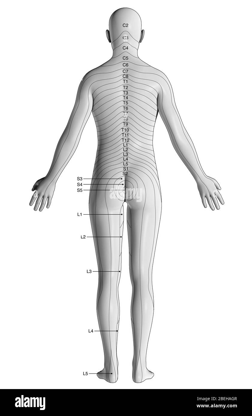 Dermatomes, Posterior View Stock Photohttps://www.alamy.com/image-license-details/?v=1https://www.alamy.com/dermatomes-posterior-view-image353194039.html
RM2BEHAGR–Dermatomes, Posterior View
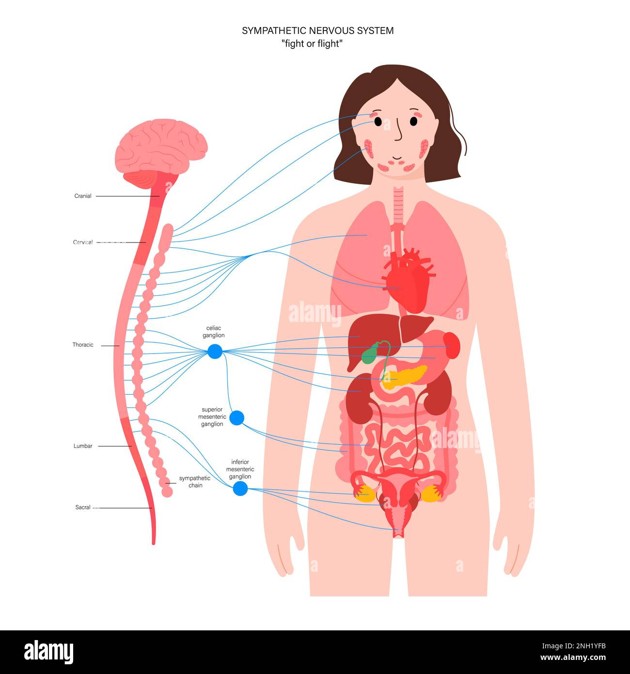 Sympathetic nervous system, illustration Stock Photohttps://www.alamy.com/image-license-details/?v=1https://www.alamy.com/sympathetic-nervous-system-illustration-image526803743.html
RF2NH1YFB–Sympathetic nervous system, illustration
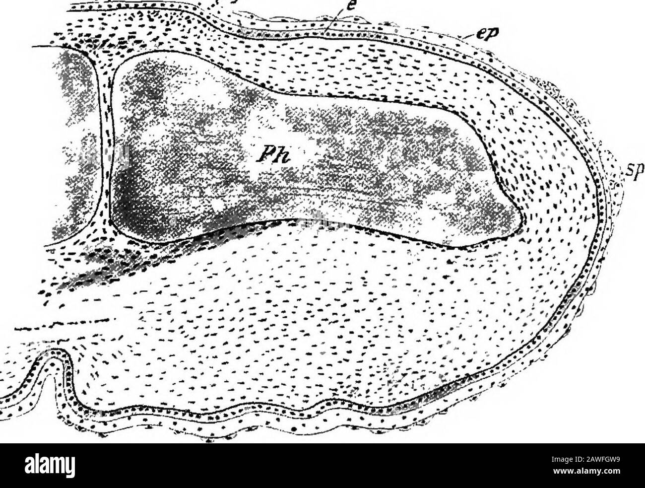 The development of the human body; a manual of human embryology . ers from the nerve which supplies the muscle. Thus, in thelower half of the abdomen, the skin at any point will be sup-plied by fibers from higher nerves than those supplying theunderlying muscles (Sherrington), and the skin of the limbsmay receive twigs from nerves which are not represented atall in the muscle-supply (second and third thoracic and thirdsacral). The Development of the Nails.—The earliest indicationsof the development of the nails have been described byZander in embryos of about nine weeks as slight thicken- THE Stock Photohttps://www.alamy.com/image-license-details/?v=1https://www.alamy.com/the-development-of-the-human-body-a-manual-of-human-embryology-ers-from-the-nerve-which-supplies-the-muscle-thus-in-thelower-half-of-the-abdomen-the-skin-at-any-point-will-be-sup-plied-by-fibers-from-higher-nerves-than-those-supplying-theunderlying-muscles-sherrington-and-the-skin-of-the-limbsmay-receive-twigs-from-nerves-which-are-not-represented-atall-in-the-muscle-supply-second-and-third-thoracic-and-thirdsacral-the-development-of-the-nailsthe-earliest-indicationsof-the-development-of-the-nails-have-been-described-byzander-in-embryos-of-about-nine-weeks-as-slight-thicken-the-image342705925.html
RM2AWFGW9–The development of the human body; a manual of human embryology . ers from the nerve which supplies the muscle. Thus, in thelower half of the abdomen, the skin at any point will be sup-plied by fibers from higher nerves than those supplying theunderlying muscles (Sherrington), and the skin of the limbsmay receive twigs from nerves which are not represented atall in the muscle-supply (second and third thoracic and thirdsacral). The Development of the Nails.—The earliest indicationsof the development of the nails have been described byZander in embryos of about nine weeks as slight thicken- THE
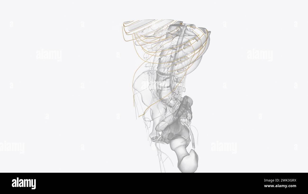 Branches of thoracic spinal 3d medical Stock Photohttps://www.alamy.com/image-license-details/?v=1https://www.alamy.com/branches-of-thoracic-spinal-3d-medical-image596910046.html
RF2WK3GRX–Branches of thoracic spinal 3d medical
 Vector illustration of where thoracic outlet syndrome occurs Stock Vectorhttps://www.alamy.com/image-license-details/?v=1https://www.alamy.com/vector-illustration-of-where-thoracic-outlet-syndrome-occurs-image605812831.html
RF2X5H4CF–Vector illustration of where thoracic outlet syndrome occurs
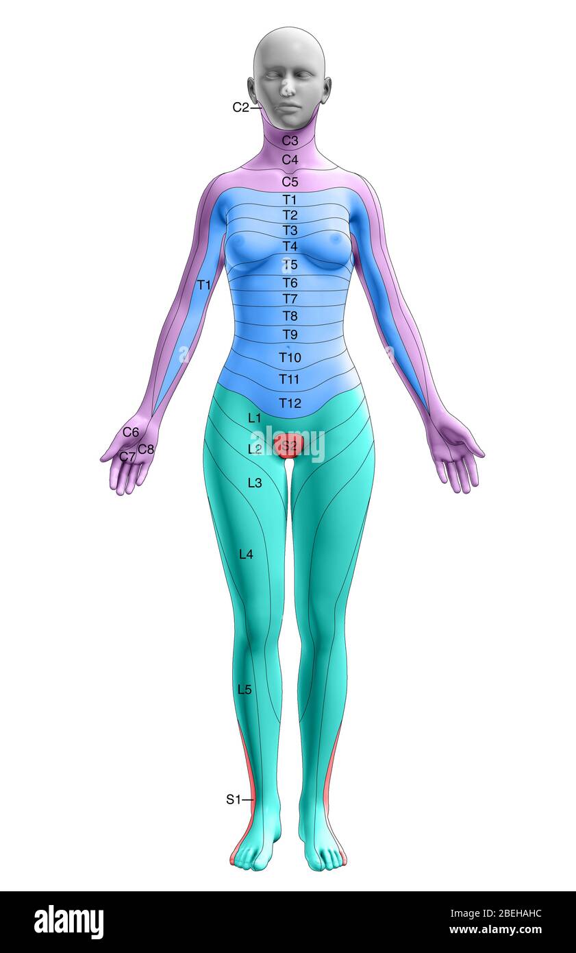 Dermatomes, Anterior View Stock Photohttps://www.alamy.com/image-license-details/?v=1https://www.alamy.com/dermatomes-anterior-view-image353194056.html
RM2BEHAHC–Dermatomes, Anterior View
 Parasympathetic nervous system, illustration Stock Photohttps://www.alamy.com/image-license-details/?v=1https://www.alamy.com/parasympathetic-nervous-system-illustration-image526803789.html
RF2NH1YH1–Parasympathetic nervous system, illustration
 A manual of anatomy . Dorsal scapular nerve of the brachial plexus(fifth cer.). THIRD LAYER The m. serratus posticus superior arises from the ligamentumnuchas and the spines of the seventh cervical and first three thoracicvertebrae. It is inserted into the second, third, fourth and fifthribs. The m. serratus posticus inferior arises from the lumbosacral THE MUSCLES OF THE BACK 163 fascia attached to the last two thoracic and first two lumbar spines;after a horizontal course it is inserted into the last four ribs. Actions.—Extensors of the vertebral column and accessory jmisclesof respiration. Stock Photohttps://www.alamy.com/image-license-details/?v=1https://www.alamy.com/a-manual-of-anatomy-dorsal-scapular-nerve-of-the-brachial-plexusfifth-cer-third-layer-the-m-serratus-posticus-superior-arises-from-the-ligamentumnuchas-and-the-spines-of-the-seventh-cervical-and-first-three-thoracicvertebrae-it-is-inserted-into-the-second-third-fourth-and-fifthribs-the-m-serratus-posticus-inferior-arises-from-the-lumbosacral-the-muscles-of-the-back-163-fascia-attached-to-the-last-two-thoracic-and-first-two-lumbar-spinesafter-a-horizontal-course-it-is-inserted-into-the-last-four-ribs-actionsextensors-of-the-vertebral-column-and-accessory-jmisclesof-respiration-image343363273.html
RM2AXHFA1–A manual of anatomy . Dorsal scapular nerve of the brachial plexus(fifth cer.). THIRD LAYER The m. serratus posticus superior arises from the ligamentumnuchas and the spines of the seventh cervical and first three thoracicvertebrae. It is inserted into the second, third, fourth and fifthribs. The m. serratus posticus inferior arises from the lumbosacral THE MUSCLES OF THE BACK 163 fascia attached to the last two thoracic and first two lumbar spines;after a horizontal course it is inserted into the last four ribs. Actions.—Extensors of the vertebral column and accessory jmisclesof respiration.

