. A laboratory manual and text-book of embryology. Embryology. 122 THE STUDY OF SIX AND TEN MILLIMETER PIG EMBRYOS the head, the shorter tail, the much smaller mesonephric region, the longer umbilical cord and the less prominent segments. The yolk-sac is pear-shaped with long slender yolk-stalk. Central Nervous System and Viscera.—Dissections show well the form and relations of the organs (Figs. 115, 116 and 117). Directions for preparing dis- sections are given in Chapter VI. Metencephalon N. trochlearis Gang. n. 5 I Mesencephalon Gang. Gang, jugula Gang, petrosa Gang. Fror Gang, nodos. n.
Image details
Contributor:
The Book Worm / Alamy Stock PhotoImage ID:
RE0745File size:
7.2 MB (310.4 KB Compressed download)Releases:
Model - no | Property - noDo I need a release?Dimensions:
1487 x 1681 px | 25.2 x 28.5 cm | 9.9 x 11.2 inches | 150dpiMore information:
This image is a public domain image, which means either that copyright has expired in the image or the copyright holder has waived their copyright. Alamy charges you a fee for access to the high resolution copy of the image.
This image could have imperfections as it’s either historical or reportage.
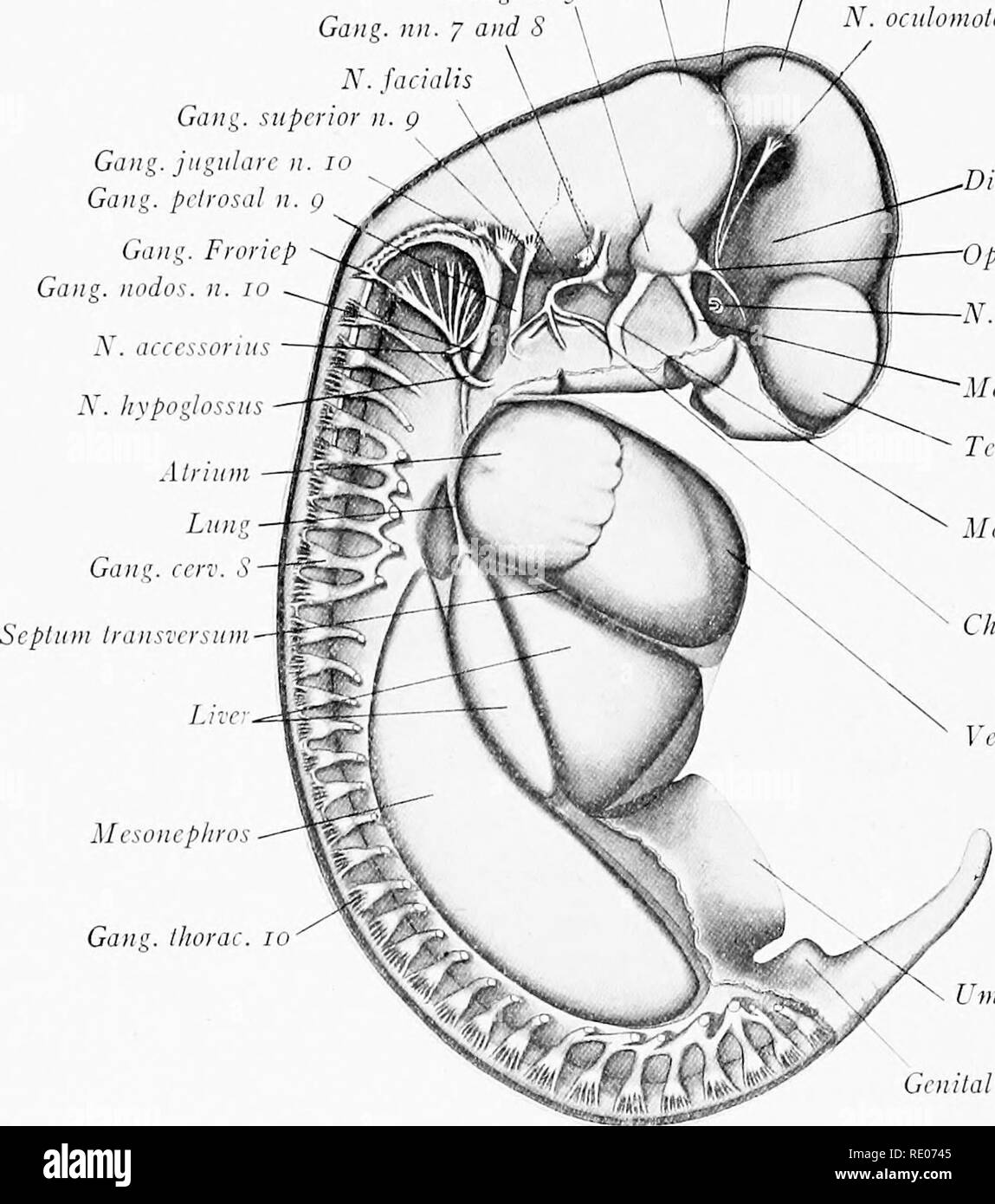
. A laboratory manual and text-book of embryology. Embryology. 122 THE STUDY OF SIX AND TEN MILLIMETER PIG EMBRYOS the head, the shorter tail, the much smaller mesonephric region, the longer umbilical cord and the less prominent segments. The yolk-sac is pear-shaped with long slender yolk-stalk. Central Nervous System and Viscera.—Dissections show well the form and relations of the organs (Figs. 115, 116 and 117). Directions for preparing dis- sections are given in Chapter VI. Metencephalon N. trochlearis Gang. n. 5 I Mesencephalon Gang. Gang, jugula Gang, petrosa Gang. Fror Gang, nodos. n. /. olonus icnccphalon Ophthalmic r. n. 5 opticus axillary r. n. 5 Icnccphalon andibnlar r. n. 5 orda lymp. n. 7 nlricle Gang, thora mbilical cord eminence Fig. 115.—Lateral dissection of a 10 mm. pig embryo, showing the viscera and nervous system from the right side. The eye has been removed and the otic vesicle is represented by a broken line. The ventral roots of the spinal nerves arc not indicated. X 10.5. 11., nerve; r., ramus. Brain.—Five distinct regions may be distinguished (Figs. 115 and 117): (1) The telencephalon with its rounded lateral outgrowths, the cerebral hemispheres. Their cavities, the lateral ventricles communicate by the interventricular foramen with the third ventricle. (2) The dienccphalon shows a laterally flattened cavity, the third ventricle. Ventro-laterally from the diencephalon pass off the optic stalks and an evagination of the mid-ventral wall is the anlage of the posterior. Please note that these images are extracted from scanned page images that may have been digitally enhanced for readability - coloration and appearance of these illustrations may not perfectly resemble the original work.. Prentiss, Charles William, 1874-1915. Philadelphia, London, W. B. Saunders