A manual of anatomy . de. The superior margin is attachedor fused to the manubrium forming an angle at the junction that isusually apDreciable to the touch. This is the sternal angle {angiilussterni). The lateral portions of this margin complete the facet forthe second costal cartilage. The inferior margin is convex and its 36 OSTEOLOGY middle part is connected to the xyphoid process. Lateral to thisthe sixth and seventh costal cartilages are attached. Each lateralmargin is thick and irregular presenting facets at the extremitiesof the ridges that accommodate the third, fourth and fifth costal
Image details
Contributor:
The Reading Room / Alamy Stock PhotoImage ID:
2AXJYMHFile size:
7.1 MB (260.3 KB Compressed download)Releases:
Model - no | Property - noDo I need a release?Dimensions:
1097 x 2278 px | 18.6 x 38.6 cm | 7.3 x 15.2 inches | 150dpiMore information:
This image is a public domain image, which means either that copyright has expired in the image or the copyright holder has waived their copyright. Alamy charges you a fee for access to the high resolution copy of the image.
This image could have imperfections as it’s either historical or reportage.
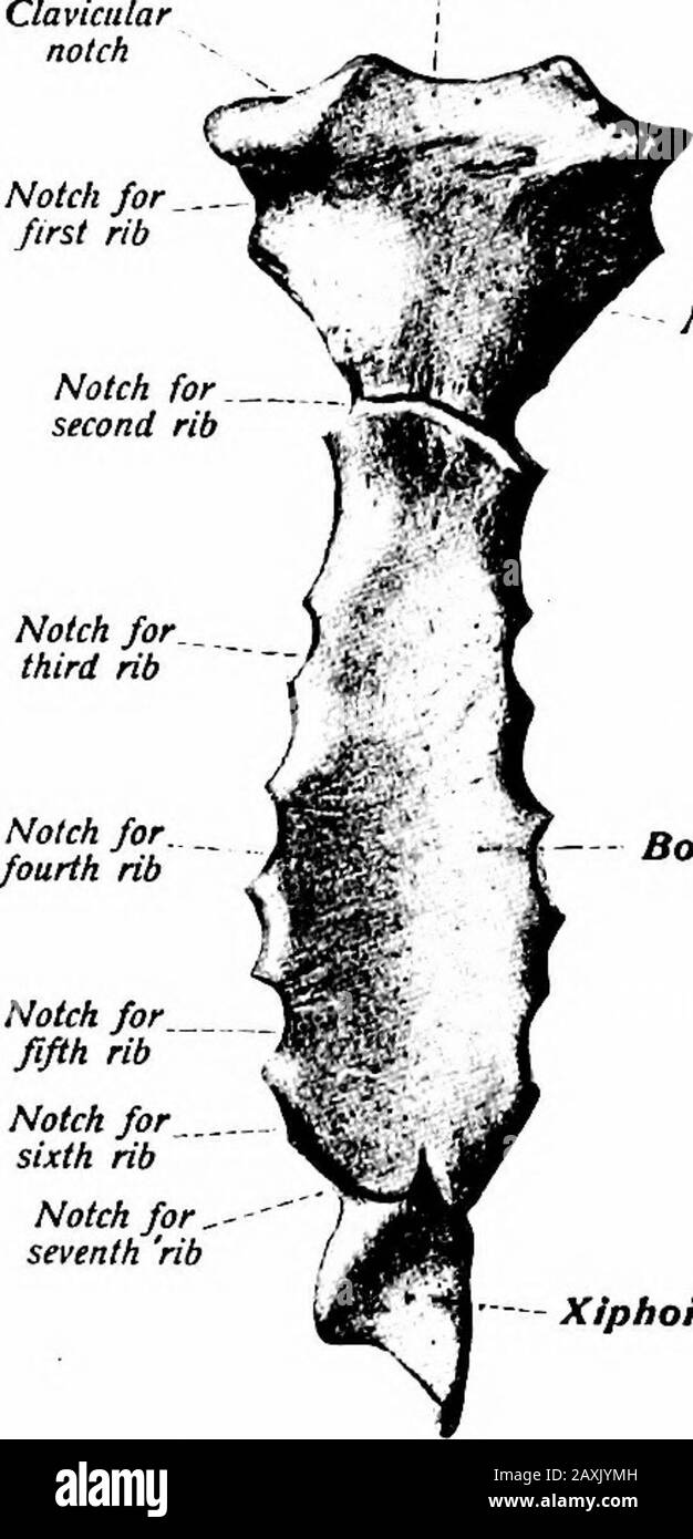
A manual of anatomy . de. The superior margin is attachedor fused to the manubrium forming an angle at the junction that isusually apDreciable to the touch. This is the sternal angle {angiilussterni). The lateral portions of this margin complete the facet forthe second costal cartilage. The inferior margin is convex and its 36 OSTEOLOGY middle part is connected to the xyphoid process. Lateral to thisthe sixth and seventh costal cartilages are attached. Each lateralmargin is thick and irregular presenting facets at the extremitiesof the ridges that accommodate the third, fourth and fifth costalcartilages. The dorsal surface is concave from above downward andmay exhibit the transverse ridges. Its superior portion affordsattachment to the m. transversus thoracis, or triangularis sterni, while the remainder is in relation with the pericardium and pleuras.The xyphoid process (processtis xyphoideus) is the smallest segmentand variable in form. Its base is attached to the body of the sternum Jugular noun Manubrium. Body of sternum Notch for fifth rib Notch for sixth rib Notch for ^seventh rib Xiphoid process Fig. IS-—The sternum seen from in front. (Sobotta and McMurrichJ} while its apex affords attachment to the linea alba of the abdomen.The process slopes dorsally forming the pit of the stomach, or infra-sternal depression. To its lateral margins the aponeuroses of theabdominal muscles are attached, while the dorsal surface affords at-tachment to a portion of the m. transversus thoracis and diaphragm.In the male the sternum is longer and broader than in the female, its direction is more oblique and variations of the different segmentsare also noted. Ossification.—The sternum is an endochondral bone and the cartilaginoussternum is formed by the fusion of two lateral bars. Ossification begins duringthe sixth month of fetal life with the appearance of the center for the manubrium. THE RIBS 37 Secondary centers may appear for the clavicular facets. The body developsfrom fo