A system of human anatomy, general and special . metimestwo ureters to one kidney. The ureter, the pelvis, the infundibula,and the calices are composed of two coats, an external or fibrouscoat, the tunica propria; and an internal mucous coat, which is con-tinuous with the mucous membrane of the bladder inferiorly, andwith the tubuli uriniferi above. Vessels and Nerves.—The renal artery is derived from the aorta;it divides into several large branches before entering the hilus, andwithin the organ ramifies in an arborescent manner, terminating innutrient twigs, and in the small inferent vessels

Image details
Contributor:
The Reading Room / Alamy Stock PhotoImage ID:
2AWFBCPFile size:
7.1 MB (202.3 KB Compressed download)Releases:
Model - no | Property - noDo I need a release?Dimensions:
1402 x 1781 px | 23.7 x 30.2 cm | 9.3 x 11.9 inches | 150dpiMore information:
This image is a public domain image, which means either that copyright has expired in the image or the copyright holder has waived their copyright. Alamy charges you a fee for access to the high resolution copy of the image.
This image could have imperfections as it’s either historical or reportage.
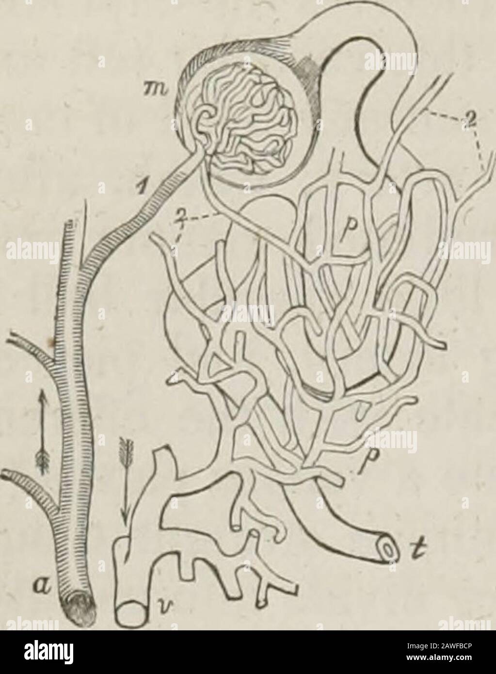
A system of human anatomy, general and special . metimestwo ureters to one kidney. The ureter, the pelvis, the infundibula, and the calices are composed of two coats, an external or fibrouscoat, the tunica propria; and an internal mucous coat, which is con-tinuous with the mucous membrane of the bladder inferiorly, andwith the tubuli uriniferi above. Vessels and Nerves.—The renal artery is derived from the aorta;it divides into several large branches before entering the hilus, andwithin the organ ramifies in an arborescent manner, terminating innutrient twigs, and in the small inferent vessels of the corpora Mal-pighiana. In the Malpighian bodies the inferent vessels divide intoseveral primary twigs, which subdivide into capillaries, and the capil-laries, after forming loops, converge to the efferent vein, which is * For this account I am indebted to the excellent paper of Mr. Bowman, On theStructure and Use of the Malpighian Bodies of the Kidney, &c, published in the Philo-sophical Transactions for 1842. 562 PELVIS. Fig. 218.*. generally smaller than the corresponding artery. The efferent veinsproceed to and form a capillary venous plexus, which surrounds the tortuous tubuli uriniferi, and from this venous plexus the blood is con-veyed by converging branches into the renalvein. Thus, remarks Mr. Bowman, there arein the kidney two perfectly distinct systems ofcapillary vessels, through both of which theblood passes in its course from the arteriesinto the veins: the first, that inserted into thedilated extremities of the uriniferous tubes andin immediate connexion with the arteries; thesecond, that enveloping the convolutions of thetubes and communicating directly with the veins. The efferentvessels of the Malpighian bodies, that carry the blood betweenthese two systems may collectively be termed the portal systemof the kidney. The inferences drawn by Mr. Bowman from hisinvestigations are peculiarly interesting; they are: that the capil-lary tufts of the Malpighian bo