. A text-book of physiology : for medical students and physicians . or fibers, accelerator fibers to heart, pupilodilatorfibers, visceromotor fibers, etc. Emerging in the anterior roots, theypass to the sympathetic chain by way of the corresponding ramuscommunicans. Having reached the chain, they end in one or otherof the ganglia, not necessarily in the ganglion with which the ramusconnects anatomically. The preganglionic fibers for the blood-vessels of the submaxillary gland, for instance, enter the firstthoracic ganglion of the sympathetic chain, but do not actuallyterminate until they reach
Image details
Contributor:
Reading Room 2020 / Alamy Stock PhotoImage ID:
2CE745AFile size:
7.1 MB (353.8 KB Compressed download)Releases:
Model - no | Property - noDo I need a release?Dimensions:
1548 x 1614 px | 26.2 x 27.3 cm | 10.3 x 10.8 inches | 150dpiMore information:
This image is a public domain image, which means either that copyright has expired in the image or the copyright holder has waived their copyright. Alamy charges you a fee for access to the high resolution copy of the image.
This image could have imperfections as it’s either historical or reportage.
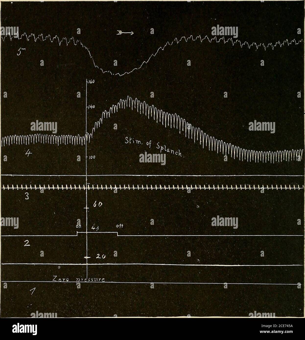
. A text-book of physiology : for medical students and physicians . or fibers, accelerator fibers to heart, pupilodilatorfibers, visceromotor fibers, etc. Emerging in the anterior roots, theypass to the sympathetic chain by way of the corresponding ramuscommunicans. Having reached the chain, they end in one or otherof the ganglia, not necessarily in the ganglion with which the ramusconnects anatomically. The preganglionic fibers for the blood-vessels of the submaxillary gland, for instance, enter the firstthoracic ganglion of the sympathetic chain, but do not actuallyterminate until they reach the superior cervical ganglion high in theneck. The postganglionic fibers arise in the ganglion in which thepreganglionic fibers terminate. Those destined to supply the skin 608 CIRCULATION OF BLOOD AND LYMPH. of the trunk and extremities pass from the ganglion to the cor-responding spinal nerve by way of the ramus communicans (grayramus) and after reaching the spinal nerve they are distributed withit to its corresponding region (Fig. 251). In the general region. Fig. 252.—Vasomotor effect of stimulation of the splanchnic nerve—peripheral end—in the dog (Dawson): 1. The line of zero pressure; 2, the line of the stimulating pen; onand off mark the beginning and end of the stimulation; 3, the time record in seconds; 4, the blood-pressure record (stimulation causes a marked rise of blood-pressure due to stimu-lation of vasoconstrictor fibers); 5, plethysmography tracing of the volume of the kidney(oncometer); stimulation of the splanchnic causes a diminution in volume of the kidneyowing to the constriction of its arterioles. under consideration (lower cervical to upper lumbar) each ramuscommunicans between a spinal nerve and a sympathetic ganglionconsists, therefore, of two parts, one (white ramus) of preganglionicfibers passing from the spinal nerve to the ganglion, the other(gray ramus) of postganglionic fibers coming from the ganglion tothe spinal nerve for distribution t