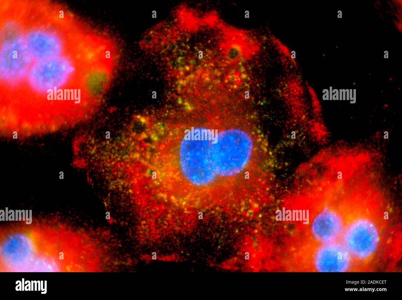
Active macrophages. Immunofluorescent light micrograph of active macrophage white blood cells producing cytokine proteins. The cells have multiple nuc
Image details
Contributor:
Science Photo Library / Alamy Stock PhotoImage ID:
2ADKCETFile size:
26.8 MB (708.6 KB Compressed download)Releases:
Model - no | Property - noDo I need a release?Dimensions:
3721 x 2516 px | 31.5 x 21.3 cm | 12.4 x 8.4 inches | 300dpiDate taken:
1 December 1999Photographer:
NANCY KEDERSHA/SCIENCE PHOTO LIBRARYMore information:
Active macrophages. Immunofluorescent light micrograph of active macrophage white blood cells producing cytokine proteins. The cells have multiple nuclei (blue). Vaults are red and TIA-1, a protein which stops cytokine overproduction, is yellow/green. Cytokines are released by one cell population in order to influence another. If too much cytokine has been made, TIA-1 stops its production by binding to the mRNA (messenger ribonucleic acid) that codes for it, forming stress bodies (yellow). These macrophages have been activated by exposure to a mock bacterial infection. Macrophages help to kill invading organisms. Magnification: x200 at 5x7cm size.