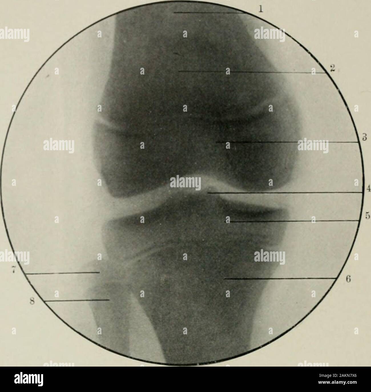
American practice of surgery : a complete system of the science and art of surgery . Fig. 168.—Lateral View of Knee Joint at Eleven Years. 1, Patella; 2, inner condyle; 3, epiphys-eal line at upper end of tibia; 4, epiphyseal line between the femur and the outer condyle; 5, outercondyle; 6, spine of tibia; 7, upper epiphysis of fibula. (Original.) somewhat like a tongue—a projection which afterward becomes the tubercleof the tibia. The antero-posterior view of the knee joint is shown in Fig. 169, which wasmade with the posterior surface of the joint next to the photographic plate.The distance

Image details
Contributor:
The Reading Room / Alamy Stock PhotoImage ID:
2AKN7X6File size:
7.1 MB (172.8 KB Compressed download)Releases:
Model - no | Property - noDo I need a release?Dimensions:
1590 x 1570 px | 26.9 x 26.6 cm | 10.6 x 10.5 inches | 150dpiMore information:
This image is a public domain image, which means either that copyright has expired in the image or the copyright holder has waived their copyright. Alamy charges you a fee for access to the high resolution copy of the image.
This image could have imperfections as it’s either historical or reportage.
American practice of surgery : a complete system of the science and art of surgery . Fig. 168.—Lateral View of Knee Joint at Eleven Years. 1, Patella; 2, inner condyle; 3, epiphys-eal line at upper end of tibia; 4, epiphyseal line between the femur and the outer condyle; 5, outercondyle; 6, spine of tibia; 7, upper epiphysis of fibula. (Original.) somewhat like a tongue—a projection which afterward becomes the tubercleof the tibia. The antero-posterior view of the knee joint is shown in Fig. 169, which wasmade with the posterior surface of the joint next to the photographic plate.The distance of the patella from the plate increases its apparent size, so thatits shadow is less distinct and can scarcely be differentiated from the lower endof the tibia. The spinous process of the tibia is ?hovm. as it projects into thejoint space. The epiphyseal lines are so distinct and regular that their appear-ance is not likely to lead to a misinterpretation.VOL. I.—38 594 AMERICAN PRACTICE OF SURGERY.. Fig. 169.—Radiograph Showing Vertical View of Knee at Eleven Years. 1, Femur; 2, patella; 3, lower epiphysis of femur; 4, spine of tibia; 5, upper epiphysis of tibia; 6, tibia; 7, upper epiphysis offibula: S, fibula. (Original.)