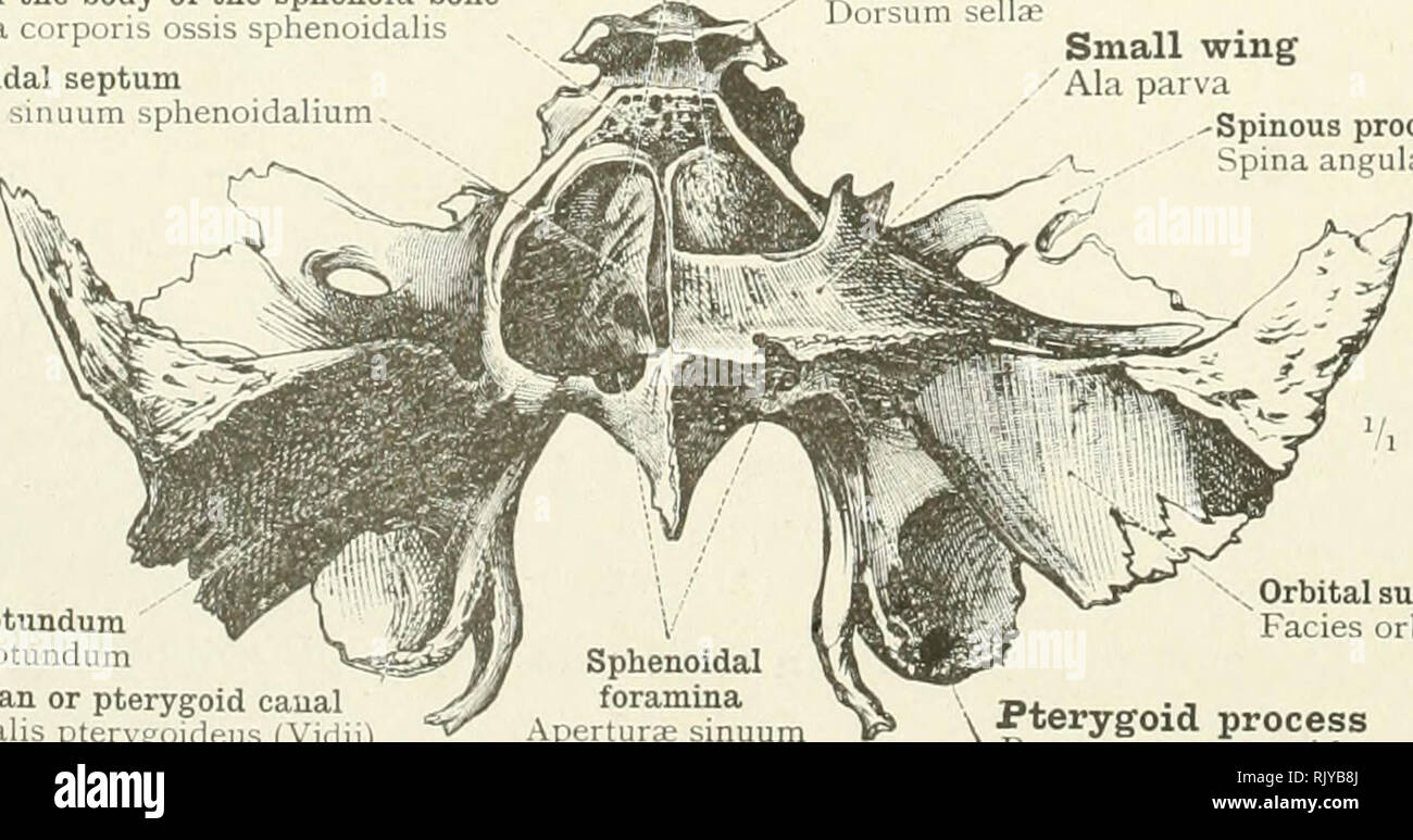
. An atlas of human anatomy for students and physicians. Anatomy. Optic foramen—Foramen opticum Anterior clinoid process Processus clinoideus anterior ^Pituitary fossa Fossa hypophyseos Dorsum sella; Dorsum sellae Body of the sphenoid bone Corpus ossis sphenoidalis Occipitosphenoidal fissure Fissura spheno-occipitalis Sphenoidal septum i Septum sinuum sphenoidalium Basilar portion or process of the occipital hone I "VI y Pars basilaris ossis occipitalis Fig. 121.—The Sphenoidal Sinuses in Median Sagittal Section, the Greater Part of the Sphenoidal Septum having been removed. Seen from the

Image details
Contributor:
Library Book Collection / Alamy Stock PhotoImage ID:
RJYB8JFile size:
7.2 MB (351 KB Compressed download)Releases:
Model - no | Property - noDo I need a release?Dimensions:
2181 x 1146 px | 36.9 x 19.4 cm | 14.5 x 7.6 inches | 150dpiMore information:
This image is a public domain image, which means either that copyright has expired in the image or the copyright holder has waived their copyright. Alamy charges you a fee for access to the high resolution copy of the image.
This image could have imperfections as it’s either historical or reportage.
. An atlas of human anatomy for students and physicians. Anatomy. Optic foramen—Foramen opticum Anterior clinoid process Processus clinoideus anterior ^Pituitary fossa Fossa hypophyseos Dorsum sella; Dorsum sellae Body of the sphenoid bone Corpus ossis sphenoidalis Occipitosphenoidal fissure Fissura spheno-occipitalis Sphenoidal septum i Septum sinuum sphenoidalium Basilar portion or process of the occipital hone I "VI y Pars basilaris ossis occipitalis Fig. 121.—The Sphenoidal Sinuses in Median Sagittal Section, the Greater Part of the Sphenoidal Septum having been removed. Seen from the Left Side. Sphenoidal sinuses—Sinus sphenoidales Cancellous portion of the body of the sphenoid bone Substantia spongiosa corporis ossis sphenoidalis Sphenoidal septum Septum sinuum sphenoidalium.^ Dorsum sellae Dorsum sellae Small wing Ala parva -Spinous process Spina angularis. Foramen rotundum Foramen rotundum Vidian or pterygoid canal Canalis pterygoideus (Vidii) Orbital surface of the great wing Facies orbi talis alae magnae Pterygoid process Processus pterygoideus Sphenoidal foramina Aperturae sinuum sphenoidalium Fig. 122.—The Sphenoidal Sinuses, exposed from Above by the Removal of the Inner Lamella of Compact Bone. The right sinus is opened from above; the left is unopened. Os sphenoidale—The sphenoid bone.. Please note that these images are extracted from scanned page images that may have been digitally enhanced for readability - coloration and appearance of these illustrations may not perfectly resemble the original work.. Toldt, Carl, 1840-1920; Dalla Rosa, Alois, b. 1848; Paul, Eden, 1865-1944, tr; Toldt, Carl, 1840-1920. Anatomischer atlas für Studierende und Ärtze. English. New York, Rebman Company