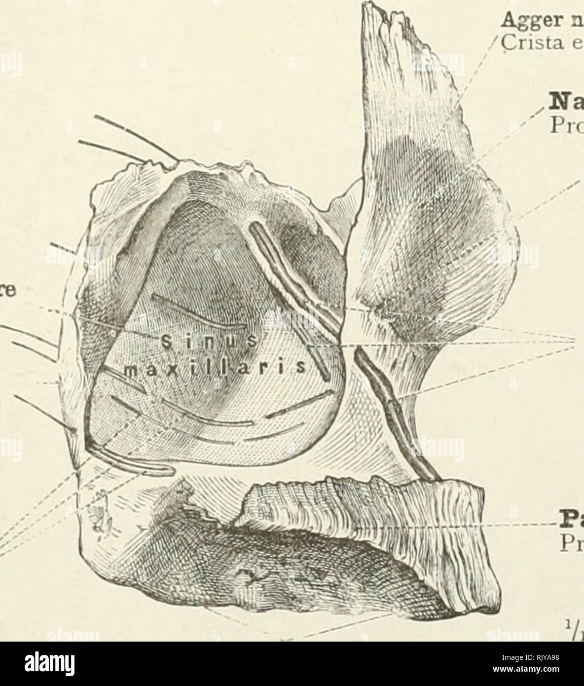
. An atlas of human anatomy for students and physicians. Anatomy. r Posterior dental foramina '' Foramina alveolaria (posteriora) -Dental canals Canales alveolares Alveolar margin Limbus alveolaris Body Corpus maxilla; Fig. 173.—The Left Superior Maxillary Bone. External Surface. The dental canals are exposed by partial removal of the superficial plate of bone, and their course is shown by means of bristles passed through them. Maxillary sinus, or antrum of Highmore Tuberosity Tuber maxillare Dental canals j^ Canales alveolares. Agger nasi, or ethmoidal crest Crista ethmoidalis Nasal process P

Image details
Contributor:
Library Book Collection / Alamy Stock PhotoImage ID:
RJYA98File size:
7.1 MB (305 KB Compressed download)Releases:
Model - no | Property - noDo I need a release?Dimensions:
1509 x 1656 px | 25.6 x 28 cm | 10.1 x 11 inches | 150dpiMore information:
This image is a public domain image, which means either that copyright has expired in the image or the copyright holder has waived their copyright. Alamy charges you a fee for access to the high resolution copy of the image.
This image could have imperfections as it’s either historical or reportage.
. An atlas of human anatomy for students and physicians. Anatomy. r Posterior dental foramina '' Foramina alveolaria (posteriora) -Dental canals Canales alveolares Alveolar margin Limbus alveolaris Body Corpus maxilla; Fig. 173.—The Left Superior Maxillary Bone. External Surface. The dental canals are exposed by partial removal of the superficial plate of bone, and their course is shown by means of bristles passed through them. Maxillary sinus, or antrum of Highmore Tuberosity Tuber maxillare Dental canals j^ Canales alveolares. Agger nasi, or ethmoidal crest Crista ethmoidalis Nasal process Processus frontalis , , Inferior turbinate crest Crista conchalis Dental canals Canales alveolares Palatine process Processus palatinus Alveolar margin Limbus alveolaris Fig. 174.—The Left Superior Maxillary Bone. Internal Surface. The foremost and the hindmost of the dental canals have been exposed by the removal of the superficial plate of bone. By means of bristles passed through the canals the situation of the respective dental foramina is indicated. Most of the inner wall of the antrum of Highmore has been cut away. Maxilla —Superior maxillary bone.. Please note that these images are extracted from scanned page images that may have been digitally enhanced for readability - coloration and appearance of these illustrations may not perfectly resemble the original work.. Toldt, Carl, 1840-1920; Dalla Rosa, Alois, b. 1848; Paul, Eden, 1865-1944, tr; Toldt, Carl, 1840-1920. Anatomischer atlas für Studierende und Ärtze. English. New York, Rebman Company