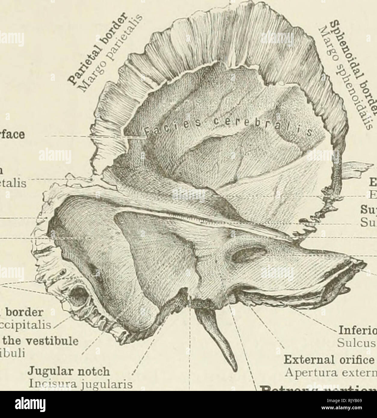
. An atlas of human anatomy for students and physicians. Anatomy. THE SKULL AND THE BONES OF THE SKULL 63 Squamous portion of the temporal bone Squama temporalis Cerebral surface Parietal notch Incisura parietalis. Mastoid portion Pars mastoidea Sigmoid sulcus Sulcus sigmoideus Mastoid foramen Foramen mastoideum Occipital border Margo occipitalis,- External orifice of the aqueduct of the vestibule Apertura externa aquasductus vestibuli Jugular notch Incisura jugularis Intrajugular process Processus intrajugularis Zygoma Processus zygomaticus Eminence of superior semicircular canal Eminentia ar

Image details
Contributor:
Library Book Collection / Alamy Stock PhotoImage ID:
RJYB69File size:
7.1 MB (353.8 KB Compressed download)Releases:
Model - no | Property - noDo I need a release?Dimensions:
1553 x 1609 px | 26.3 x 27.2 cm | 10.4 x 10.7 inches | 150dpiMore information:
This image is a public domain image, which means either that copyright has expired in the image or the copyright holder has waived their copyright. Alamy charges you a fee for access to the high resolution copy of the image.
This image could have imperfections as it’s either historical or reportage.
. An atlas of human anatomy for students and physicians. Anatomy. THE SKULL AND THE BONES OF THE SKULL 63 Squamous portion of the temporal bone Squama temporalis Cerebral surface Parietal notch Incisura parietalis. Mastoid portion Pars mastoidea Sigmoid sulcus Sulcus sigmoideus Mastoid foramen Foramen mastoideum Occipital border Margo occipitalis, - External orifice of the aqueduct of the vestibule Apertura externa aquasductus vestibuli Jugular notch Incisura jugularis Intrajugular process Processus intrajugularis Zygoma Processus zygomaticus Eminence of superior semicircular canal Eminentia arcuata Superior petrosal sulcus Sulcus petrosus superior Flcccular fossa, or hiatus subarcuatus Frssa subarcuata Internal auditory aperture Porus acusticus interims Apex of the petrous portion Apex pyramidis Vi -Inferior petrosal sulcus Sulcus petrosus inferior External orifice of the aqueduct of the cochlea Apertura externa canaliculi cochlear Petrous portion Pars petrosa (Pyramis) Fig. 129.—The Left Temporal Bone seen from Within (Cerebral Surface). Parietal notch Incisura parietalis Petrosquamous fissure Fissura petrosquamosa Eminence of the superior semicircular canal Eminentia arcuata Superior petrosal sulcus Sulcus petrosus superior Hiatus Fallopii—Hiatus canalis facialis Groove of the great superficial petrosal nerve Groove of the small superficial petrosal nerve Sulcus nervi petrosi superficialis minoris Fossa of the Gtasserian ganglion—- Impressio trigemini Apex of the petrous portion..._ Apex pyramidis Carotid canal ' Canalis caroticus Eustachian canal and canal for the tensor tympani muscle Canalis musculotubarius. Please note that these images are extracted from scanned page images that may have been digitally enhanced for readability - coloration and appearance of these illustrations may not perfectly resemble the original work.. Toldt, Carl, 1840-1920; Dalla Rosa, Alois, b. 1848; Paul, Eden, 1865-1944, tr; Toldt, Carl, 1840-1920. Anatomischer atlas für Stu