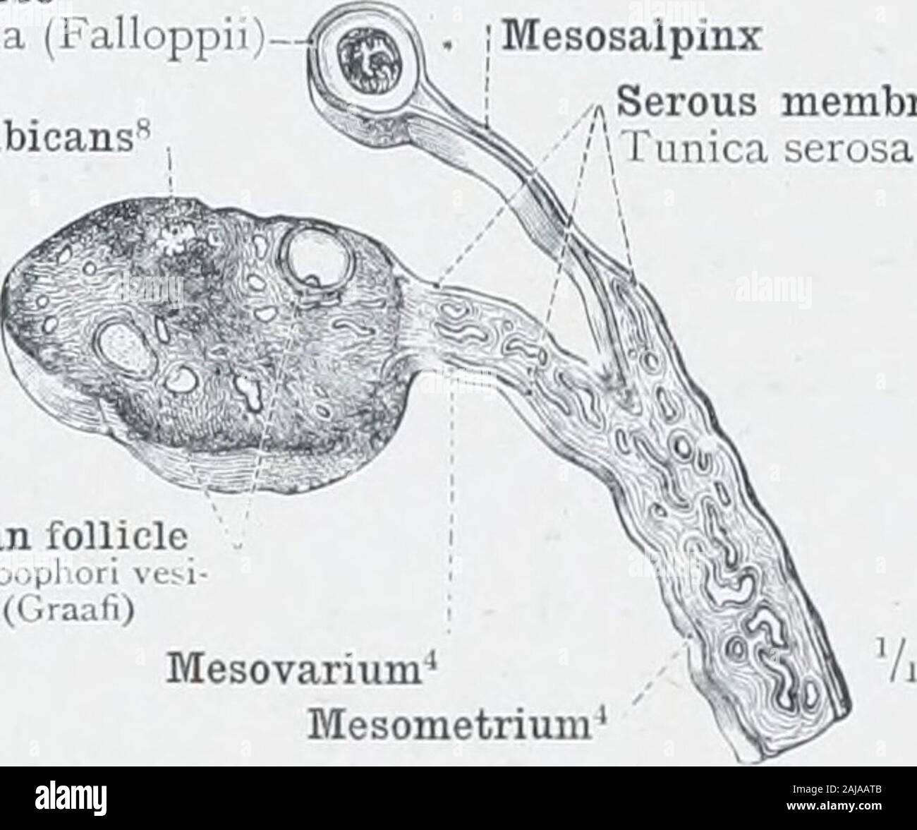
An atlas of human anatomy for students and physicians . Hydatid of Morgagni ^pi cndicsiLuiosa(Morgagnii)Uterovagmal venous plexus 3 PleM.s (vencu*) uteroaginalis Ovarian or pampiniform venous plexus (,,) HUum of the ovary-HilMesometriumUterine artery—A. uterinaRight lateral border of the uterus—Margo lateralis uteriVaginal fornixFornix vaginae (2) Osi abdominale tub;e (3) Plexus (V Fig. S75.—The Uterus and the Right Fallopian Tube, opened from Behind. Epoophoron, Parovarium, OR Organ of Rosenmuller. Fallopian tube Tuba uterina (Falloppiil •Corpus albicans The posterior layer of ihe broad li
Image details
Contributor:
The Reading Room / Alamy Stock PhotoImage ID:
2AJAATBFile size:
7.1 MB (232.5 KB Compressed download)Releases:
Model - no | Property - noDo I need a release?Dimensions:
1729 x 1445 px | 29.3 x 24.5 cm | 11.5 x 9.6 inches | 150dpiMore information:
This image is a public domain image, which means either that copyright has expired in the image or the copyright holder has waived their copyright. Alamy charges you a fee for access to the high resolution copy of the image.
This image could have imperfections as it’s either historical or reportage.
An atlas of human anatomy for students and physicians . Hydatid of Morgagni ^pi_cndicsiLuiosa(Morgagnii)Uterovagmal venous plexus 3 PleM.s (vencu*) uteroaginalis Ovarian or pampiniform venous plexus (, , ) HUum of the ovary-HilMesometriumUterine artery—A. uterinaRight lateral border of the uterus—Margo lateralis uteriVaginal fornixFornix vaginae (2) Osi abdominale tub;e (3) Plexus (V Fig. S75.—The Uterus and the Right Fallopian Tube, opened from Behind. Epoophoron, Parovarium, OR Organ of Rosenmuller. Fallopian tube Tuba uterina (Falloppiil •Corpus albicans The posterior layer of ihe broad ligament of the uterus has been removed. Mesosalpinx Serous membrane 1 unica seiusa. Corpus luteum Graafian follicle I 111 I phori vesiculos Graafian follicle Follicuho5phorne iculosi (Graafi) Mesovarium^ Mesometnum Fig. 876.—Lig omentum Latum Uteri, the Broad Liga-ment OF the Uterus, with the Mesovarium theMesosalpinx, the Ovary, and the Fallopian Tube, IN Transverse Section. Fibrous capsuleof the follicle Theca folliculi Primitive Graafian Stroma of the ovary follicle—FoUiculus Stroma ovarii / .?: oophori primarin .Jli/iiaiifV ?? ???./