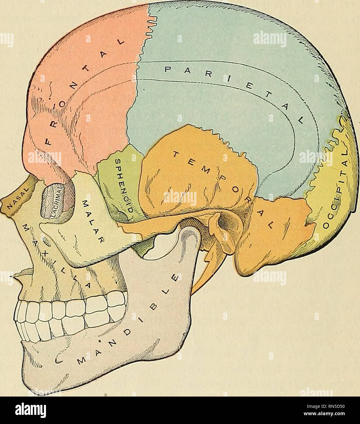
. Anatomy, descriptive and applied. Anatomy. 132 SPECIAL ANATOMY OF THE SKELETON opisthion. On either side of each condyle anteriorly is the anterior condylar fossa, continued as the anterior condylar foramen, for the passage of the hypoglossal nerve and often a meningeal branch of the ascending pharyngeal artery. Behind each condyle is the posterior condylar fossa, continued as the posterior condylar foramen, for the transmission of a vein to the lateral sinus. Behind the foramen magnum is the external occipital crest, terminating above at the external occipital protuberance, while on each si
Image details
Contributor:
Library Book Collection / Alamy Stock PhotoImage ID:
RN5D50File size:
7.1 MB (372.4 KB Compressed download)Releases:
Model - no | Property - noDo I need a release?Dimensions:
1507 x 1657 px | 25.5 x 28.1 cm | 10 x 11 inches | 150dpiMore information:
This image is a public domain image, which means either that copyright has expired in the image or the copyright holder has waived their copyright. Alamy charges you a fee for access to the high resolution copy of the image.
This image could have imperfections as it’s either historical or reportage.
. Anatomy, descriptive and applied. Anatomy. 132 SPECIAL ANATOMY OF THE SKELETON opisthion. On either side of each condyle anteriorly is the anterior condylar fossa, continued as the anterior condylar foramen, for the passage of the hypoglossal nerve and often a meningeal branch of the ascending pharyngeal artery. Behind each condyle is the posterior condylar fossa, continued as the posterior condylar foramen, for the transmission of a vein to the lateral sinus. Behind the foramen magnum is the external occipital crest, terminating above at the external occipital protuberance, while on each side are seen the superior and inferior curved lines; these, as well as the surfaces of bone between them, are rough for the attachment of the muscles, which are enumerated on pages 70 and 71. The Lateral Region of the Skull.—The norma lateralis is of a somewhat triangular form, the base of the triangle being formed by a line extending from the external angular process of the frontal bone along the temporal ridge backward to the outer extremity of the superior curved line of the occiput; and the sides by two lines, the one drawn downward and backward from the external angular process of the frontal bone to the angle of the mandible, the other from the angle of the mandible upward and backward to the outer extremity of the superior curved line. This region is divisible into three portions—temporal fossa, mastoid portion, and zygomatic or infratemporal fossa.. Fig. 99.—Lateral aspect of the skull. The Temporal Fossa (fossa temporalis).—The temporal fossa is bounded above and behind by the temporal ridges, which extend from the extei-nal angular process of the frontal upward and backward across the frontal and parietal bones, curving downward behind to terminate in the posterior root of the zygomatic process. In front it is bounded by the frontal, malar, and greater wing of the sphenoid; externally by the zygomatic arch formed conjointly by the malar and temporal bones; helo