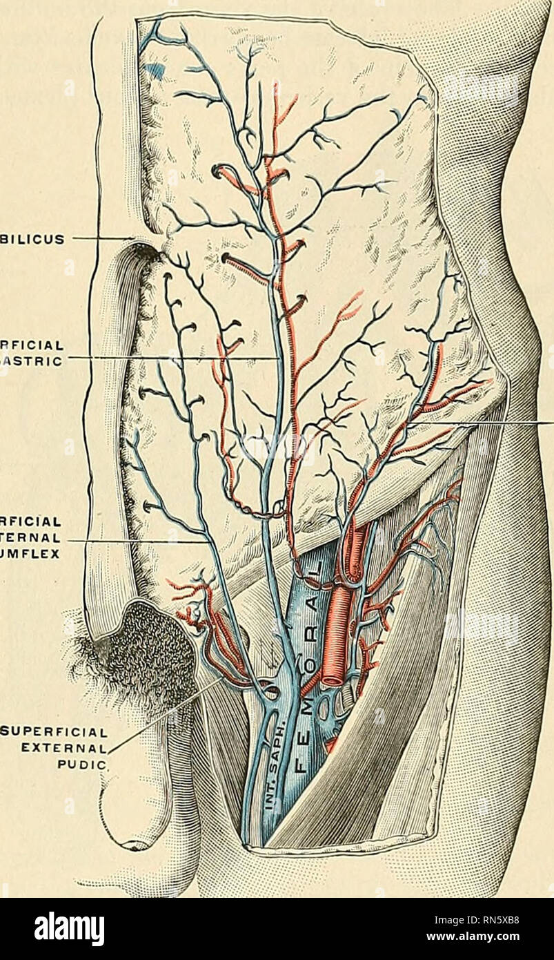
. Anatomy, descriptive and applied. Anatomy. THE BEEP VETNS OF THE LOWER EXTREMITY 74.3 and joins the external iliac vein about three-quarters of an inch above Poupart's ligament. The pubic vein communicates with the obturator em in the obturator fora- men, and ascends on the back of the pubis to terminate in the external iliac -ein. The internal iliac vein {v. hypoc/astrica) commences near the upper part of the great sacrosciatic foramen, passes upward behind and slightly to the inner side of the internal iliac artery, and at the brim of the pelvis joins with the external iliac to form the
Image details
Contributor:
Library Book Collection / Alamy Stock PhotoImage ID:
RN5XB8File size:
7.2 MB (556.5 KB Compressed download)Releases:
Model - no | Property - noDo I need a release?Dimensions:
1243 x 2011 px | 21 x 34.1 cm | 8.3 x 13.4 inches | 150dpiMore information:
This image is a public domain image, which means either that copyright has expired in the image or the copyright holder has waived their copyright. Alamy charges you a fee for access to the high resolution copy of the image.
This image could have imperfections as it’s either historical or reportage.
. Anatomy, descriptive and applied. Anatomy. THE BEEP VETNS OF THE LOWER EXTREMITY 74.3 and joins the external iliac vein about three-quarters of an inch above Poupart's ligament. The pubic vein communicates with the obturator em in the obturator fora- men, and ascends on the back of the pubis to terminate in the external iliac -ein. The internal iliac vein {v. hypoc/astrica) commences near the upper part of the great sacrosciatic foramen, passes upward behind and slightly to the inner side of the internal iliac artery, and at the brim of the pelvis joins with the external iliac to form the common iliac. UMBfLICUS. SUPERFICIAL EXTERNAL CIRCUMFLEX SUPERFICIAL INTERNAL CIRCUMFLEX Fig. 520.—The femoral Dtl its tributaries. (Poirier and Charpy.) Tributaries.—With the exception of the fetal umbilical vein, which passes up- ward and backward from the umbilicus to the liver, and the iliolumbar vein which usually joins the common iliac vein, the tributaries of the internal iliac vein corre- spond with the branches of the internal iliac artery. It receives (a) the gluteal, sciatic, internal pudic, and obturator veins, which have their origins outside the pelvis; (b) the lateral sacral veins, which lie in front of the sacrum; and (c) the middle hemorrhoidal, vesical, uterine, and vaginal veins, which originate in venous plexuses connected with the pelvic viscera. I. The gluteal veins (vv. glutaeae super lores) or venae comites of the gluteal artery, receive tributaries from the buttock corresponding with the branches of. Please note that these images are extracted from scanned page images that may have been digitally enhanced for readability - coloration and appearance of these illustrations may not perfectly resemble the original work.. Gray, Henry, 1825-1861; Spitzka, Edward Anthony, 1876-1922. Philadelphia, New York, Lea & Febiger