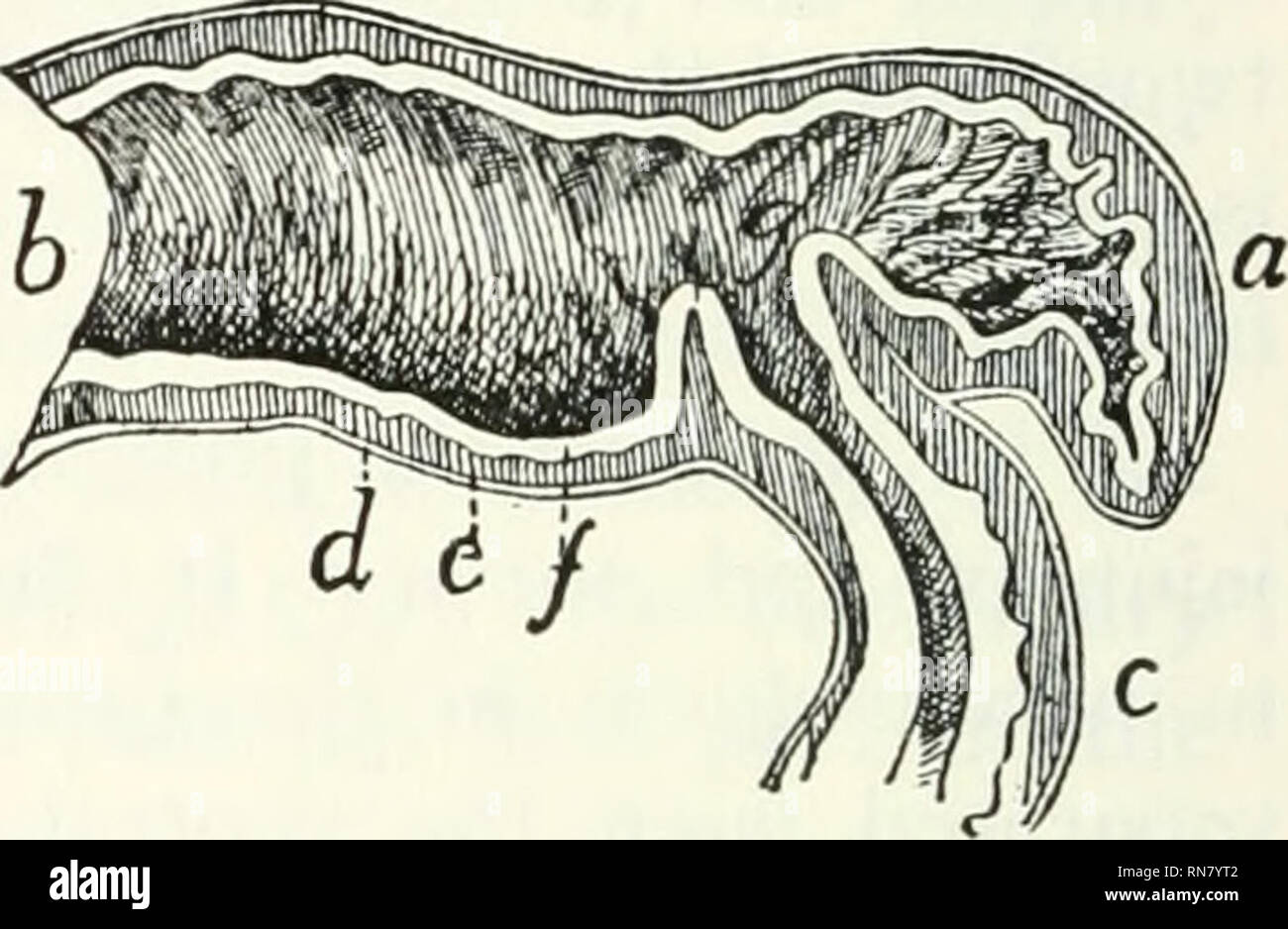
. Anatomy of the cat. Cats; Mammals. Fig. 98.—Junction of Small and Large Intestine. Fig. 99.—Section ok the Ileo- colic Valve. Fig. 98.—a, ileum; i>, ascending colon; c, coecum; d, position of ileocolic valve. Fig. 99.—a, cajcum; b, colon; c, ileum; J, longitudinal muscle layer; e, trans- verse muscle layer; /, mucosa; g, ileocolic valve (opened, as when material is pass- ing into the colon). blind pouch thus formed by the cranial end of the colon is the caecum (Fig. 98, c Fig. 99, a). The caecum ends in a slight conical projection which may be considered as the rudiment of a vermiform ap
Image details
Contributor:
Library Book Collection / Alamy Stock PhotoImage ID:
RN7YT2File size:
7.1 MB (280.7 KB Compressed download)Releases:
Model - no | Property - noDo I need a release?Dimensions:
1957 x 1277 px | 33.1 x 21.6 cm | 13 x 8.5 inches | 150dpiMore information:
This image is a public domain image, which means either that copyright has expired in the image or the copyright holder has waived their copyright. Alamy charges you a fee for access to the high resolution copy of the image.
This image could have imperfections as it’s either historical or reportage.
. Anatomy of the cat. Cats; Mammals. Fig. 98.—Junction of Small and Large Intestine. Fig. 99.—Section ok the Ileo- colic Valve. Fig. 98.—a, ileum; i>, ascending colon; c, coecum; d, position of ileocolic valve. Fig. 99.—a, cajcum; b, colon; c, ileum; J, longitudinal muscle layer; e, trans- verse muscle layer; /, mucosa; g, ileocolic valve (opened, as when material is pass- ing into the colon). blind pouch thus formed by the cranial end of the colon is the caecum (Fig. 98, c Fig. 99, a). The caecum ends in a slight conical projection which may be considered as the rudiment of a vermiform appendix. The colon lies at first on the right side and passes at first craniad; then transversely to the left, then caudad, lying nearly in the middle line and next to the dorsal abdominal wall. The colon may thus be distinguished accord- ing to its direction into ascending, transverse, and descending colon. At its caudal end the colon passes without sharp limit into the rectum. At the bottom of the caecum on its inner surface is seen a collection of solitary glands forming one of the agminated glands of Peyer, or Peyer's patches. The mucous membrane is without villi. It presents a few considerable elevations, probably solitary glands.. Please note that these images are extracted from scanned page images that may have been digitally enhanced for readability - coloration and appearance of these illustrations may not perfectly resemble the original work.. Reighard, Jacob Ellsworth, 1861-1942; Jennings, H. S. (Herbert Spencer), 1868-1947. [Austin, TX] : BookLab, Inc.