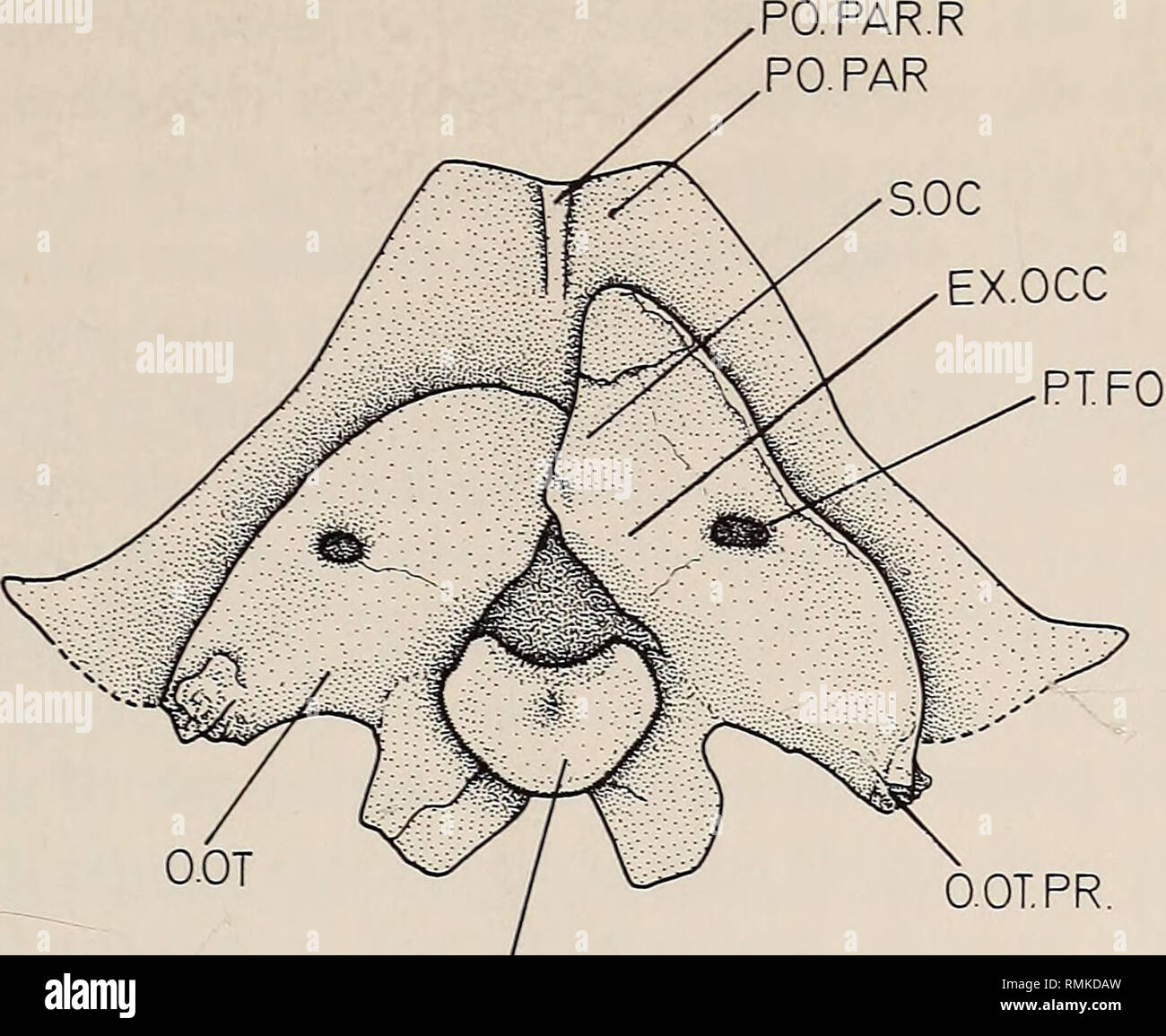
. Annals of the South African Museum = Annale van die Suid-Afrikaanse Museum. Natural history. A NEW DICYNODONT ANCESTOR FROM THE UPPER ECCA 133. O.OT.PR. QC Fig. 13. Eodicynodon oosthuizeni. Occipital view. from the general Dicynodon condition. The borders of the supraoccipital are only faintly recognizable but it would seem that its ventro-medial border forms the dorsal rim of the foramen magnum in the usual way. From here it fans out dorsally and laterally to meet the postparietal and tabulars. The line of fusion between the supraoccipital and exoccipital cannot be traced. The exoccipital f
Image details
Contributor:
Library Book Collection / Alamy Stock PhotoImage ID:
RMKDAWFile size:
7.1 MB (337.5 KB Compressed download)Releases:
Model - no | Property - noDo I need a release?Dimensions:
1746 x 1431 px | 29.6 x 24.2 cm | 11.6 x 9.5 inches | 150dpiMore information:
This image is a public domain image, which means either that copyright has expired in the image or the copyright holder has waived their copyright. Alamy charges you a fee for access to the high resolution copy of the image.
This image could have imperfections as it’s either historical or reportage.
. Annals of the South African Museum = Annale van die Suid-Afrikaanse Museum. Natural history. A NEW DICYNODONT ANCESTOR FROM THE UPPER ECCA 133. O.OT.PR. QC Fig. 13. Eodicynodon oosthuizeni. Occipital view. from the general Dicynodon condition. The borders of the supraoccipital are only faintly recognizable but it would seem that its ventro-medial border forms the dorsal rim of the foramen magnum in the usual way. From here it fans out dorsally and laterally to meet the postparietal and tabulars. The line of fusion between the supraoccipital and exoccipital cannot be traced. The exoccipital forms the lateral rim of the foramen magnum and extends outward to the post-temporal fossa. From its base a flat roughly triangu- lar flange extends to cover the ventro-medial portion of the opisthotic. Although the sutures are indistinct it seems that the exoccipitals also form the lateral borders of the foramen magnum. The basioccipital forms the ventro-medial portion of the occipital condyle, it is continued anteriorly in the midline from where two ventro-laterally directed triangular extensions develop to cover the posterior surface of the prominent tubera, which form the bony casing of the fenestra ovalis and for the cochlear recess of the internal ear. In most dicyno- donts the tubera are described as being formed by the basioccipitals, but in Eodicynodon each structure is quite clearly formed by the basioccipital and the basisphenoid, with the opisthotic forming the postero-lateral rim. The opisthotic, similarly, is a roughly triangular plate. Its dorsal boundary extends from the ventro-lateral corner of the foramen magnum to the ventral rim of the post-temporal fossa. A ventro-medially directed flange forms the postero-dorso-lateral rim of the prominent tuber. As in Pristerodon (Barry 1967) the opisthotic carries a strongly developed ridge-like opisthotic process which laterally tapers into a posteriorly directed point (Fig. 13). As stated in the afore- mentioned paper i