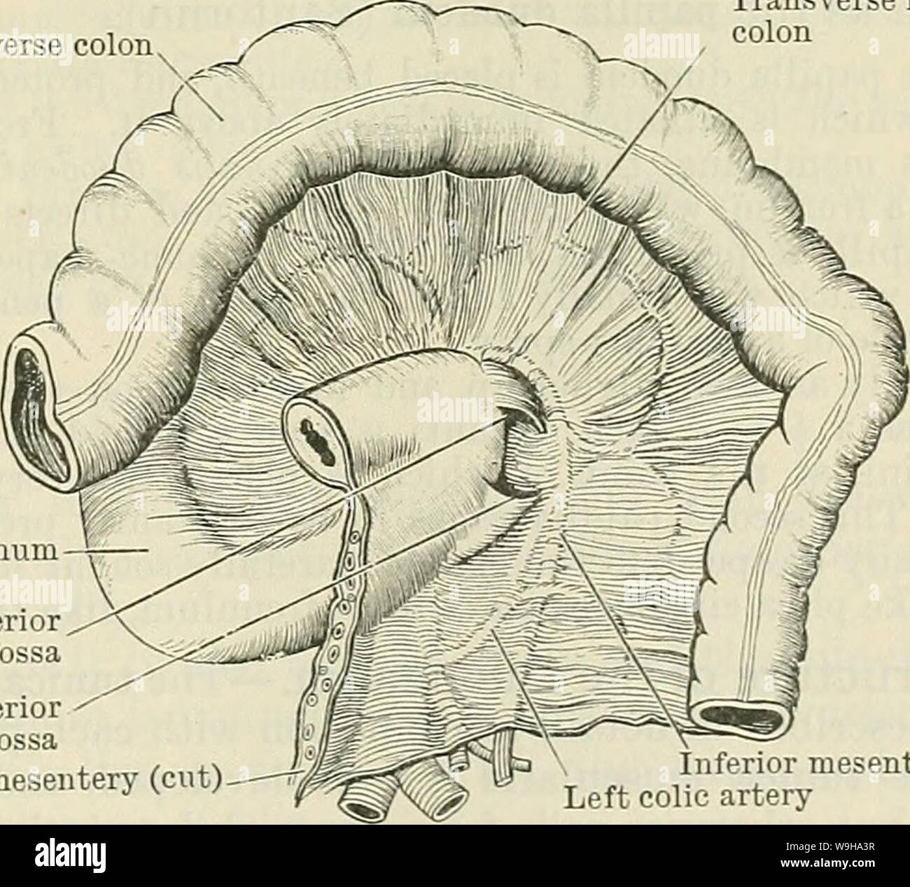
Archive image from page 1218 of Cunningham's Text-book of anatomy (1914). Cunningham's Text-book of anatomy cunninghamstextb00cunn Year: 1914 ( THE DUODENUM. 1185 The bile-duct, after passing down behind the superior part of the duodenum, descends between the head of the pancreas and the descending part, nearly as far as its middle ; here it is joined by the pancreatic duct, and the two, piercing the wall of the duodenum obliquely, open by a common orifice on its inner aspect, about 3 to 4 inches (8-7 to 10 cm.) beyond the pylorus. Pars Inferior.—The inferior part (O.T. third portion) begins

Image details
Contributor:
Bookive / Alamy Stock PhotoImage ID:
W9HA3RFile size:
5.7 MB (303.7 KB Compressed download)Releases:
Model - no | Property - noDo I need a release?Dimensions:
1486 x 1346 px | 25.2 x 22.8 cm | 9.9 x 9 inches | 150dpiMore information:
This image is a public domain image, which means either that copyright has expired in the image or the copyright holder has waived their copyright. Alamy charges you a fee for access to the high resolution copy of the image.
This image could have imperfections as it’s either historical or reportage.
Archive image from page 1218 of Cunningham's Text-book of anatomy (1914). Cunningham's Text-book of anatomy cunninghamstextb00cunn Year: 1914 ( THE DUODENUM. 1185 The bile-duct, after passing down behind the superior part of the duodenum, descends between the head of the pancreas and the descending part, nearly as far as its middle ; here it is joined by the pancreatic duct, and the two, piercing the wall of the duodenum obliquely, open by a common orifice on its inner aspect, about 3 to 4 inches (8-7 to 10 cm.) beyond the pylorus. Pars Inferior.—The inferior part (O.T. third portion) begins at the right side of the third or fourth lumbar vertebra. It is described in two parts, pars horizontalis, transverse in direction, and pars ascenclens, and it shows that arrangement in Fig. 933. The pars horizontalis runs more or less transversely to the left across the inferior vena cava (Fig. 933) for one or two inches, and the pars ascenclens passes very obliquely, or even vertically, upwards in front of the aorta and left psoas major muscle. Finally, having reached the inferior surface of the pancreas, it bends forwards, and passes into the jejunum. Anteriorly, it is crossed (about the junction of its two divisions) by the superior mesenteric vessels, and also by the root of the mesentery (Fig. 933). On each side of this it is covered by coils of small intestine. Posteriorly, the pars horizontalis lies over the vena cava inferior ; the pars ascendens on the aorta, the left renal vein and occasionally also the artery, and the left psoas major muscle, all of which separate it from the vertebral column. Above, it is closely applied in its whole extent to the head of the pancreas. The left side of the pars ascendens, which is free, lies in contact with some coils of the small intestine. Peritoneal Relations.—The inferior part of the duodenum is covered by peritoneum on its anterior surface throughout, except where it is crossed by the superior mesenteric vessels and the ro