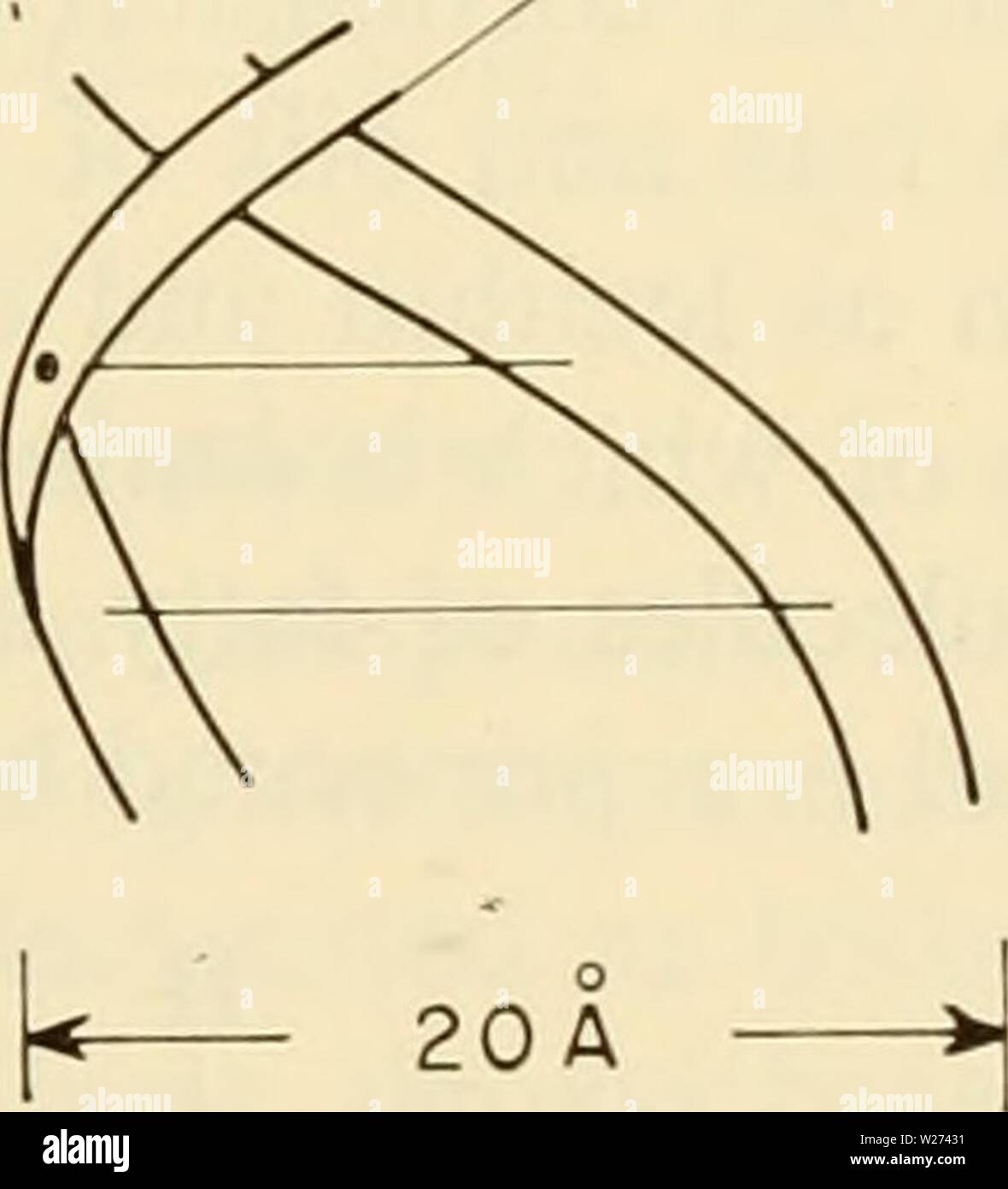
Archive image from page 38 of Cytology (1961). Cytology cytology00wils Year: 1961 ,SâT ⢠H â AâS P P 'SâC- H-G-S Figure 2-5. Schematic Representation of the Watson-Crick Model of the DNA Molecule. The molecule consists of two sugar-phosphate chains (-P-S-P-) entwined to form a helix which is held together by hydrogen bonds (â¢H) between the companion bases of the two chains. A, adenine; T, thy- mine; G, guanine; C, cytosine. The horizontal solid lines represent comple- mentary bases held together by hydrogen bonding in other parts of the helical molecule. (Redrawn from Watson, J. D., and
Image details
Contributor:
Bookive / Alamy Stock PhotoImage ID:
W27431File size:
5.7 MB (85.7 KB Compressed download)Releases:
Model - no | Property - noDo I need a release?Dimensions:
1348 x 1484 px | 22.8 x 25.1 cm | 9 x 9.9 inches | 150dpiMore information:
This image is a public domain image, which means either that copyright has expired in the image or the copyright holder has waived their copyright. Alamy charges you a fee for access to the high resolution copy of the image.
This image could have imperfections as it’s either historical or reportage.
Archive image from page 38 of Cytology (1961). Cytology cytology00wils Year: 1961 , SâT ⢠H â AâS P P 'SâC- H-G-S Figure 2-5. Schematic Representation of the Watson-Crick Model of the DNA Molecule. The molecule consists of two sugar-phosphate chains (-P-S-P-) entwined to form a helix which is held together by hydrogen bonds (â¢H) between the companion bases of the two chains. A, adenine; T, thy- mine; G, guanine; C, cytosine. The horizontal solid lines represent comple- mentary bases held together by hydrogen bonding in other parts of the helical molecule. (Redrawn from Watson, J. D., and Crick, F. H. C, 1953. 'Ge- netical Implications of the Structure of Deoxyribonucleic Acid, ' Nature, 171. Fig. 2, p. 965.) gen bonds (Figure 2-5). Each turn of the helix is about 34 A and there are about 10 base pairs for every gyre. The diameter is about 20 A, this dimension corresponding to the minimal space required to fit in the base pairs, which on the basis of these measurements would be adenine- GENERAL MORPHOLOGY AND CHEMISTRY OF THE CELL / 19