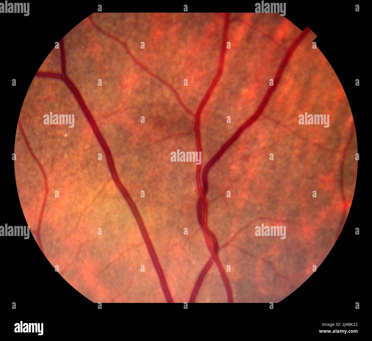
Arteriovenous nicking, fundoscopy

RMID:Image ID:2J4BK22
Image details
Contributor:
Science Photo Library / Alamy Stock PhotoImage ID:
2J4BK22File size:
30.6 MB (780.4 KB Compressed download)Releases:
Model - no | Property - noDo I need a release?Dimensions:
3550 x 3011 px | 30.1 x 25.5 cm | 11.8 x 10 inches | 300dpiDate taken:
5 April 2022Photographer:
ALAN FROHLICHSTEIN/SCIENCE PHOTO LIBRARYMore information:
Fundoscopy image of the retina of the eye showing arteriovenous nicking (in blood vessels down centre right). This is where a retinal venule is compressed by a retinal arteriole that has become stiffened by disease. The walls of the arteriole have also thickened, given them the appearance of a copper wire. Both of these changes are caused by high blood pressure (hypertension) and are a sign that the patient has chronic systemic hypertension.