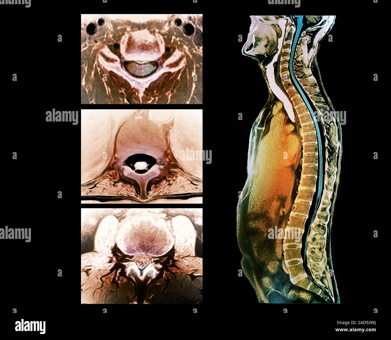
Backbone and spinal cord anatomy. Coloured magnetic resonance imaging (MRI) scans of the vertebral column and spinal cord in a 36-year-old man. The sc
Image details
Contributor:
Science Photo Library / Alamy Stock PhotoImage ID:
2AD5XWJFile size:
50 MB (1 MB Compressed download)Releases:
Model - no | Property - noDo I need a release?Dimensions:
4673 x 3739 px | 39.6 x 31.7 cm | 15.6 x 12.5 inches | 300dpiDate taken:
23 January 2017Photographer:
ZEPHYR/SCIENCE PHOTO LIBRARYMore information:
Backbone and spinal cord anatomy. Coloured magnetic resonance imaging (MRI) scans of the vertebral column and spinal cord in a 36-year-old man. The scan at right shows the spine in sagittal section, with the front of the body at left. The three scans at left show the spinal cord in axial cross-section at different levels and vertebrae within the vertebral column. At top left is a section at the C5-C6 cervical level. At centre left is a section at the T9 thoracic level. At lower left is a section at the L2-L3 lumbar level. The spine runs from the skull to the pelvis. It is a flexible column of compact bones called vertebrae, that enclose and protect the spinal cord. The vertebrae are separated by intervertebral discs (discs of fibrocartilage). In an adult, there are 26 vertebrae.