···
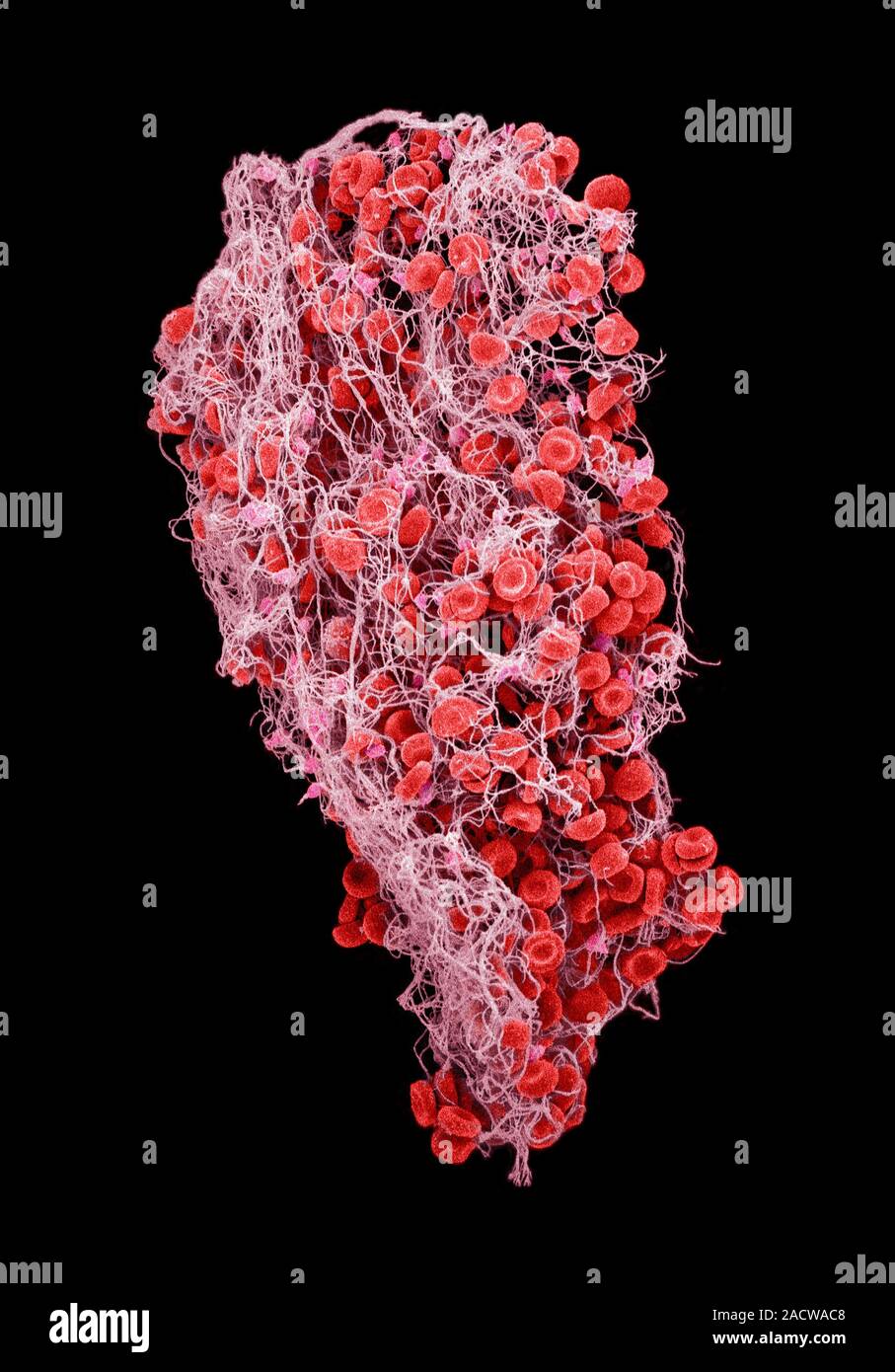
Blood clot, coloured scanning electron micrograph (SEM). Red blood cells (erythrocytes) are trapped within a fibrin protein mesh (light pink). The fib Image details File size:
36.7 MB (1.4 MB Compressed download)
Open your image file to the full size using image processing software.
Dimensions:
2990 x 4285 px | 25.3 x 36.3 cm | 10 x 14.3 inches | 300dpi
Date taken:
22 January 2014
More information:
Blood clot, coloured scanning electron micrograph (SEM). Red blood cells (erythrocytes) are trapped within a fibrin protein mesh (light pink). The fibrin mesh is formed in response to chemicals secreted by platelets (bright pink), fragments of white blood cells. Clots are formed in response to cardiovascular disease or injuries to blood vessels.
Search stock photos by tags
Similar stock images White blood cells and a platelet. Coloured scanning electron micrograph (SEM) of white blood cells and a single platelet (blue). Platelets are fragments of white blood cells that under normal circumstances are small and biconcave in form. However, if there is a break in the surface of a blood vessel the platelets come into contact with molecules they are not used to and become activated. Magnification: x3000 when printed at 10 centimetres wide. Stock Photo https://www.alamy.com/image-license-details/?v=1 https://www.alamy.com/white-blood-cells-and-a-platelet-coloured-scanning-electron-micrograph-sem-of-white-blood-cells-and-a-single-platelet-blue-platelets-are-fragments-of-white-blood-cells-that-under-normal-circumstances-are-small-and-biconcave-in-form-however-if-there-is-a-break-in-the-surface-of-a-blood-vessel-the-platelets-come-into-contact-with-molecules-they-are-not-used-to-and-become-activated-magnification-x3000-when-printed-at-10-centimetres-wide-image220702048.html RF PR1RM0 – White blood cells and a platelet. Coloured scanning electron micrograph (SEM) of white blood cells and a single platelet (blue). Platelets are fragments of white blood cells that under normal circumstances are small and biconcave in form. However, if there is a break in the surface of a blood vessel the platelets come into contact with molecules they are not used to and become activated. Magnification: x3000 when printed at 10 centimetres wide. Activated platelets, coloured scanning electron micrograph (SEM). Platelets are blood cell fragments that play an essential role in blood clotting and wound repair, and can also activate certain immune responses. They are formed in the red bone marrow, lungs, and spleen by fragmentation of very large cells known as megakaryocytes. Platelets in the blood are small oval disks and are termed non-activated platelets or thrombocytes. They are the body's first line of defence against excessive blood loss. Magnification: x2,000 at 10 cm wide. Stock Photo https://www.alamy.com/image-license-details/?v=1 https://www.alamy.com/activated-platelets-coloured-scanning-electron-micrograph-sem-platelets-are-blood-cell-fragments-that-play-an-essential-role-in-blood-clotting-and-wound-repair-and-can-also-activate-certain-immune-responses-they-are-formed-in-the-red-bone-marrow-lungs-and-spleen-by-fragmentation-of-very-large-cells-known-as-megakaryocytes-platelets-in-the-blood-are-small-oval-disks-and-are-termed-non-activated-platelets-or-thrombocytes-they-are-the-bodys-first-line-of-defence-against-excessive-blood-loss-magnification-x2000-at-10-cm-wide-image220702406.html RF PR1T4P – Activated platelets, coloured scanning electron micrograph (SEM). Platelets are blood cell fragments that play an essential role in blood clotting and wound repair, and can also activate certain immune responses. They are formed in the red bone marrow, lungs, and spleen by fragmentation of very large cells known as megakaryocytes. Platelets in the blood are small oval disks and are termed non-activated platelets or thrombocytes. They are the body's first line of defence against excessive blood loss. Magnification: x2,000 at 10 cm wide. Blood clot. Coloured scanning electron micrograph (SEM) of red blood cells (erythrocytes) trapped in a fibrin mesh. The production of fibrin is triggered by cells called platelets, activated when a blood vessel is damaged. The fibrin binds the various blood cells together, forming a solid structure called a blood clot. A blood clot is a normal response, preventing an excessive loss of blood. However, inappropriate clotting is a major cause of heart attacks and strokes. Magnification x 2000 at 10cm wide. Stock Photo https://www.alamy.com/image-license-details/?v=1 https://www.alamy.com/blood-clot-coloured-scanning-electron-micrograph-sem-of-red-blood-cells-erythrocytes-trapped-in-a-fibrin-mesh-the-production-of-fibrin-is-triggered-by-cells-called-platelets-activated-when-a-blood-vessel-is-damaged-the-fibrin-binds-the-various-blood-cells-together-forming-a-solid-structure-called-a-blood-clot-a-blood-clot-is-a-normal-response-preventing-an-excessive-loss-of-blood-however-inappropriate-clotting-is-a-major-cause-of-heart-attacks-and-strokes-magnification-x-2000-at-10cm-wide-image622879745.html RF 2Y5AHD5 – Blood clot. Coloured scanning electron micrograph (SEM) of red blood cells (erythrocytes) trapped in a fibrin mesh. The production of fibrin is triggered by cells called platelets, activated when a blood vessel is damaged. The fibrin binds the various blood cells together, forming a solid structure called a blood clot. A blood clot is a normal response, preventing an excessive loss of blood. However, inappropriate clotting is a major cause of heart attacks and strokes. Magnification x 2000 at 10cm wide. Blood clot. Coloured scanning electron micrograph (SEM) of red blood cells (erythrocytes) trapped in a fibrin mesh. The production of fibrin is triggered by cells called platelets, activated when a blood vessel is damaged. The fibrin binds the various blood cells together, forming a solid structure called a blood clot. A blood clot is a normal response, preventing an excessive loss of blood. However, inappropriate clotting is a major cause of heart attacks and strokes. Magnification x 2000 at 10cm wide. Stock Photo https://www.alamy.com/image-license-details/?v=1 https://www.alamy.com/blood-clot-coloured-scanning-electron-micrograph-sem-of-red-blood-cells-erythrocytes-trapped-in-a-fibrin-mesh-the-production-of-fibrin-is-triggered-by-cells-called-platelets-activated-when-a-blood-vessel-is-damaged-the-fibrin-binds-the-various-blood-cells-together-forming-a-solid-structure-called-a-blood-clot-a-blood-clot-is-a-normal-response-preventing-an-excessive-loss-of-blood-however-inappropriate-clotting-is-a-major-cause-of-heart-attacks-and-strokes-magnification-x-2000-at-10cm-wide-image622879887.html RF 2Y5AHJ7 – Blood clot. Coloured scanning electron micrograph (SEM) of red blood cells (erythrocytes) trapped in a fibrin mesh. The production of fibrin is triggered by cells called platelets, activated when a blood vessel is damaged. The fibrin binds the various blood cells together, forming a solid structure called a blood clot. A blood clot is a normal response, preventing an excessive loss of blood. However, inappropriate clotting is a major cause of heart attacks and strokes. Magnification x 2000 at 10cm wide. Crenated blood clot, coloured scanning electron micrograph (SEM). Red blood cells are red and fibrin proteinstrands are green Stock Photo https://www.alamy.com/image-license-details/?v=1 https://www.alamy.com/stock-photo-crenated-blood-clot-coloured-scanning-electron-micrograph-sem-red-21201609.html RF B6DPT9 – Crenated blood clot, coloured scanning electron micrograph (SEM). Red blood cells are red and fibrin proteinstrands are green Crenated blood clot, coloured scanning electron micrograph (SEM). Red blood cells are red and fibrin proteinstrands are green Stock Photo https://www.alamy.com/image-license-details/?v=1 https://www.alamy.com/stock-photo-crenated-blood-clot-coloured-scanning-electron-micrograph-sem-red-21201610.html RF B6DPTA – Crenated blood clot, coloured scanning electron micrograph (SEM). Red blood cells are red and fibrin proteinstrands are green Blood clot, coloured scanning electron micrograph (SEM). Stock Photo https://www.alamy.com/image-license-details/?v=1 https://www.alamy.com/stock-photo-blood-clot-coloured-scanning-electron-micrograph-sem-19413251.html RF B3G9PB – Blood clot, coloured scanning electron micrograph (SEM). Blood clot. Coloured scanning electron micrograph (SEM) of red blood cells clumped together with fibrin to form a blood clot. Stock Photo https://www.alamy.com/image-license-details/?v=1 https://www.alamy.com/stock-photo-blood-clot-coloured-scanning-electron-micrograph-sem-of-red-blood-21207763.html RF B6E2M3 – Blood clot. Coloured scanning electron micrograph (SEM) of red blood cells clumped together with fibrin to form a blood clot. Blood clot. Coloured scanning electron micrograph (SEM) of red blood cells clumped together with fibrin to form a blood clot. Stock Photo https://www.alamy.com/image-license-details/?v=1 https://www.alamy.com/stock-photo-blood-clot-coloured-scanning-electron-micrograph-sem-of-red-blood-21207765.html RF B6E2M5 – Blood clot. Coloured scanning electron micrograph (SEM) of red blood cells clumped together with fibrin to form a blood clot. White blood cells and a platelet. Coloured scanning electron micrograph (SEM) of white blood cells and a single platelet (blue). Platelets are fragments of white blood cells that under normal circumstances are small and biconcave in form. However, if there is a break in the surface of a blood vessel the platelets come into contact with molecules they are not used to and become activated. Magnification: x3000 when printed at 10 centimetres wide. Stock Photo https://www.alamy.com/image-license-details/?v=1 https://www.alamy.com/white-blood-cells-and-a-platelet-coloured-scanning-electron-micrograph-sem-of-white-blood-cells-and-a-single-platelet-blue-platelets-are-fragments-of-white-blood-cells-that-under-normal-circumstances-are-small-and-biconcave-in-form-however-if-there-is-a-break-in-the-surface-of-a-blood-vessel-the-platelets-come-into-contact-with-molecules-they-are-not-used-to-and-become-activated-magnification-x3000-when-printed-at-10-centimetres-wide-image220702026.html RF PR1RK6 – White blood cells and a platelet. Coloured scanning electron micrograph (SEM) of white blood cells and a single platelet (blue). Platelets are fragments of white blood cells that under normal circumstances are small and biconcave in form. However, if there is a break in the surface of a blood vessel the platelets come into contact with molecules they are not used to and become activated. Magnification: x3000 when printed at 10 centimetres wide. White blood cells and a platelet. Coloured scanning electron micrograph (SEM) of white blood cells and a single platelet (blue). Platelets are fragments of white blood cells that under normal circumstances are small and biconcave in form. However, if there is a break in the surface of a blood vessel the platelets come into contact with molecules they are not used to and become activated. Magnification: x3000 when printed at 10 centimetres wide. Stock Photo https://www.alamy.com/image-license-details/?v=1 https://www.alamy.com/white-blood-cells-and-a-platelet-coloured-scanning-electron-micrograph-sem-of-white-blood-cells-and-a-single-platelet-blue-platelets-are-fragments-of-white-blood-cells-that-under-normal-circumstances-are-small-and-biconcave-in-form-however-if-there-is-a-break-in-the-surface-of-a-blood-vessel-the-platelets-come-into-contact-with-molecules-they-are-not-used-to-and-become-activated-magnification-x3000-when-printed-at-10-centimetres-wide-image220702012.html RF PR1RJM – White blood cells and a platelet. Coloured scanning electron micrograph (SEM) of white blood cells and a single platelet (blue). Platelets are fragments of white blood cells that under normal circumstances are small and biconcave in form. However, if there is a break in the surface of a blood vessel the platelets come into contact with molecules they are not used to and become activated. Magnification: x3000 when printed at 10 centimetres wide. Red blood cells and platelets. Coloured scanning electon micrograph (SEM) of human erythrocytes (red blood cells) and a platelet aggregate (yellow). Platelets are fragments of white blood cells that under normal circumstances are small and biconcave in form. However, if there is a break in the surface of a blood vessel the platelets come into contact with molecules they are not used to and become activated. They become amorphous in form, with long projections (pseudopodia) that help them adhere to other cells and each other, forming a clot. Magnification: x3000 when printed at 10 centimetres Stock Photo https://www.alamy.com/image-license-details/?v=1 https://www.alamy.com/stock-photo-red-blood-cells-and-platelets-coloured-scanning-electon-micrograph-102520732.html RF FXP66M – Red blood cells and platelets. Coloured scanning electon micrograph (SEM) of human erythrocytes (red blood cells) and a platelet aggregate (yellow). Platelets are fragments of white blood cells that under normal circumstances are small and biconcave in form. However, if there is a break in the surface of a blood vessel the platelets come into contact with molecules they are not used to and become activated. They become amorphous in form, with long projections (pseudopodia) that help them adhere to other cells and each other, forming a clot. Magnification: x3000 when printed at 10 centimetres Red blood cells and platelets. Coloured scanning electon micrograph (SEM) of human erythrocytes (red blood cells) and a platelet aggregate (orange). Platelets are fragments of white blood cells that under normal circumstances are small and biconcave in form. However, if there is a break in the surface of a blood vessel the platelets come into contact with molecules they are not used to and become activated. They become amorphous in form, with long projections (pseudopodia) that help them adhere to other cells and each other, forming a clot. Magnification: x3000 when printed at 10 centimetres Stock Photo https://www.alamy.com/image-license-details/?v=1 https://www.alamy.com/stock-photo-red-blood-cells-and-platelets-coloured-scanning-electon-micrograph-102520725.html RF FXP66D – Red blood cells and platelets. Coloured scanning electon micrograph (SEM) of human erythrocytes (red blood cells) and a platelet aggregate (orange). Platelets are fragments of white blood cells that under normal circumstances are small and biconcave in form. However, if there is a break in the surface of a blood vessel the platelets come into contact with molecules they are not used to and become activated. They become amorphous in form, with long projections (pseudopodia) that help them adhere to other cells and each other, forming a clot. Magnification: x3000 when printed at 10 centimetres 