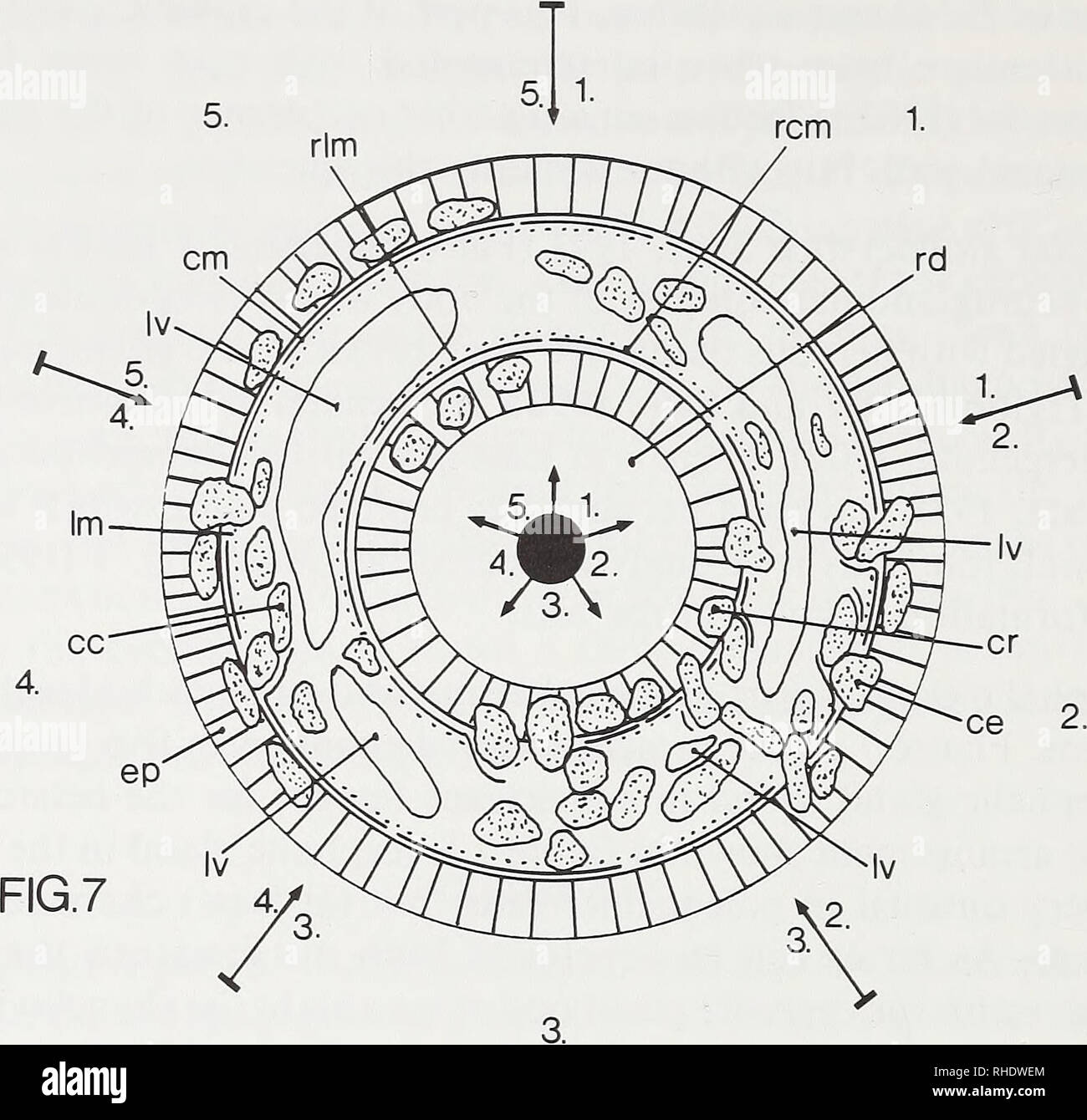
. Bonner zoologische Beiträge : Herausgeber: Zoologisches Forschungsinstitut und Museum Alexander Koenig, Bonn. Biology; Zoology. 84 W. Senz. Fig. 7: Schematic presentation of observed arrangements of the cephahc gland in nemerteans (excluding heteronemerteans). Sector 1: Cephahc gland restricted to the typical position; Sec- tor 2: Cephahc gland present in the typical position as well as rhynchodaeal wall and epider- mis (proximal part); Sector 3: Cephalic gland present in the typical position and the rhyn- chodaeal wall; Sector 4: Cephahc gland present in the typical position and the epiderm
Image details
Contributor:
Library Book Collection / Alamy Stock PhotoImage ID:
RHDWEMFile size:
7.1 MB (321.6 KB Compressed download)Releases:
Model - no | Property - noDo I need a release?Dimensions:
1612 x 1550 px | 27.3 x 26.2 cm | 10.7 x 10.3 inches | 150dpiMore information:
This image is a public domain image, which means either that copyright has expired in the image or the copyright holder has waived their copyright. Alamy charges you a fee for access to the high resolution copy of the image.
This image could have imperfections as it’s either historical or reportage.
. Bonner zoologische Beiträge : Herausgeber: Zoologisches Forschungsinstitut und Museum Alexander Koenig, Bonn. Biology; Zoology. 84 W. Senz. Fig. 7: Schematic presentation of observed arrangements of the cephahc gland in nemerteans (excluding heteronemerteans). Sector 1: Cephahc gland restricted to the typical position; Sec- tor 2: Cephahc gland present in the typical position as well as rhynchodaeal wall and epider- mis (proximal part); Sector 3: Cephalic gland present in the typical position and the rhyn- chodaeal wall; Sector 4: Cephahc gland present in the typical position and the epidermis (proximal part); Sector 5: Cephahc gland present in the epidermis (proximal part) and rhyn- chodaeal wah solely; cc — cephahc gland package in typical position, ce — cephalic gland package in the epidermis, cm — circular muscle layer of the body wall, cr — cephalic gland package in the rhynchodaeal wall, ep — epidermis, Im — longitudinal muscle layer of the body wah, Iv — lateral vessel, rem — rhynchodaeal circular musculature, rd — rhyn- chodaeum, rim — rhynchodaeal longitudinal musculature. Acknowledgements I am very grateful to Dr. J. Moore (Cambridge) and Prof. Dr. L. v. Salvini-Plawen (Vienna) for reading and criticing drafts of the manuscript in a most helpful way. I also thank the staff of the Museum of Natural History Stockholm (Sweden) for providing me with the Carinina, Gallinera, Tubulanus theeli and Cephalotrix arenaria material. Zusammenfassung In den meisten Paleonemertinen ist die Kopfdrüse nicht auf den Bereich zwischen der Körper- wand und dem Rhynchodaeum beschränkt, sondern tritt auch in der preseptalen Epidermis (proximaler Bereich) und der Rhynchodealwand auf. Bei einigen Arten (z. B. Carinina arenaria und Tubulanus theeli) ist die Kopfdrüse sogar auf diese Organe beschränkt. Von den übrigen Nemertinen (Heteronemertini und Enopla) ist dies zumindest (abgesehen von zwei Hoplonemertinen-Arten) nicht bekannt. Dies, wie auch der Um