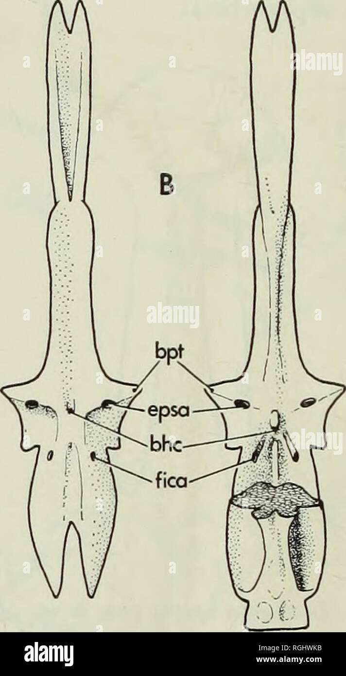
. Bulletin of the British Museum (Natural History), Geology. 236 TWO UPPER CRETACEOUS SALMONIFORM basipterygoid processes articulated with the palate is not possible to discover, but in skulls preserved in lateral view (Fig. 19) the metapterygoid lies very close to the basipterygoid process and such an articulation is Ukely. Between the bases of the basipterygoid processes there is a very small bucco-hypophysial canal (bhc), patent in the single specimen where both sides of the parasphenoid are visible. The ascending processes of the parasphenoid are long and high, forming the lower part of th

Image details
Contributor:
Book Worm / Alamy Stock PhotoImage ID:
RGHWKBFile size:
7.1 MB (195.8 KB Compressed download)Releases:
Model - no | Property - noDo I need a release?Dimensions:
1170 x 2135 px | 19.8 x 36.2 cm | 7.8 x 14.2 inches | 150dpiMore information:
This image is a public domain image, which means either that copyright has expired in the image or the copyright holder has waived their copyright. Alamy charges you a fee for access to the high resolution copy of the image.
This image could have imperfections as it’s either historical or reportage.
. Bulletin of the British Museum (Natural History), Geology. 236 TWO UPPER CRETACEOUS SALMONIFORM basipterygoid processes articulated with the palate is not possible to discover, but in skulls preserved in lateral view (Fig. 19) the metapterygoid lies very close to the basipterygoid process and such an articulation is Ukely. Between the bases of the basipterygoid processes there is a very small bucco-hypophysial canal (bhc), patent in the single specimen where both sides of the parasphenoid are visible. The ascending processes of the parasphenoid are long and high, forming the lower part of the somewhat inflated otoUth chambers. The ascending process is penetrated by the internal carotid in the usual way (fica). Posteriorly the parasphenoid ends just in front of the occipital condyle (Fig. 18). Probably themyodome opened posteriorly, as in Gaudryella.. Fig. 17. Humbertia operta gen. & sp. nov. a, isolated vomer, P.51265, in dorsal (left) and ventral view, b, parasphenoid in ventral view (left) and in dorsal view with basioccipital in position (right), restorations based on P.51266 and AM 3783. All from Hajula, Leb- anon, bhc, bucco-hypophysial canal ; explanation of other abbreviations p. 296. The endocranium is partially visible in several transfer preparations, and parts of the otic and occipital regions are preserved in two dissociated individuals on AM 3783 (Fig. 18). In comparison with Gaudryella the endocranium is rather poorly ossified, with cartilagenous interspaces between many of the bones so that they become more or less disarticulated during fossilisation : this break-up of the underlying endocran- ium is responsible for the poor preservation of the posterior part of the skull roof in this species. As in Gaudryella, the occipital condyle is formed by the basioccipital (Boc) alone. The wall of the saccular recess is more inflated than in Gaudryella but there is no fenestra at the junction of the prootic (Pro), exoccipital (Exo) and basi- occipital. Th