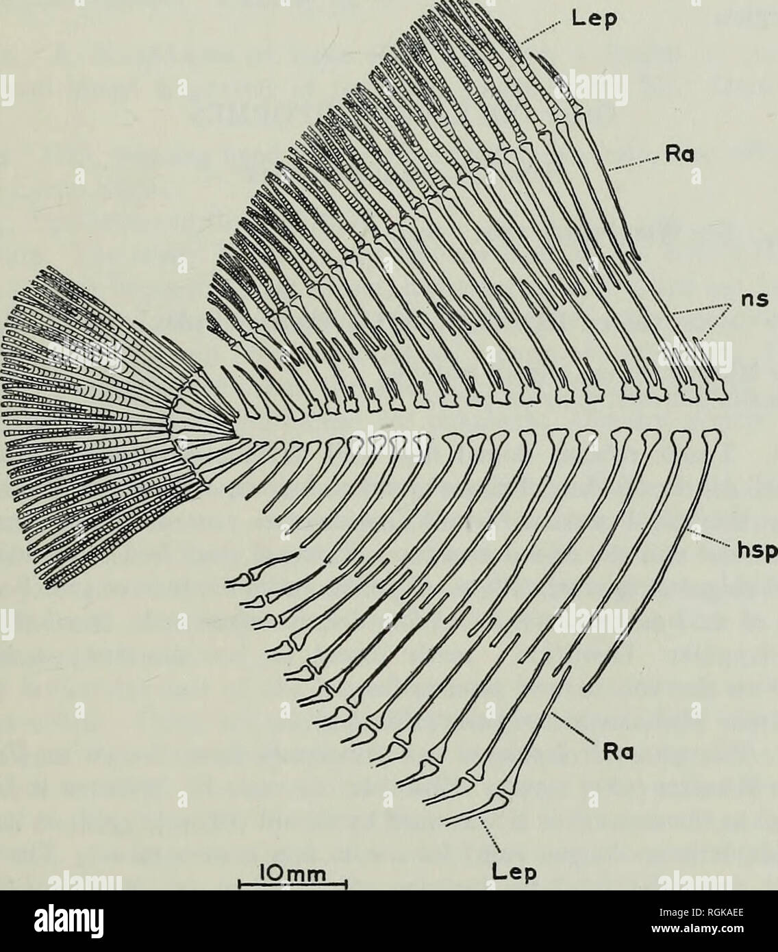
. Bulletin of the British Museum (Natural History), Geology. REVISION OF ACTINOPTERYGIAN AND COELACANTH FISHES 325 The caudal fin has between eighteen and twenty lepidotrichia and its posterior margin is convex. Axial skeleton. The notochord is persistent and the neural and haemal arches are well ossified. The neural and haemal spines of the vertebrae do not reach the dorsal border of the body, there being a set of radials interposed between the ends of the spines and the base of the fins. Neither the haemals nor the neurals bear laminar expansions in the caudal region. However, except near th

Image details
Contributor:
Book Worm / Alamy Stock PhotoImage ID:
RGKAEEFile size:
7.2 MB (357.2 KB Compressed download)Releases:
Model - no | Property - noDo I need a release?Dimensions:
1478 x 1691 px | 25 x 28.6 cm | 9.9 x 11.3 inches | 150dpiMore information:
This image is a public domain image, which means either that copyright has expired in the image or the copyright holder has waived their copyright. Alamy charges you a fee for access to the high resolution copy of the image.
This image could have imperfections as it’s either historical or reportage.
. Bulletin of the British Museum (Natural History), Geology. REVISION OF ACTINOPTERYGIAN AND COELACANTH FISHES 325 The caudal fin has between eighteen and twenty lepidotrichia and its posterior margin is convex. Axial skeleton. The notochord is persistent and the neural and haemal arches are well ossified. The neural and haemal spines of the vertebrae do not reach the dorsal border of the body, there being a set of radials interposed between the ends of the spines and the base of the fins. Neither the haemals nor the neurals bear laminar expansions in the caudal region. However, except near the tail the neural spines. Fig. 54. Eomesodon liassicus (Egerton). Restoration of the endoskeleton of the trunk. beneath the dorsal fin are double, each having a short slightly curved rod arising from the anterior portion of the neural arch, followed by the long backwardly directed neural spine proper. Squamation. The squamation is complete over the whole of the trunk in advance of the median fins. The scales are largest in the nuchal region and are ornamented with coarse tubercles, somewhat finer than those on the parietals and frontals.. Please note that these images are extracted from scanned page images that may have been digitally enhanced for readability - coloration and appearance of these illustrations may not perfectly resemble the original work.. British Museum (Natural History). London : BM(NH)