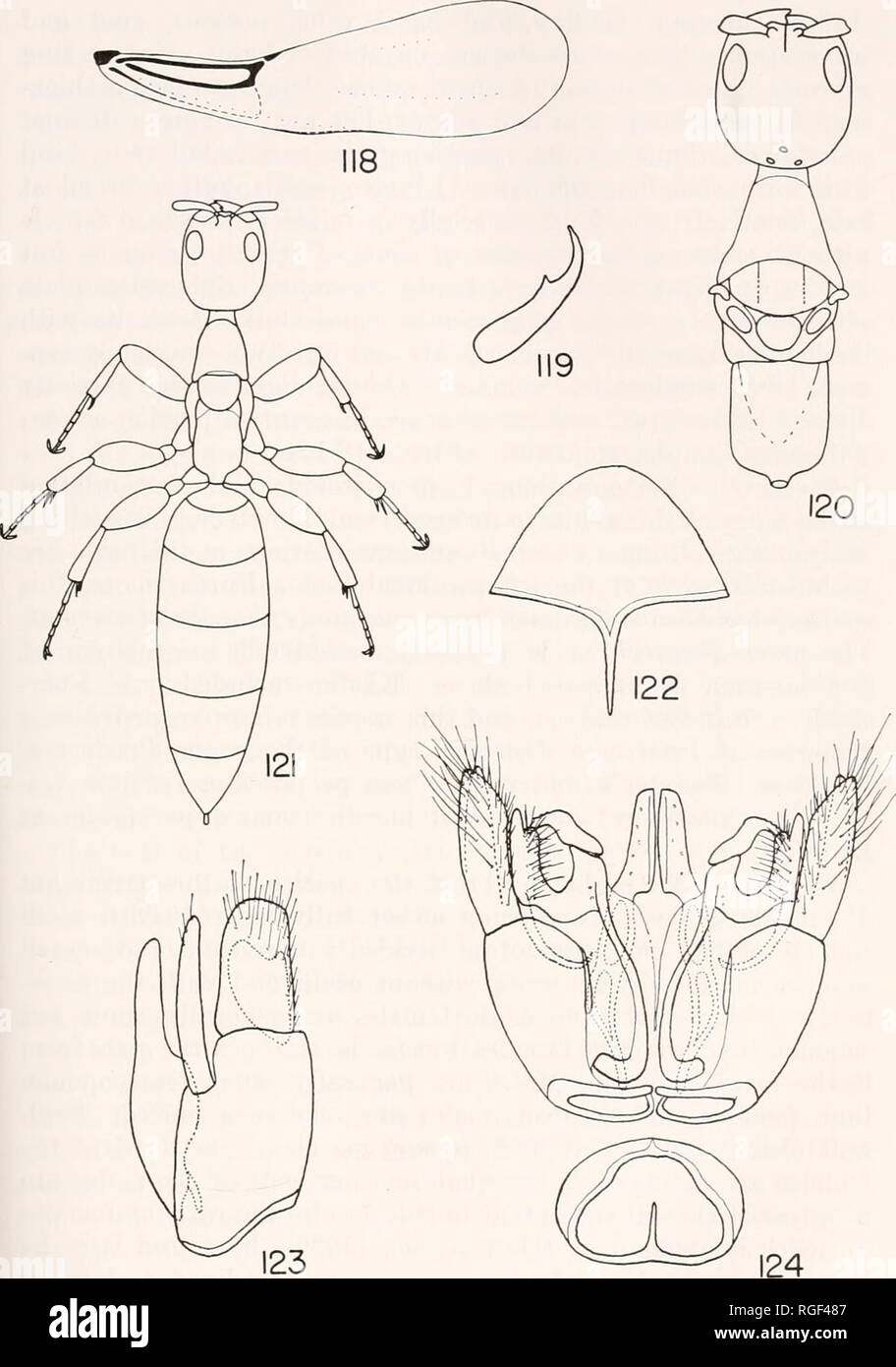
. Bulletin of the Museum of Comparative Zoology at Harvard College. Zoology. EVANS: AMERICAN BETHYLIDAE 175. Scleroderma macrogaster (Ashmead). Fig. 118. Fore wing of alate female. Fig. 119. Hind tarsal claw of alate female. Fig. 120. Head and thorax of alate female. Fig. 121. Apterous female. Fig. 122. Male sub- genital plate. Fig. 123. Male genitalia, lateral aspect, dorsal surface (aedoeagus) at left. Fig. 124. Male genitalia, ventral aspect with basal ring detached from genital capsule.. Please note that these images are extracted from scanned page images that may have been digitally enhan

Image details
Contributor:
Book Worm / Alamy Stock PhotoImage ID:
RGF487File size:
7.1 MB (209.2 KB Compressed download)Releases:
Model - no | Property - noDo I need a release?Dimensions:
1324 x 1887 px | 22.4 x 32 cm | 8.8 x 12.6 inches | 150dpiMore information:
This image is a public domain image, which means either that copyright has expired in the image or the copyright holder has waived their copyright. Alamy charges you a fee for access to the high resolution copy of the image.
This image could have imperfections as it’s either historical or reportage.
. Bulletin of the Museum of Comparative Zoology at Harvard College. Zoology. EVANS: AMERICAN BETHYLIDAE 175. Scleroderma macrogaster (Ashmead). Fig. 118. Fore wing of alate female. Fig. 119. Hind tarsal claw of alate female. Fig. 120. Head and thorax of alate female. Fig. 121. Apterous female. Fig. 122. Male sub- genital plate. Fig. 123. Male genitalia, lateral aspect, dorsal surface (aedoeagus) at left. Fig. 124. Male genitalia, ventral aspect with basal ring detached from genital capsule.. Please note that these images are extracted from scanned page images that may have been digitally enhanced for readability - coloration and appearance of these illustrations may not perfectly resemble the original work.. Harvard University. Museum of Comparative Zoology. Cambridge, Mass. : The Museum