. Cephalopoda. Cephalopoda. FIGURE 27. Diagrammatic frontal sections of the development of the right eye of Cephalopoda: a — Nautilus. The stalk bears the optic nerve (n); the retina (ret) is actually many-layered with the rods pointed toward the light; other elements are the primary pupil (pp) and the ring fold (ir) which forms a wide circle and probably corresponds to the iris of Dibranchiata; b — e — typical stages of the embryonic eye of Sepia. The eye chamber is completely closed. The double epithelial lamella (pc), which becomes thinner later, forms the lens (1) at the site of the primar
Image details
Contributor:
Library Book Collection / Alamy Stock PhotoImage ID:
RJC100File size:
7.1 MB (324 KB Compressed download)Releases:
Model - no | Property - noDo I need a release?Dimensions:
2391 x 1045 px | 40.5 x 17.7 cm | 15.9 x 7 inches | 150dpiMore information:
This image is a public domain image, which means either that copyright has expired in the image or the copyright holder has waived their copyright. Alamy charges you a fee for access to the high resolution copy of the image.
This image could have imperfections as it’s either historical or reportage.
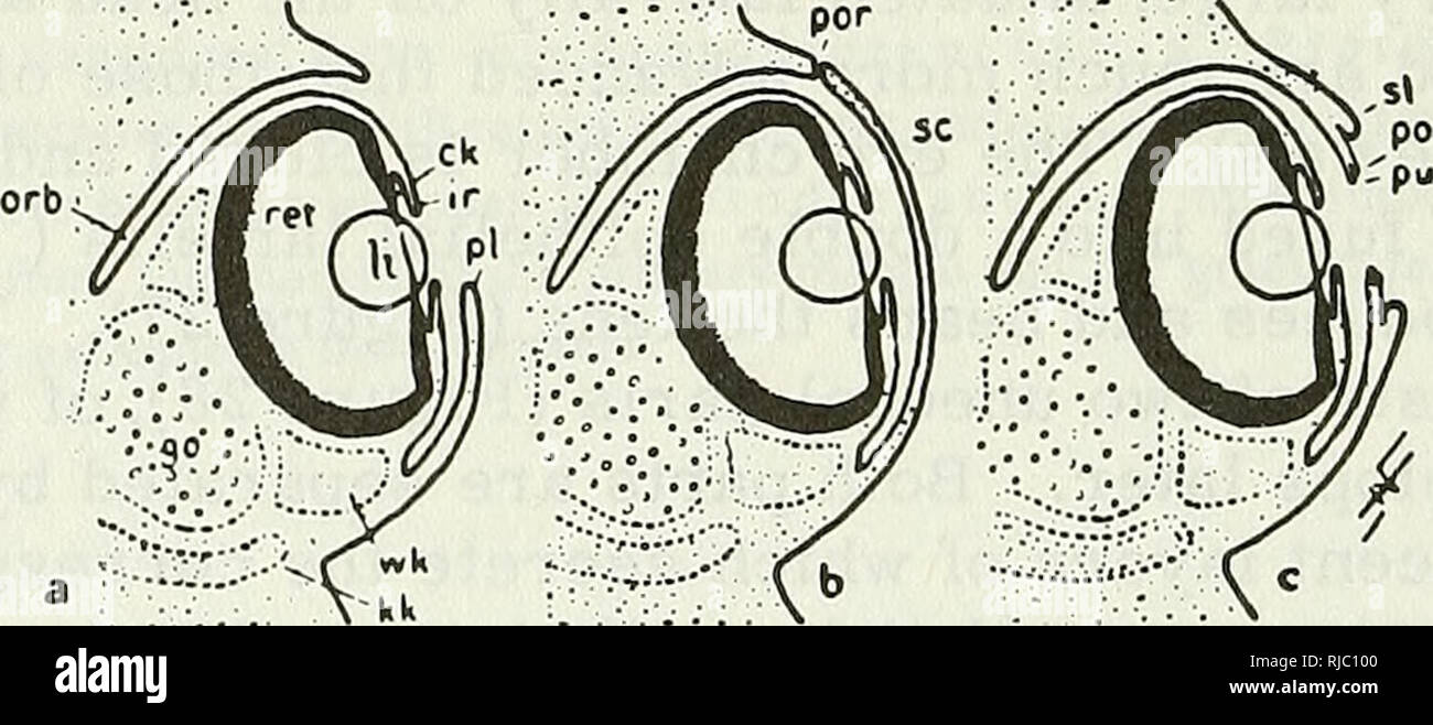
. Cephalopoda. Cephalopoda. FIGURE 27. Diagrammatic frontal sections of the development of the right eye of Cephalopoda: a — Nautilus. The stalk bears the optic nerve (n); the retina (ret) is actually many-layered with the rods pointed toward the light; other elements are the primary pupil (pp) and the ring fold (ir) which forms a wide circle and probably corresponds to the iris of Dibranchiata; b — e — typical stages of the embryonic eye of Sepia. The eye chamber is completely closed. The double epithelial lamella (pc), which becomes thinner later, forms the lens (1) at the site of the primary pupil; only the inner segment of the lens is formed at first. (Compare description of development in Volume II.) Close around the lens develops the iris fold (ir). The resulting eyeball (e) is comparable to that of Nautilus in the following aspects: it projects from the head, and is borne in the embryo on a more or less distinct stalk which is much thicker here because the organs it contains are more developed than those of Nautilus (optic ganglion, white body; see Figure 28).. FIGURE 28. Frontal sections through the right eye of differ- ent Dibranchiata (diagrammatic), a) Typical form, found in Oegopsida. The orbit (orb) is wide open; the posterior margin (pi) of the orbital opening forms a primary lid which can cover the lens. This protective closure becomes permanent in Loligo(b), inwhich the primary lid folddevel- ops into a cornea (sc) and thecommunicationof the orbit with the exterior is reduced to a narrow pore (por). In Octopoda (c) (q.v.) the primary lid margin becomes complicated and a secondary lid fold appears at its periphery: go — optic ganglion; wk — white body; po — primary upper margin of the primary lid; pu — primary lower mar- gin of the primary lid, overlapping in Octopoda. 5110/1 90. Please note that these images are extracted from scanned page images that may have been digitally enhanced for readability - coloration and appearance of these i