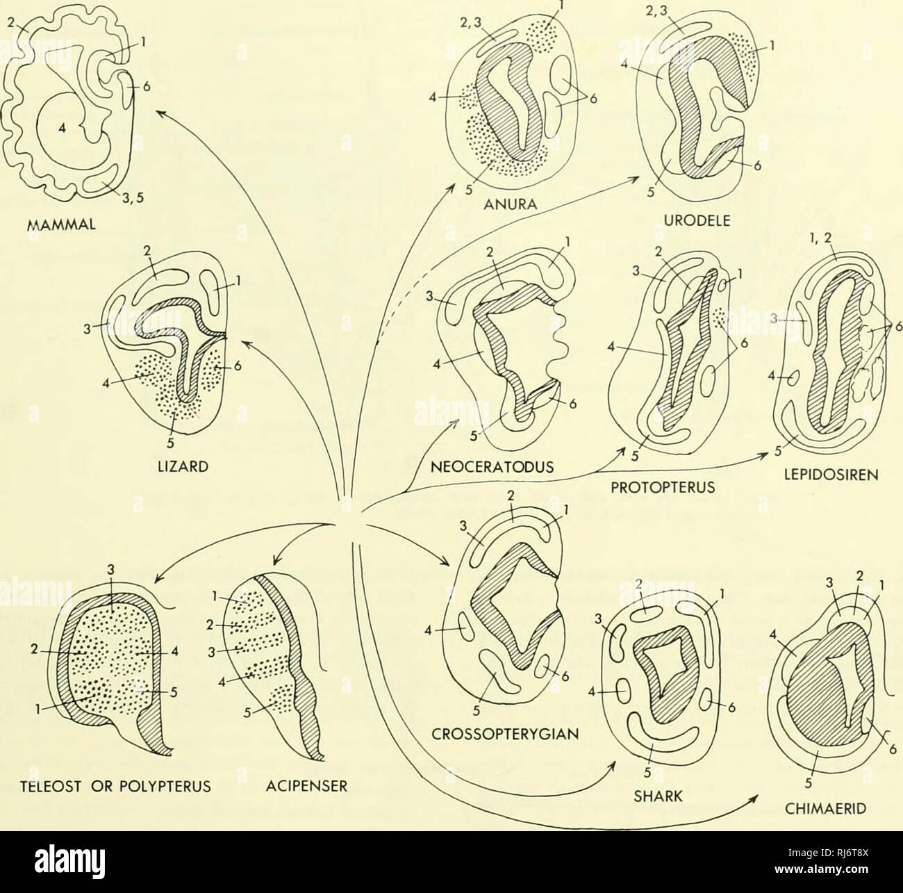
. Chordate morphology. Morphology (Animals); Chordata. TELEOST OR POLYPTERUS ACIPENSER Figure 13-5. Cross sections of cerebral hemispheres of different vertebrates with suggested path- ways of evolution. 1, hippocampus; 2, general pallium (neopallium); 3, pyriformts; 4, lateral olfac- tory nucleus (striate body); 5 tuberculum olfactorium (polaeopollium); 6, septal nuclei; ventricular layer of nuclei cross hatched. (Moinly after Rudebeck, 1945) CHIMAERID fore the cerebral lobes. Basically the cerebral lobes are ol- factory lobes, but have incipient higher centers. The diencephalon has a pineal
Image details
Contributor:
Library Book Collection / Alamy Stock PhotoImage ID:
RJ6T8XFile size:
7.1 MB (302.2 KB Compressed download)Releases:
Model - no | Property - noDo I need a release?Dimensions:
1648 x 1516 px | 27.9 x 25.7 cm | 11 x 10.1 inches | 150dpiMore information:
This image is a public domain image, which means either that copyright has expired in the image or the copyright holder has waived their copyright. Alamy charges you a fee for access to the high resolution copy of the image.
This image could have imperfections as it’s either historical or reportage.
. Chordate morphology. Morphology (Animals); Chordata. TELEOST OR POLYPTERUS ACIPENSER Figure 13-5. Cross sections of cerebral hemispheres of different vertebrates with suggested path- ways of evolution. 1, hippocampus; 2, general pallium (neopallium); 3, pyriformts; 4, lateral olfac- tory nucleus (striate body); 5 tuberculum olfactorium (polaeopollium); 6, septal nuclei; ventricular layer of nuclei cross hatched. (Moinly after Rudebeck, 1945) CHIMAERID fore the cerebral lobes. Basically the cerebral lobes are ol- factory lobes, but have incipient higher centers. The diencephalon has a pineal evagination which generally lacks an eye at its tip; a pineal nerve is lacking. In the frog a pineal eye is present and the pineal sac has in part lost con- nection with the brain (Figure 13-7). Anterior to the pos- terior commissure is a saccus dorsalis which in front culmi- nates in a paraphysis. Paraphysis and roof are much thickened as a chorioid plexus from which fingers of tissue extend down into the third ventricle and through the foramina of Monroe into the telencoels. A distinct velum transversum is lacking. The optic nerves decussate in entering the brain and some fibers of each optic tract synapse at a lateral geniculate nucleus with neurons leading to the cerebrum. The reduced eyes o{ Necturus are reflected by the relatively smaller optic lobes, as compared with the frog. The amphibian brain is peculiar in having a greatly re- duced cerebellum which is no more than a transverse band of tissue anterior to the chorioid plexus in the roof of the medulla. The cranial nerves of the amphibian resemble those of the preceding groups (Figure 13-8). The seventh has a super- ficial ophthalmic branch in salamanders but not in anu- THE CONDUCTING AND INTEGRATING SYSTEM • 389. Please note that these images are extracted from scanned page images that may have been digitally enhanced for readability - coloration and appearance of these illustrations may not perfectly resemble the