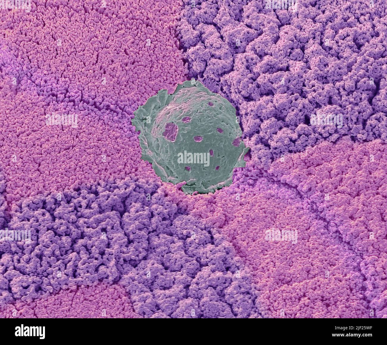
Colon. Colour scanning electron micrograph (SEM) of the wall of the large intestine (colon), showing mucous secretion (cyan). Goblet cells are mucous

RMID:Image ID:2JF25WF
Image details
Contributor:
Science Photo Library / Alamy Stock PhotoImage ID:
2JF25WFFile size:
50 MB (2.8 MB Compressed download)Releases:
Model - no | Property - noDo I need a release?Dimensions:
4603 x 3797 px | 39 x 32.1 cm | 15.3 x 12.7 inches | 300dpiPhotographer:
***DEPENDS ON PIC***/SCIENCE PHOTO LIBRARYMore information:
Colon. Colour scanning electron micrograph (SEM) of the wall of the large intestine (colon), showing mucous secretion (cyan). Goblet cells are mucous secreting cells. Large numbers micro- villi of absorptive cells are visible. The colon functions to absorb water and remaining nutrients from digested food faeces. Microvilli on absorptive cells increase the colon surface area for this purpose. The mucous secreted by goblet cells lubricates the passage of faeces, protecting the colon wall from mechanical damage; it also enables faeces to pass closely over absorptive cells to assist with absorption. Magnification: x4500 when printed 10 centimetres wide.