···
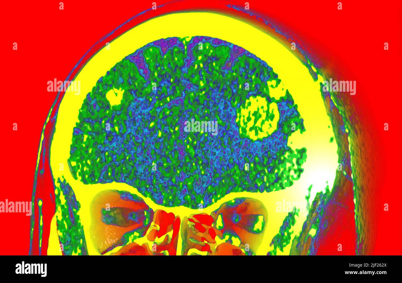
Coloured computed tomography (CT) scan showing secondary malignant (cancerous) tumours (yellow) in a patient's brain. The cancer has spread (metastasised) from the site of a primary cancer. Image details File size:
30 MB (891.7 KB Compressed download)
Open your image file to the full size using image processing software.
Dimensions:
4066 x 2579 px | 34.4 x 21.8 cm | 13.6 x 8.6 inches | 300dpi
Search stock photos by tags
Similar stock images Brain in dementia. Coloured computed tomography (CT) scan of a section through the brain of an 89-year-old male patient with dementia. The brain has atrophied (shrunk), shown by the enlarged central ventricles (dark blue) and deep indentations around the brain's edges. Stock Photo https://www.alamy.com/image-license-details/?v=1 https://www.alamy.com/brain-in-dementia-coloured-computed-tomography-ct-scan-of-a-section-through-the-brain-of-an-89-year-old-male-patient-with-dementia-the-brain-has-atrophied-shrunk-shown-by-the-enlarged-central-ventricles-dark-blue-and-deep-indentations-around-the-brains-edges-image231023270.html RF RBT0F2 – Brain in dementia. Coloured computed tomography (CT) scan of a section through the brain of an 89-year-old male patient with dementia. The brain has atrophied (shrunk), shown by the enlarged central ventricles (dark blue) and deep indentations around the brain's edges. Coloured CTscan of the upper chest showing a tumor in the lung. Stock Photo https://www.alamy.com/image-license-details/?v=1 https://www.alamy.com/stock-image-coloured-ctscan-of-the-upper-chest-showing-a-tumor-in-the-lung-168550373.html RM KP63M5 – Coloured CTscan of the upper chest showing a tumor in the lung. CT scan of head showing acute subdural hematoma Stock Photo https://www.alamy.com/image-license-details/?v=1 https://www.alamy.com/ct-scan-of-head-showing-acute-subdural-hematoma-image65495857.html RF DPFGHN – CT scan of head showing acute subdural hematoma Brain in dementia. Coloured computed tomography (CT) scan of a section through the brain of an 89-year-old male patient with dementia. The brain has atrophied (shrunk), shown by the enlarged central ventricles (dark green) and deep indentations around the brain's edges. Stock Photo https://www.alamy.com/image-license-details/?v=1 https://www.alamy.com/brain-in-dementia-coloured-computed-tomography-ct-scan-of-a-section-through-the-brain-of-an-89-year-old-male-patient-with-dementia-the-brain-has-atrophied-shrunk-shown-by-the-enlarged-central-ventricles-dark-green-and-deep-indentations-around-the-brains-edges-image231023269.html RF RBT0F1 – Brain in dementia. Coloured computed tomography (CT) scan of a section through the brain of an 89-year-old male patient with dementia. The brain has atrophied (shrunk), shown by the enlarged central ventricles (dark green) and deep indentations around the brain's edges. Coloured CTscan of the upper chest showing a tumor in the lung. Stock Photo https://www.alamy.com/image-license-details/?v=1 https://www.alamy.com/stock-image-coloured-ctscan-of-the-upper-chest-showing-a-tumor-in-the-lung-168550360.html RM KP63KM – Coloured CTscan of the upper chest showing a tumor in the lung. CT scan showing kidney stone Stock Photo https://www.alamy.com/image-license-details/?v=1 https://www.alamy.com/ct-scan-showing-kidney-stone-image65488072.html RF DPF6KM – CT scan showing kidney stone Brain in dementia. Coloured computed tomography (CT) scan of a section through the brain of an 89-year-old male patient with dementia. The brain has atrophied (shrunk), shown by the enlarged central ventricles (dark red) and deep indentations around the brain's edges. Stock Photo https://www.alamy.com/image-license-details/?v=1 https://www.alamy.com/brain-in-dementia-coloured-computed-tomography-ct-scan-of-a-section-through-the-brain-of-an-89-year-old-male-patient-with-dementia-the-brain-has-atrophied-shrunk-shown-by-the-enlarged-central-ventricles-dark-red-and-deep-indentations-around-the-brains-edges-image231023272.html RF RBT0F4 – Brain in dementia. Coloured computed tomography (CT) scan of a section through the brain of an 89-year-old male patient with dementia. The brain has atrophied (shrunk), shown by the enlarged central ventricles (dark red) and deep indentations around the brain's edges. Coloured CTscan of the upper chest showing a tumor in the lungs ( in green). Stock Photo https://www.alamy.com/image-license-details/?v=1 https://www.alamy.com/stock-photo-coloured-ctscan-of-the-upper-chest-showing-a-tumor-in-the-lungs-in-79765082.html RM EHNH4X – Coloured CTscan of the upper chest showing a tumor in the lungs ( in green). Glioblastoma brain cancer. Coloured computed tomography (CT) scan of a section through the brain of an 84-year-old female patient with glioblastoma (dark, left). Glioblastoma is the most aggressive form of brain cancer. Treatment involves surgery, after which chemotherapy and radiation therapy are used. However, the cancer usually reoccurs despite treatment and the most common length of survival after diagnosis is 12-15 months. Without treatment, survival is typically 3 months. Stock Photo https://www.alamy.com/image-license-details/?v=1 https://www.alamy.com/glioblastoma-brain-cancer-coloured-computed-tomography-ct-scan-of-a-section-through-the-brain-of-an-84-year-old-female-patient-with-glioblastoma-dark-left-glioblastoma-is-the-most-aggressive-form-of-brain-cancer-treatment-involves-surgery-after-which-chemotherapy-and-radiation-therapy-are-used-however-the-cancer-usually-reoccurs-despite-treatment-and-the-most-common-length-of-survival-after-diagnosis-is-12-15-months-without-treatment-survival-is-typically-3-months-image231023275.html RF RBT0F7 – Glioblastoma brain cancer. Coloured computed tomography (CT) scan of a section through the brain of an 84-year-old female patient with glioblastoma (dark, left). Glioblastoma is the most aggressive form of brain cancer. Treatment involves surgery, after which chemotherapy and radiation therapy are used. However, the cancer usually reoccurs despite treatment and the most common length of survival after diagnosis is 12-15 months. Without treatment, survival is typically 3 months. Coloured CTscan of the upper chest showing a tumor in the lung. Stock Photo https://www.alamy.com/image-license-details/?v=1 https://www.alamy.com/stock-image-coloured-ctscan-of-the-upper-chest-showing-a-tumor-in-the-lung-168550372.html RM KP63M4 – Coloured CTscan of the upper chest showing a tumor in the lung. Glioblastoma brain cancer. Coloured computed tomography (CT) scan of a section through the brain of an 84-year-old female patient with glioblastoma (dark, left). Glioblastoma is the most aggressive form of brain cancer. Treatment involves surgery, after which chemotherapy and radiation therapy are used. However, the cancer usually reoccurs despite treatment and the most common length of survival after diagnosis is 12-15 months. Without treatment, survival is typically 3 months. Stock Photo https://www.alamy.com/image-license-details/?v=1 https://www.alamy.com/glioblastoma-brain-cancer-coloured-computed-tomography-ct-scan-of-a-section-through-the-brain-of-an-84-year-old-female-patient-with-glioblastoma-dark-left-glioblastoma-is-the-most-aggressive-form-of-brain-cancer-treatment-involves-surgery-after-which-chemotherapy-and-radiation-therapy-are-used-however-the-cancer-usually-reoccurs-despite-treatment-and-the-most-common-length-of-survival-after-diagnosis-is-12-15-months-without-treatment-survival-is-typically-3-months-image231023273.html RF RBT0F5 – Glioblastoma brain cancer. Coloured computed tomography (CT) scan of a section through the brain of an 84-year-old female patient with glioblastoma (dark, left). Glioblastoma is the most aggressive form of brain cancer. Treatment involves surgery, after which chemotherapy and radiation therapy are used. However, the cancer usually reoccurs despite treatment and the most common length of survival after diagnosis is 12-15 months. Without treatment, survival is typically 3 months. Coloured CTscan of the upper chest showing a tumor in the lung. Stock Photo https://www.alamy.com/image-license-details/?v=1 https://www.alamy.com/stock-image-coloured-ctscan-of-the-upper-chest-showing-a-tumor-in-the-lung-168550396.html RM KP63N0 – Coloured CTscan of the upper chest showing a tumor in the lung. Healthy lungs, CT scan Stock Photo https://www.alamy.com/image-license-details/?v=1 https://www.alamy.com/healthy-lungs-ct-scan-image569663355.html RF 2T2PBBR – Healthy lungs, CT scan 