. Comparative anatomy of the vegetative organs of the phanerogams and ferns. Plant anatomy; Phanerogams; Ferns. 90 CELLULAR TISSUE. and treated with dissolving reagentsâe. g. alcohol or etherâis intercalated between cell-membrane and cuticle. The same phenomenon, in the main, occurs in the multicellular heads of the bladder-like glandular hairs, villi, and scales. The thickening by the secretion begins in these cases at a point more or less near the apex of the whole (not on each or on several single cells) and extends from this point centrifugally, varying greatly in extent and bulk according
Image details
Contributor:
The Book Worm / Alamy Stock PhotoImage ID:
REFHH3File size:
7.2 MB (163.7 KB Compressed download)Releases:
Model - no | Property - noDo I need a release?Dimensions:
2279 x 1097 px | 38.6 x 18.6 cm | 15.2 x 7.3 inches | 150dpiMore information:
This image is a public domain image, which means either that copyright has expired in the image or the copyright holder has waived their copyright. Alamy charges you a fee for access to the high resolution copy of the image.
This image could have imperfections as it’s either historical or reportage.
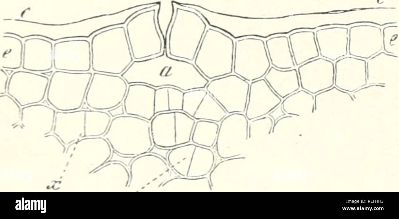
. Comparative anatomy of the vegetative organs of the phanerogams and ferns. Plant anatomy; Phanerogams; Ferns. 90 CELLULAR TISSUE. and treated with dissolving reagentsâe. g. alcohol or etherâis intercalated between cell-membrane and cuticle. The same phenomenon, in the main, occurs in the multicellular heads of the bladder-like glandular hairs, villi, and scales. The thickening by the secretion begins in these cases at a point more or less near the apex of the whole (not on each or on several single cells) and extends from this point centrifugally, varying greatly in extent and bulk according to the special cases. The outer walls of the .single cells thus form with one another either a smooth, even, or domed surface, or they arch outwards like papillae into the secretory mass which overlies them, and is in its turn bounded by the cuticle. The raised cuticle itself is usually homogeneous and structureless, in other cases (shield-Hke scales of Humulus, Ribes nigrum) it is marked off into areas corresponding to the lateral limits of the cells. As already intimated the glandular structure in hairs is not always restricted to the head; it may also occur on the lateral wall of capitate hairs, and of such as are not capitate as in Cistus. (Fig. 36.) From the point of insertion of glandular hairs in the buds of Rumex, Rheum, Cunonia, Coffea, Alnus, Carpinus, Corylus, &c., the glandular structure of the walls extends over the smooth epidermis (Hans- tein, Bot. Ztg. 1868). This attains thereby the properties of the glandular surface. The same occurs in exquisite form on the sticky young shoots of Betula alba (Fig. 35), where the glandular nature of the wall of the shield-like glandular scales extends over the whole epidermis, as far FIG. 3S.-Transverse .section through a young intemode of aS the ridgC Of Cntry of the StOmata, Betula alba (375). câf epidennis; a respiratory cavity under the , . rv^y .⢠^ j stoma; c the cuticle raised from e, by a secretory layer, as