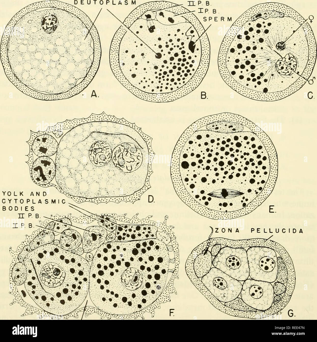
. Comparative embryology of the vertebrates; with 2057 drawings and photos. grouped as 380 illus. Vertebrates -- Embryology; Comparative embryology. 236 FERTILIZATION DEUTOPLASM. PERIVITELLiN E Fig. 118. Fertilization in the guinea pig. (After Lams, Arch. Biol., Paris, 28, figures slightly modified.) (A) Spindle of first maturation division. (B) Second maturation division completed; head of sperm in cytoplasm beginning to swell. (C) Sperm pro- nucleus, with tail still attached, greatly enlarged; female pronucleus small. (D) Pronuclei ready to fuse; chromatin material (chromosomes) evident with

Image details
Contributor:
The Book Worm / Alamy Stock PhotoImage ID:
REE47NFile size:
7.2 MB (540.3 KB Compressed download)Releases:
Model - no | Property - noDo I need a release?Dimensions:
1576 x 1586 px | 26.7 x 26.9 cm | 10.5 x 10.6 inches | 150dpiMore information:
This image is a public domain image, which means either that copyright has expired in the image or the copyright holder has waived their copyright. Alamy charges you a fee for access to the high resolution copy of the image.
This image could have imperfections as it’s either historical or reportage.
. Comparative embryology of the vertebrates; with 2057 drawings and photos. grouped as 380 illus. Vertebrates -- Embryology; Comparative embryology. 236 FERTILIZATION DEUTOPLASM. PERIVITELLiN E Fig. 118. Fertilization in the guinea pig. (After Lams, Arch. Biol., Paris, 28, figures slightly modified.) (A) Spindle of first maturation division. (B) Second maturation division completed; head of sperm in cytoplasm beginning to swell. (C) Sperm pro- nucleus, with tail still attached, greatly enlarged; female pronucleus small. (D) Pronuclei ready to fuse; chromatin material (chromosomes) evident within. (E) First cleavage spindle. (F) First cleavage completed. Observe deutoplasmic and cytoplasmic globules which have been exuded into the space between the blastomeres and the zona pellucida. (G) Four-cell cleavage stage. Observe that the zona pellucida encloses the four blasto- meres and the cytoplasmic globules which have been exuded. The zona functions to keep the entire mass intact. the emission of fluid is present in the amphibia and the egg thus is enabled to revolve within a relatively thick vitelline membrane. The latter membrane expands gradually during development, and is associated intimately with the surrounding jelly membranes secreted by the oviduct. In the reptiles and birds, the separation of the egg from the vitelline membrane or zona radiata and. Please note that these images are extracted from scanned page images that may have been digitally enhanced for readability - coloration and appearance of these illustrations may not perfectly resemble the original work.. Nelsen, Olin E. (Olin Everett), b. 1898. New York, Blakiston