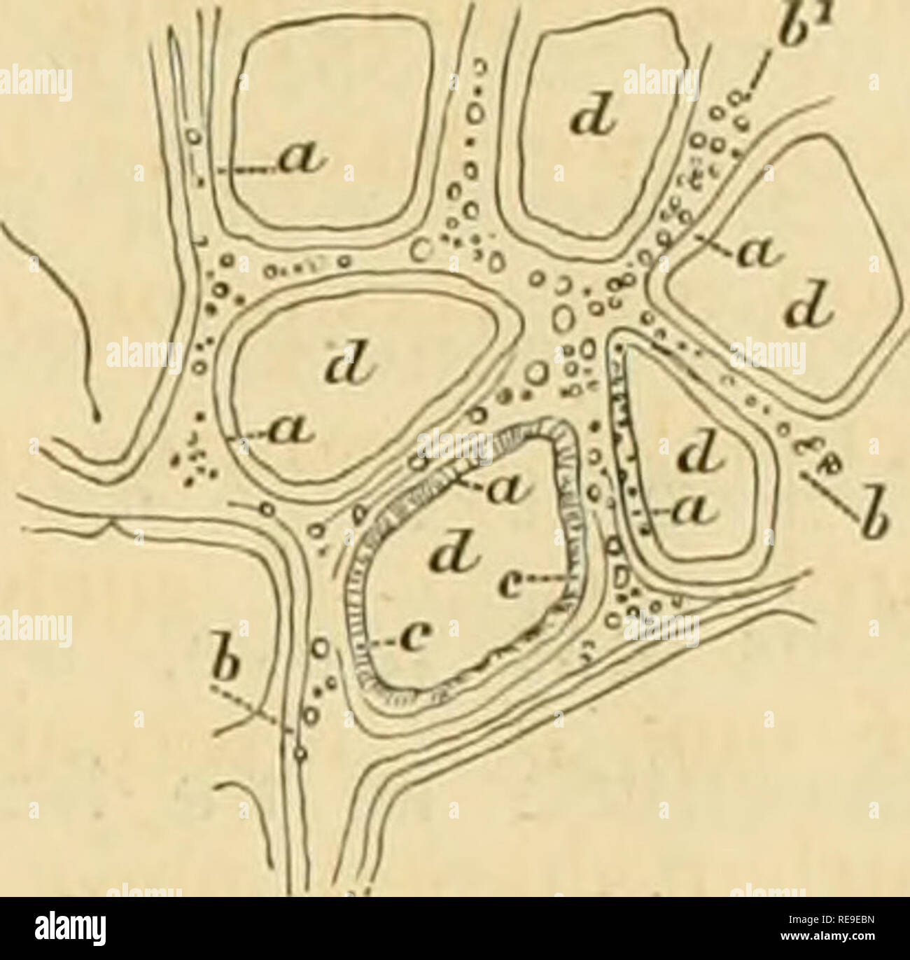
. Contributions to the natural history of the United States of America. Zoology; Chelonia (Genus); Ctenophora; Cnidaria; Animals. Chap. III. HYDRACTINIA POLYCLINA. 231 Fig. 34.. Tlie retiform stolon of Hy- dractinia polyclina. From na- ture, by H. J. Clark. The inner wall {Fig. 3, h) is very thick, and constitutes the greater bulk of the body; it has the same extent as the outer wall, and is in more intimate connection with the active functions of the Avhole colony, forming the immediate lining of the digestive cavity {d), which receives the chymiferous fluid, in common with the other hydroids
Image details
Contributor:
The Book Worm / Alamy Stock PhotoImage ID:
RE9EBNFile size:
7.1 MB (203.3 KB Compressed download)Releases:
Model - no | Property - noDo I need a release?Dimensions:
1592 x 1569 px | 27 x 26.6 cm | 10.6 x 10.5 inches | 150dpiMore information:
This image is a public domain image, which means either that copyright has expired in the image or the copyright holder has waived their copyright. Alamy charges you a fee for access to the high resolution copy of the image.
This image could have imperfections as it’s either historical or reportage.
. Contributions to the natural history of the United States of America. Zoology; Chelonia (Genus); Ctenophora; Cnidaria; Animals. Chap. III. HYDRACTINIA POLYCLINA. 231 Fig. 34.. Tlie retiform stolon of Hy- dractinia polyclina. From na- ture, by H. J. Clark. The inner wall {Fig. 3, h) is very thick, and constitutes the greater bulk of the body; it has the same extent as the outer wall, and is in more intimate connection with the active functions of the Avhole colony, forming the immediate lining of the digestive cavity {d), which receives the chymiferous fluid, in common with the other hydroids. Its inner surface is lined with an irregular layer of browni.sh-red, coarse granules, of the same nature as those {Figs. 5% g, and 5", c, wood-cut 34, b^; PI. XXVI. Fig. 18, i'. Vol. IV.) seen in the ramifying canals and the sterile hydroids. The same may be said of the cells of this layer as of those of Coryne mirabilis (p. 205). From the IQtli of December, 1855, to the 30th of April. outer wau in pronie, at the edge ^ of tbe depressious ((/). — b inner 1856, the fertile hydroids on our coast were free from medusa?- waiiiioiiowe.iby thechjmiferous " canals.—61 granules circulating buds, but from July to September, 1854, they were buddina: m 6.-^ ceiis of a in prosie.- ' '^ ^ d depressions in the outer wall, copiously. At Charleston, South Carolina, they were found "^^'^ "pp™"" «««»' "« budding from December, 1851, to February, 1852. During the unproductive season, we have found the sterile hydroids just as fully developed as at any other time. We have never known any instances in which the tentacles appeared to be resorbing, or indefinite in outline, except when the colony was attached to a shell which was cast ashore by the tide, or dragged about by the Hermit-crabs. In tide-pools, among the rocks to which they are attached, they flourish most luxuriantly, and do not exhibit any signs of unhealthiness. Each medusa-bud arises sin