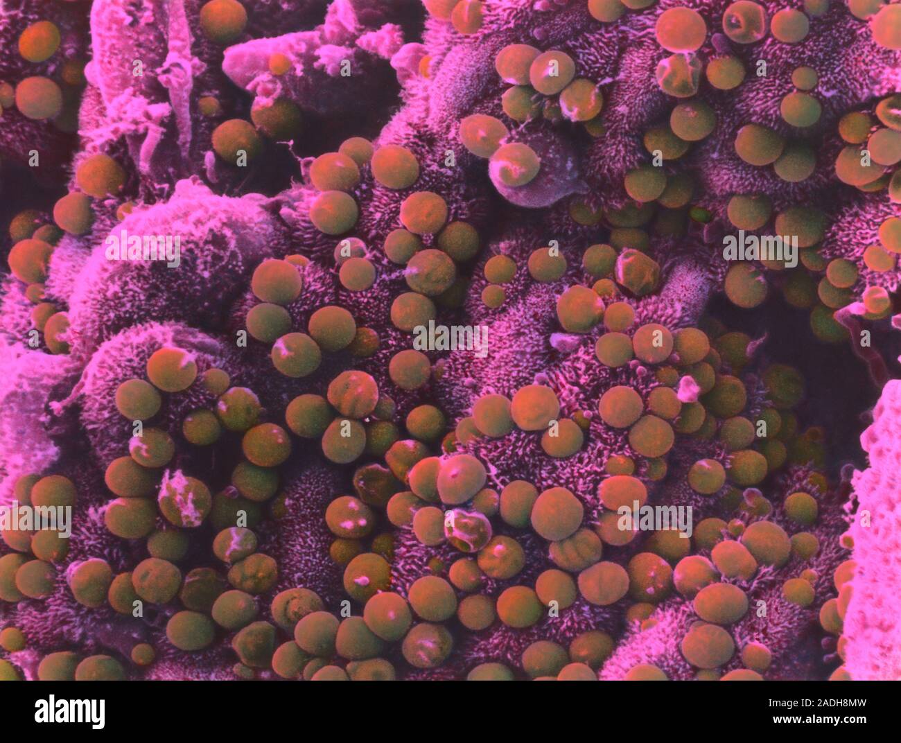
Cryptosporidium parvum. Coloured scanning electron micrograph (SEM) of the surface of the small intestine infected with Cryptosporidium parvum parasit

RMID:Image ID:2ADH8MW
Image details
Contributor:
Science Photo Library / Alamy Stock PhotoImage ID:
2ADH8MWFile size:
40.2 MB (1.5 MB Compressed download)Releases:
Model - no | Property - noDo I need a release?Dimensions:
4311 x 3258 px | 36.5 x 27.6 cm | 14.4 x 10.9 inches | 300dpiDate taken:
11 May 2001Photographer:
MOREDUN SCIENTIFIC LTD/SCIENCE PHOTO LIBRARYMore information:
Cryptosporidium parvum. Coloured scanning electron micrograph (SEM) of the surface of the small intestine infected with Cryptosporidium parvum parasites (red), cause of cryptosporidiosis. The parasite develops in the protrusions (microvilli) of epithelial cells that line the intestinal wall. Severe infection causes the folds of the intestinal wall to fuse and atrophy. Infection typically produces mild symptoms of diarrhoea, fever and headache. However, in the immuno- compromised, such as those with AIDS (acquired immune deficiency syndrome), infection can be fatal. Magnification: x8625 at 6x7cm size. x3000 at 7x9.5 inch size.