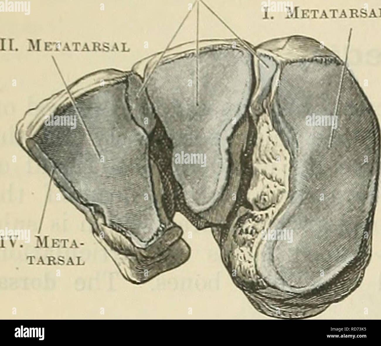
. Cunningham's Text-book of anatomy. Anatomy. 262 OSTEOLOGY. 11. METATARSAL Metatarsal III. Metatarsal. Fig. 262.âDistal Surfaces of the three Cuneiform Boxes of the Right Foot. II. Metatarsal Second cuneiform directed towards the plantar aspect; further, the vertical diameter of the bone is not the same throughout, but is much increased at its anterior or distal end. The dorsal and medial surfaces are confluent, and form a convexity from above downwards, which is most pronounced inferiorly, where it is turned to become continuous with the plantar or inferior aspect, which is rough and irregul

Image details
Contributor:
The Book Worm / Alamy Stock PhotoImage ID:
RD73K5File size:
7.2 MB (258 KB Compressed download)Releases:
Model - no | Property - noDo I need a release?Dimensions:
1724 x 1450 px | 29.2 x 24.6 cm | 11.5 x 9.7 inches | 150dpiMore information:
This image is a public domain image, which means either that copyright has expired in the image or the copyright holder has waived their copyright. Alamy charges you a fee for access to the high resolution copy of the image.
This image could have imperfections as it’s either historical or reportage.
. Cunningham's Text-book of anatomy. Anatomy. 262 OSTEOLOGY. 11. METATARSAL Metatarsal III. Metatarsal. Fig. 262.âDistal Surfaces of the three Cuneiform Boxes of the Right Foot. II. Metatarsal Second cuneiform directed towards the plantar aspect; further, the vertical diameter of the bone is not the same throughout, but is much increased at its anterior or distal end. The dorsal and medial surfaces are confluent, and form a convexity from above downwards, which is most pronounced inferiorly, where it is turned to become continuous with the plantar or inferior aspect, which is rough and irregular round the plantar side of the foot. On the distal part of the medial aspect of the bone there is usually a distinct oval impression, which indicates the surface of insertion of a portion of the tendon of the tibialis anterior muscle. Elsewhere this surface is rough for ligamentous attachments. The lateral surface of the bone, quadrilateral in shape, is directed towards the second cunei- form ; but as it exceeds it in length, it also comes in contact with the medial side of the base of the second metatarsal bone. Eunning along the proximal and dorsal edges of this area is an reshaped articular surface, the distal and dorsal part of which is for the base of the second metatarsal bone, the remainder articulating with the medial side of the second cuneiform. The non-articular part of this aspect of the bone is rough for the attachment of the strong inter- osseous ligaments which bind it to the second cunei- form and second metatarsal bones, respectively. The proximal end of the bone is provided with a piriform facet which fits on the medial articular area of the navicular. Here the wedge-shaped form of the bone is best displayed. Distally the vertical diameter of the bone is much increased, and the facet for the base of the metatarsal bone of the great toe is consequently much larger than that for the navicular. This metatarsal facet is usually of semilunar form, but not infreq