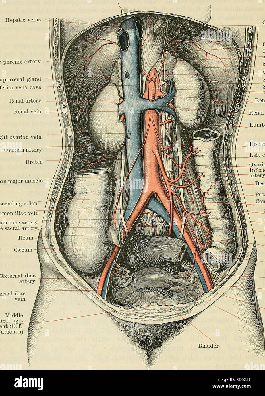
. Cunningham's Text-book of anatomy. Anatomy. 934 THE VASCULAB SYSTEM. From their origins the lumbar arteries pass laterally and posteriorly, on the front and sides of the bodies of the upper four lumbar vertebras, to the intervals between the adjacent transverse processes, beyond which they are continued into the lateral part of the abdominal wall. Each artery lies on the body of the corresponding lumbar vertebra. In its back- Hepatic veins Inferior phrenic artery Suprarenal gland Inferior vena cava Renal artery Renal vein Right ovarian vein Ovarian artery Ureter Psoas major muscle. (Esophagu

Image details
Contributor:
The Book Worm / Alamy Stock PhotoImage ID:
RD5X3TFile size:
7.1 MB (685.6 KB Compressed download)Releases:
Model - no | Property - noDo I need a release?Dimensions:
1338 x 1866 px | 22.7 x 31.6 cm | 8.9 x 12.4 inches | 150dpiMore information:
This image is a public domain image, which means either that copyright has expired in the image or the copyright holder has waived their copyright. Alamy charges you a fee for access to the high resolution copy of the image.
This image could have imperfections as it’s either historical or reportage.
. Cunningham's Text-book of anatomy. Anatomy. 934 THE VASCULAB SYSTEM. From their origins the lumbar arteries pass laterally and posteriorly, on the front and sides of the bodies of the upper four lumbar vertebras, to the intervals between the adjacent transverse processes, beyond which they are continued into the lateral part of the abdominal wall. Each artery lies on the body of the corresponding lumbar vertebra. In its back- Hepatic veins Inferior phrenic artery Suprarenal gland Inferior vena cava Renal artery Renal vein Right ovarian vein Ovarian artery Ureter Psoas major muscle. (Esophagus Crus of diaphragm Inferior phrenic artery Suprarenal gland Coeliac artery Suprarenal vein Superior mesenteric artery Renal artery Renal vein Lumbar arteries Ureter _ Left colic artery _Ovariau artery Inferior mesenteric artery Descending colon Ascending colon Common iliac vein Common iliac artery" Middle sacral artery- External iliac vein Middle umbilical ligaj ment (O.T. urachus) Psoas major muscle Common iliac artery ^- Sigmoid artery Common iliac vein Superior hsemor- rhoidal artery Iliac colon Pelvic colon External iliac -artery ^External iliac vein Uterine tube Uterus Fig. 773.—The Abdominal Aorta and its Branches. ward course, and while still in relation with the vertebral body, it is crossed by the sympathetic trunk, and then, after passing medial to and being protected by the fibrous arches from which the psoas major muscle arises, it runs behind the muscle and the lumbar plexus. The upper two arteries, on each side, also pass posterior to the crura of the diaphragm. Beyond the interval between the trans- verse processes of the vertebras each artery turns laterally and crosses the quadratus lumborum—the last usually passing anterior to, and the others posterior to the muscle; it then pierces the aponeurosis of origin of the trans- versa, and proceeds forwards in the lateral abdominal wall, in the interval between the transversus and internal oblique muscles.