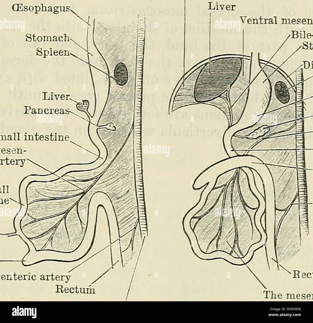
. Cunningham's Text-book of anatomy. Anatomy. DEVELOPMENT OF THE LIVEE AND PANCEEAS. 1255 (Esophagus. Stomach Spleen. Small intestine Superior mesen- teric artery Small intestine Vitelline duct Caecum Inferior mesenteric artery Rectum Ventral mesentery Liver Ventral mesentery Bile-duct Stomach. jDiaphragm Spleen Line crosses mesogastrium Pancreas Superior mesen- teric artery Duodenum Colon Inferior mesen- teric artery The mesentery Aorta Fig. 977.- -Two Diagrams to illustrate the Development of the Intestinal Canal. The figure to the right shows the rotation of the intestinal loop around the s
Image details
Contributor:
The Book Worm / Alamy Stock PhotoImage ID:
RD5HGNFile size:
7.1 MB (358.8 KB Compressed download)Releases:
Model - no | Property - noDo I need a release?Dimensions:
1610 x 1551 px | 27.3 x 26.3 cm | 10.7 x 10.3 inches | 150dpiMore information:
This image is a public domain image, which means either that copyright has expired in the image or the copyright holder has waived their copyright. Alamy charges you a fee for access to the high resolution copy of the image.
This image could have imperfections as it’s either historical or reportage.
. Cunningham's Text-book of anatomy. Anatomy. DEVELOPMENT OF THE LIVEE AND PANCEEAS. 1255 (Esophagus. Stomach Spleen. Small intestine Superior mesen- teric artery Small intestine Vitelline duct Caecum Inferior mesenteric artery Rectum Ventral mesentery Liver Ventral mesentery Bile-duct Stomach. jDiaphragm Spleen Line crosses mesogastrium Pancreas Superior mesen- teric artery Duodenum Colon Inferior mesen- teric artery The mesentery Aorta Fig. 977.- -Two Diagrams to illustrate the Development of the Intestinal Canal. The figure to the right shows the rotation of the intestinal loop around the superior mesenteric artery. In both figures the parts are supposed to be viewed from the left side. constitute the bile-ducts within the liver. Adjacent trabeculse become arranged into the form of a lobule, each with a vascular channel in its interior, which communicates with the vascular network in the surface of the lobule by capillary intervals between adjacent trabecule. The central vein becomes a tributary of a hepatic vein, and the capillary network becomes the terminal distribution of branches of the portal vein. The proximal portion of the original hollow diverticulum becomes the bile-duct, and the gall-bladder and cystic duct are formed by an evagination from it. As the liver increases in size, it begins to pro- ject down from the in- ferior part of the septum transversum into the ventral mesentery, so that now, instead of being situated within the septum, it looks like an appendage of its inferior surface. In other words, the septum begins to differentiate into two parts—an inferior, the liver, and a superior, which constitutes the greater portion of the diaphragm, both of these having been at first one continuous mass. In the course of development the separation of the two becomes more marked, and finally is complete everywhere except at the coronary and lateral ligaments behind, and at the falciform ligament in front, where they are still connected. As the liver se