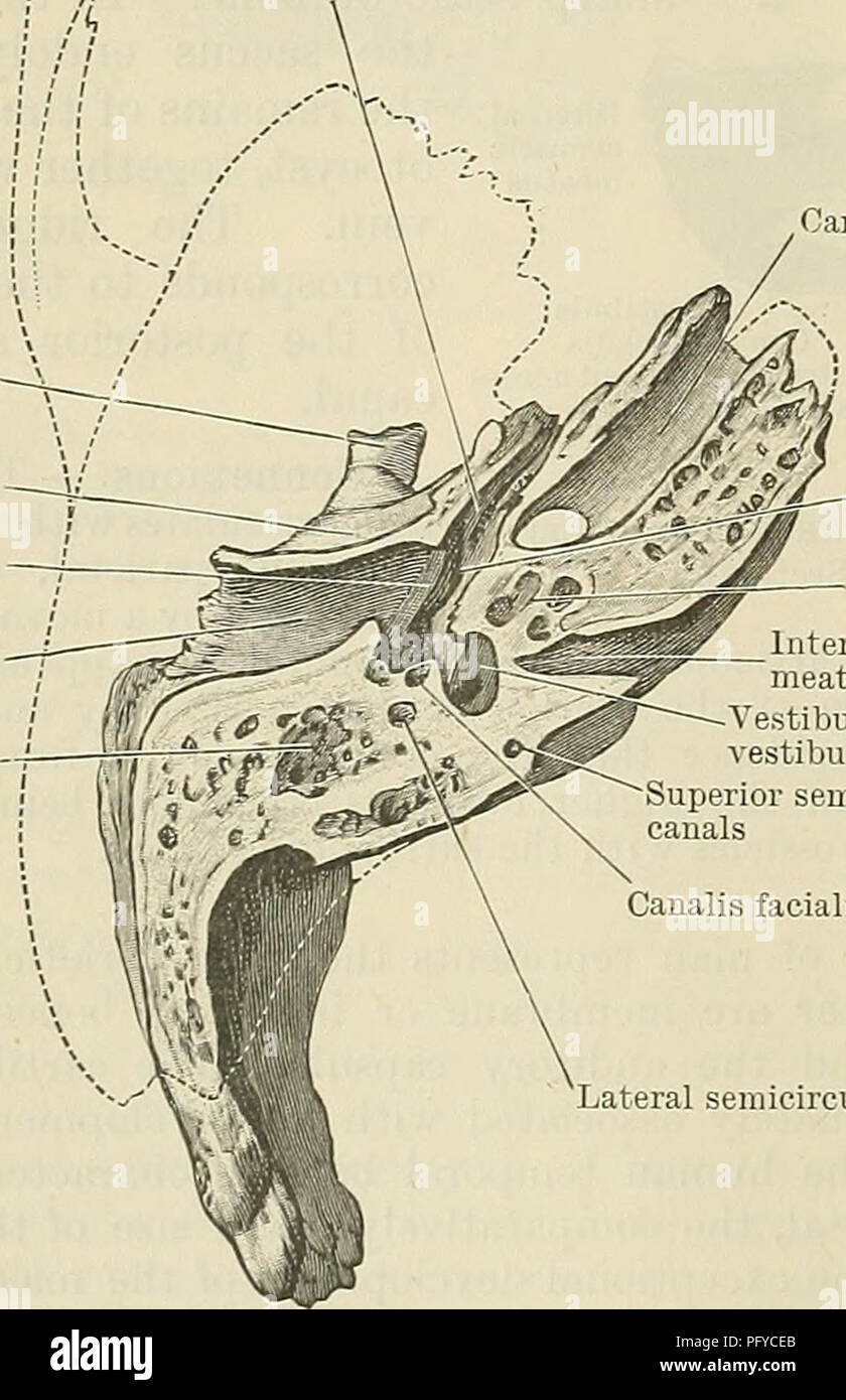
. Cunningham's Text-book of anatomy. Anatomy. Opening leading into tympanic antrum Canalis facialis Canalis stapedii Tympanum External acoustic meatus Fig, 141.—Vertical Transverse Section through the Left Temporal Bone (Posterior Half of Section). A*? Osseous part of tlie auditory tube Styloid process broken off Mandibular fossa Groove for membrana tympani External acoustic meatus Mastoid air-cells. Fig. 142. in the base of the petrous part, and envelop the posterior and lateral semi- circular canals. It is by ex- tension from this part that the mastoid process is ulti- mately developed. The
Image details
Contributor:
Central Historic Books / Alamy Stock PhotoImage ID:
PFYCEBFile size:
7.1 MB (296.7 KB Compressed download)Releases:
Model - no | Property - noDo I need a release?Dimensions:
1271 x 1966 px | 21.5 x 33.3 cm | 8.5 x 13.1 inches | 150dpiMore information:
This image is a public domain image, which means either that copyright has expired in the image or the copyright holder has waived their copyright. Alamy charges you a fee for access to the high resolution copy of the image.
This image could have imperfections as it’s either historical or reportage.
. Cunningham's Text-book of anatomy. Anatomy. Opening leading into tympanic antrum Canalis facialis Canalis stapedii Tympanum External acoustic meatus Fig, 141.—Vertical Transverse Section through the Left Temporal Bone (Posterior Half of Section). A*? Osseous part of tlie auditory tube Styloid process broken off Mandibular fossa Groove for membrana tympani External acoustic meatus Mastoid air-cells. Fig. 142. in the base of the petrous part, and envelop the posterior and lateral semi- circular canals. It is by ex- tension from this part that the mastoid process is ulti- mately developed. The styloid process, an inde- pendent development from the upper end of the carti- lage of the second visceral arch, is ossified from two centres. The upper or basal appears before birth, and rapidly unites with the petro- mastoid, the tympanic plate encircling it in front. This represents the tympanohyal of comparative anatonry. At birth, or subsequent to it, another centre appears in the cartilage below the above: this is the stylohyal. Anky- losis usually occurs in adult life between the tympanohyal and stylohyal, the union of the two constituting the so- called styloid process of human anatomy. The centre from which the squamo - zygomatic de velops appears in membrane about Situated near the root of the zygoma, it extends forwards Carotid canal Tympanum Cochlea Internal acoustic meatus 'estibule, fenestra estibuli cut across Superior semicircular canals Canalis facialis Lateral semicircular canal -Horizontal Section through the Left Temporal Bone (Lower Half of Section). the end of the second month. and laterally into that process, medially to form the floor of the"infra-temporal fossa, and upwards into the squamosal. From this latter there is a downward and backward exten-. Please note that these images are extracted from scanned page images that may have been digitally enhanced for readability - coloration and appearance of these illustrations may not perfectly resembl