. Cunningham's Text-book of anatomy. Anatomy. THE TEMPORAL BONES. 127 bevelled at the expense of its inner table, except in front, where the margin is thick and stout. Here it articulates with the great wing of the sphenoid, its union with that bone extending to near the anterior part of the summit of the curve, behind which it is united to the parietal, overlapping the squamous border of that bone: posteriorly the free margin of the squamous part ends at an angle formed between it and the mastoid process called the incisura parietalis. Pars Tympanica.âThe tympanic part of the temporal bone fo
Image details
Contributor:
The Book Worm / Alamy Stock PhotoImage ID:
RD60Y5File size:
7.1 MB (333.5 KB Compressed download)Releases:
Model - no | Property - noDo I need a release?Dimensions:
2227 x 1122 px | 37.7 x 19 cm | 14.8 x 7.5 inches | 150dpiMore information:
This image is a public domain image, which means either that copyright has expired in the image or the copyright holder has waived their copyright. Alamy charges you a fee for access to the high resolution copy of the image.
This image could have imperfections as it’s either historical or reportage.
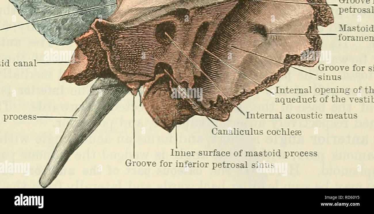
. Cunningham's Text-book of anatomy. Anatomy. THE TEMPORAL BONES. 127 bevelled at the expense of its inner table, except in front, where the margin is thick and stout. Here it articulates with the great wing of the sphenoid, its union with that bone extending to near the anterior part of the summit of the curve, behind which it is united to the parietal, overlapping the squamous border of that bone: posteriorly the free margin of the squamous part ends at an angle formed between it and the mastoid process called the incisura parietalis. Pars Tympanica.âThe tympanic part of the temporal bone forms the anterior, lower, and part of the posterior wall of the external acoustic meatus. Bounded in front and above by the petrotympanic fissure, it forms the posterior wall of the non-articular part of the mandibular fossa. Fused medially with the petrous part, its lower edge, sharp and weU defined medially, splits to enclose the root of the projecting styloid process, and is hence called the vagina processus styloidei (sheath of the styloid process). Laterally it unites with the anterior part of the A/ V r Groove for middle k meningeal artery "â ^fj Arcuate eminence or eminence of superior / semicircular canal Zy<, â¢Parietal notch Petro-squamous suture Carotid canal. Styloid process .SS^, ^ Groove for superior etrosal sinus Groove for sigmoid sinus internal opening of the iqueduct of the vestibule Internal acoustic meatus Canaliculus cochleae Inner surface of mastoid process Groove for inferior petrosal sinus Fig. 137.âThe Right Temporal Bone (Cerebral aspect). The squamo-zygomatic part is coloured blue ; the petro-mastoid part, red. The styloid process is left uncoloured. mastoid process, and higher up with the descending process of the squamous part, from both of which it is separated by the tympano-mastoid fissure, through which the auricular branch of the vagus escapes. Its free border, which forms the anterior, lower, and part of the posterior border of the