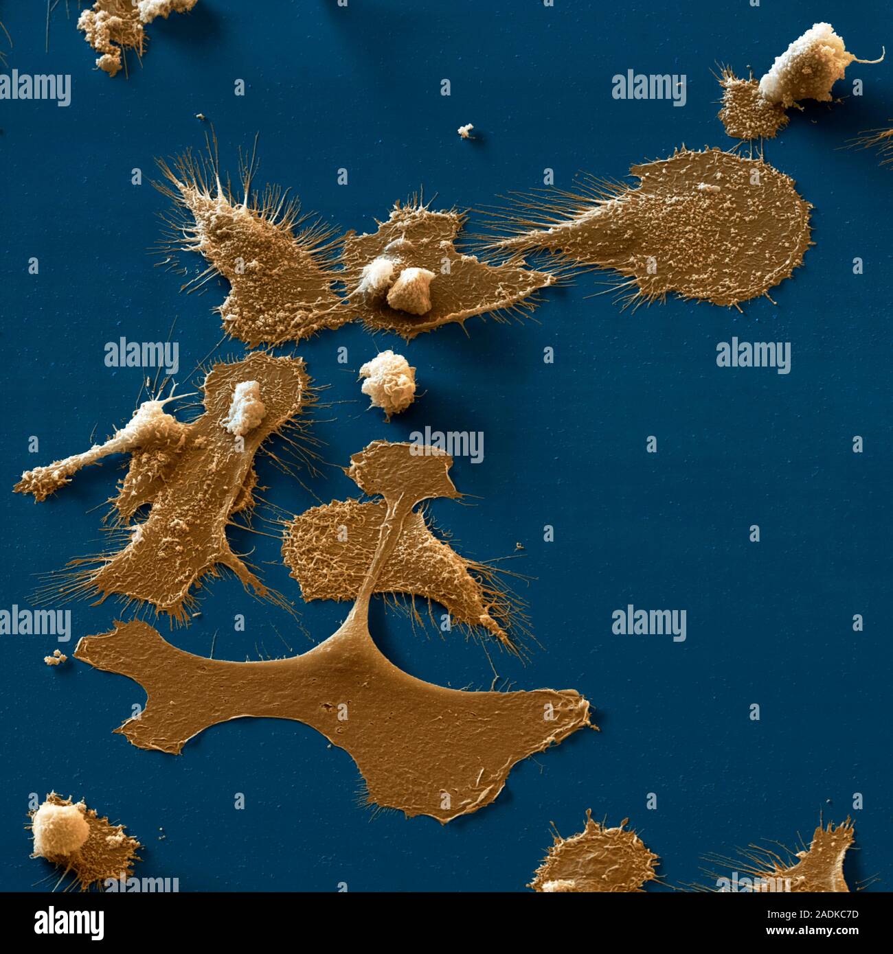
Dendritic cells. Coloured scanning electron micrograph (SEM) of dendritic cells, part of the human immune system. These are macrophage cells that are
RMID:Image ID:2ADKC7D
Image details
Contributor:
Science Photo Library / Alamy Stock PhotoImage ID:
2ADKC7DFile size:
45.8 MB (1.4 MB Compressed download)Releases:
Model - no | Property - noDo I need a release?Dimensions:
4000 x 4000 px | 33.9 x 33.9 cm | 13.3 x 13.3 inches | 300dpiDate taken:
18 May 2004Photographer:
EYE OF SCIENCE/SCIENCE PHOTO LIBRARYMore information:
Dendritic cells. Coloured scanning electron micrograph (SEM) of dendritic cells, part of the human immune system. These are macrophage cells that are found in the body's tissues. The long projections seen on the cells' surface help them to move. The cells recognise and engulf foreign cells. They may then display the foreign antigen as a warning of the infection to other cells. Dendritic cells found in the upper layer of the skin (the epidermis) are known as histiocytes or Langerhans cells. In the central nervous system dendritic cells are known as microglia, and in the liver as Kupffer cells. Magnification: x675 when printed 10 cm wide.