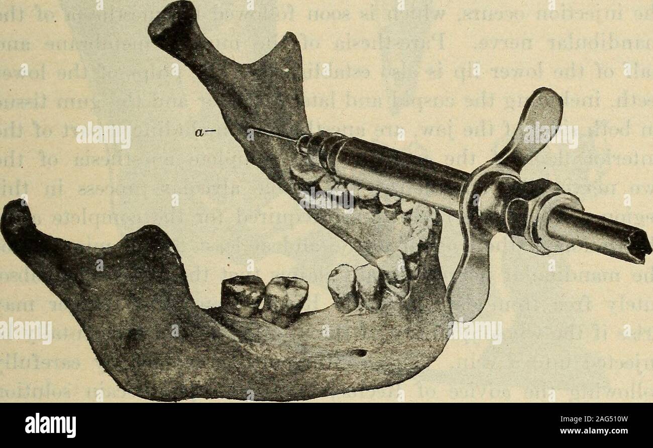
. Dental materia medica and therapeutics; with special reference to the rational application of remedial measures to dental diseases ... is-tinct bony ridge and covered with mucous membrane. As thereis no anatomic name attached to this space, Braun has called itthe retromolar triangle (trigonum retromolare). In the closedmouth it is located at the side of the upper third molar, and inthe open mouth it is found midway between the upper and lowerteeth. Immediately back of the mesial border of this triangle, TECHNIQUE OF THE INJECTION. 485 directly beneath the mucous membrane, lies the lingual ne

Image details
Contributor:
The Reading Room / Alamy Stock PhotoImage ID:
2AG510WFile size:
7.1 MB (433.8 KB Compressed download)Releases:
Model - no | Property - noDo I need a release?Dimensions:
2012 x 1242 px | 34.1 x 21 cm | 13.4 x 8.3 inches | 150dpiMore information:
This image is a public domain image, which means either that copyright has expired in the image or the copyright holder has waived their copyright. Alamy charges you a fee for access to the high resolution copy of the image.
This image could have imperfections as it’s either historical or reportage.
. Dental materia medica and therapeutics; with special reference to the rational application of remedial measures to dental diseases ... is-tinct bony ridge and covered with mucous membrane. As thereis no anatomic name attached to this space, Braun has called itthe retromolar triangle (trigonum retromolare). In the closedmouth it is located at the side of the upper third molar, and inthe open mouth it is found midway between the upper and lowerteeth. Immediately back of the mesial border of this triangle, TECHNIQUE OF THE INJECTION. 485 directly beneath the mucous membrane, lies the lingual nerveand about three-eighths of an inch farther back the mandibularnerve is to be found. This last nerve lies close to the bone, andenters into the mandibular foramen, which is partially coveredby the mandibular spine. Before starting the injection the patient should be cautionedto rest his head quietly on the headrest of the chair, as any sud-den movement or interference with the hand of the operator maybe the cause of breaking the needle in the tissues. The syringe, provided with a one-inch needle, is held in a horizontal position, . Figure 93.Position of Needle for Injecting- the Mandibular Nerve. resting on the occluding surfaces of the teeth from the cuspidbackward and slightly toward the median line. The needle is tobe inserted three-eighths of an inch above and the same distanceback of the occluding surface of the third lower molar, the needleopening facing the bone. This position will insure the correctdirection of the needle point so as to reach the tissues imme-diately surrounding the nerves, and not lose the injection in theadjacent thick muscle tissue. The needle must always be in closetouch with the bone, and is now slowly pushed forward, deposit-ing a few drops of fluid on its way until the ridge (Figure 93, a) 486 LOCAL ANESTHESIA. is reached. About five drops of fluid are injected in this, imme-diate neighborhood for the purpose of anesthetizing the lingualnerve. Th