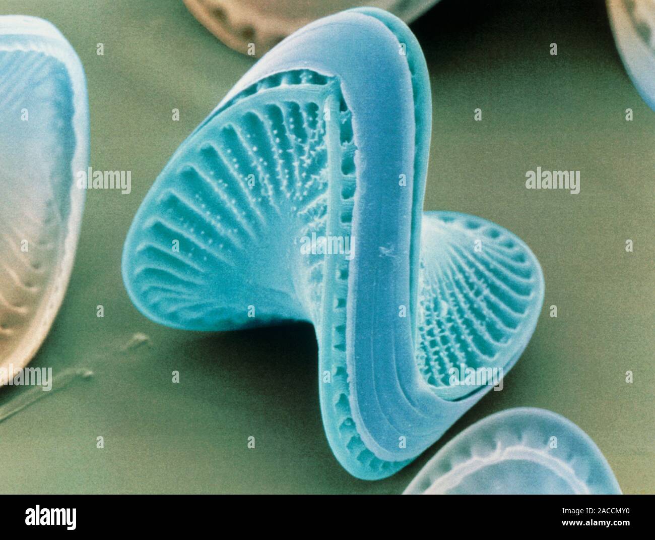
Diatoms. Coloured scanning electron micrograph of the diatom Campylodiscus hibernicus. The diatoms are a distinctive group of single-celled algae cont
Image details
Contributor:
Science Photo Library / Alamy Stock PhotoImage ID:
2ACCMY0File size:
27.1 MB (1.1 MB Compressed download)Releases:
Model - no | Property - noDo I need a release?Dimensions:
3543 x 2674 px | 30 x 22.6 cm | 11.8 x 8.9 inches | 300dpiDate taken:
29 March 1995Photographer:
POWER AND SYRED/SCIENCE PHOTO LIBRARYMore information:
Diatoms. Coloured scanning electron micrograph of the diatom Campylodiscus hibernicus. The diatoms are a distinctive group of single-celled algae containing about 10, 000 species. They form a large part of the plankton at the base of the marine and freshwater food chains. The characteristic feature of diatoms is their intricately patterned, glass- like cell wall, or frustule, which is impregnated with silica. The frustule consists of 2 halves which fit together like the lid & bottom of a box. It is often decorated with rows of tiny holes, known as striae, which are arranged either radially or on the 2 sides of a central axis. Magnification: x500 at 5x7cm size. x1475 at 8x10ins